My Boards
View Pictures
  • My Boards

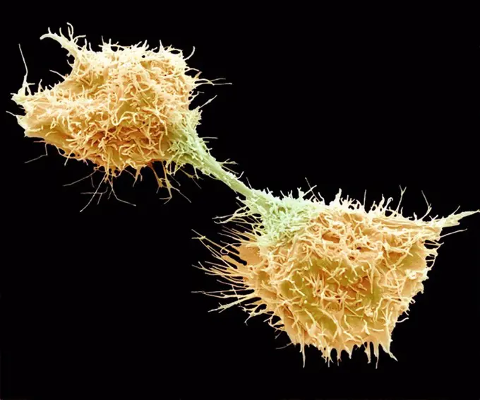
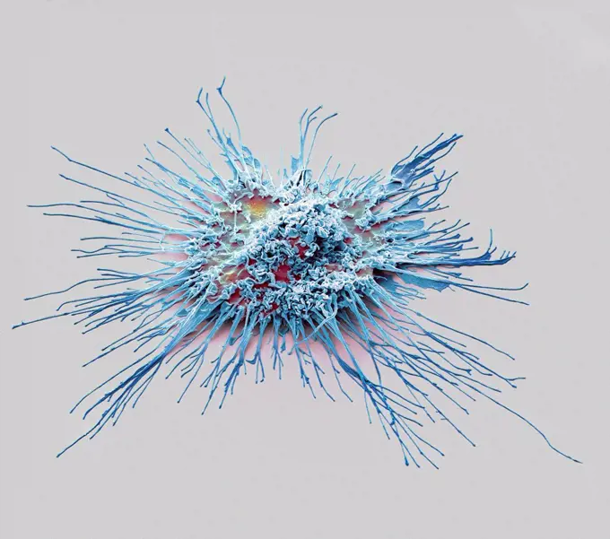
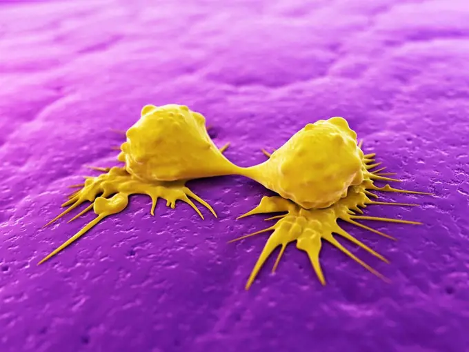
Cellular Biology Illustrations

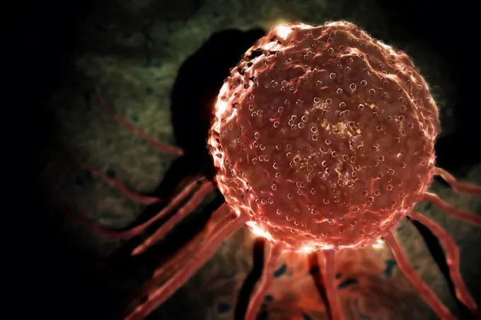
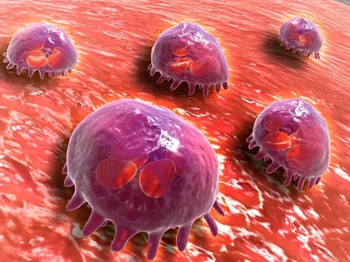
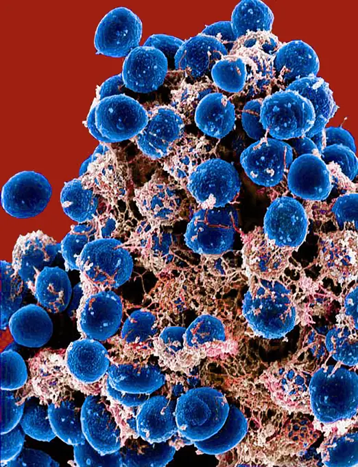
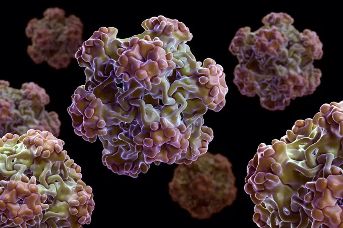
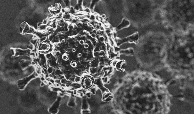
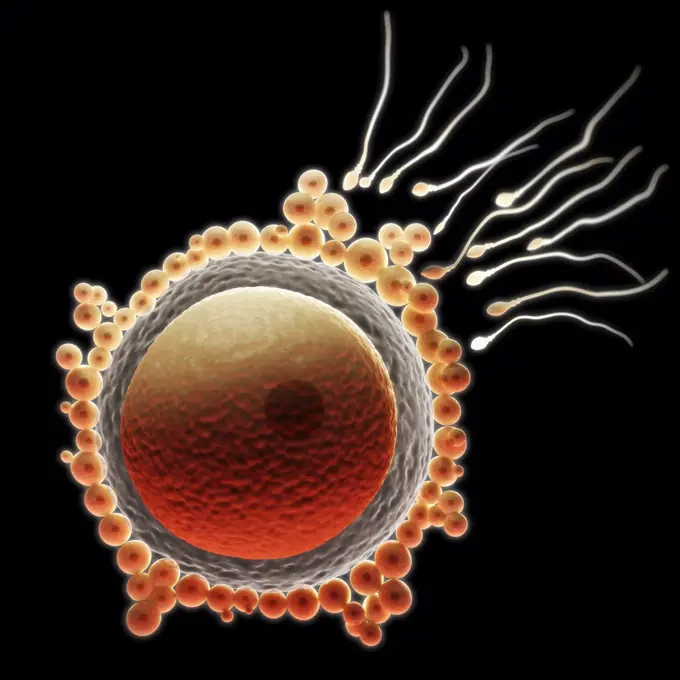
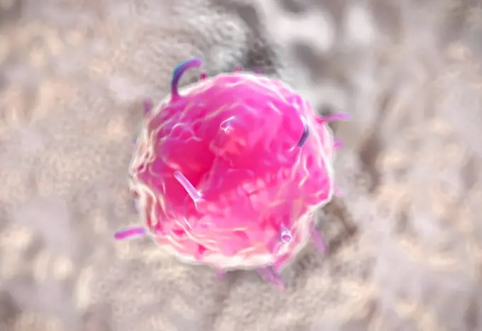
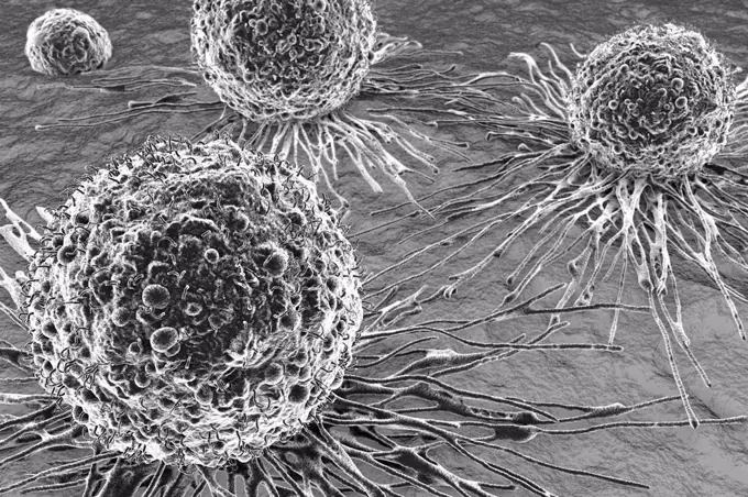
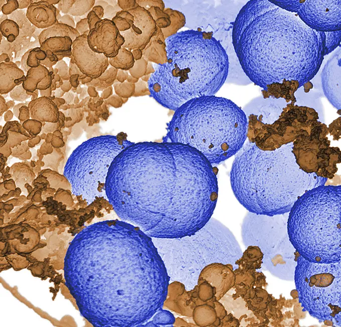
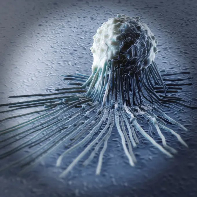
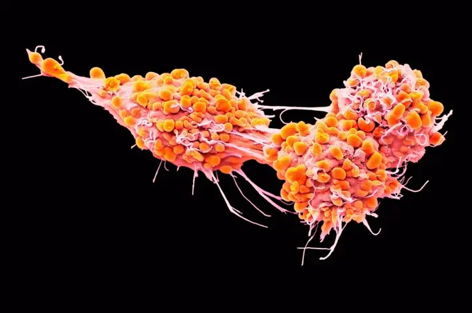
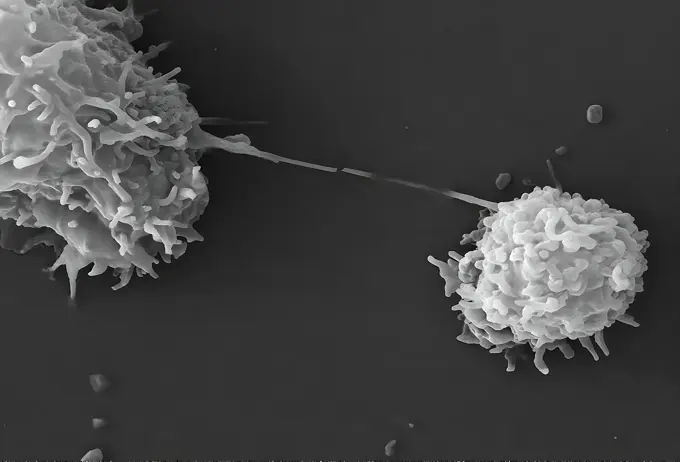
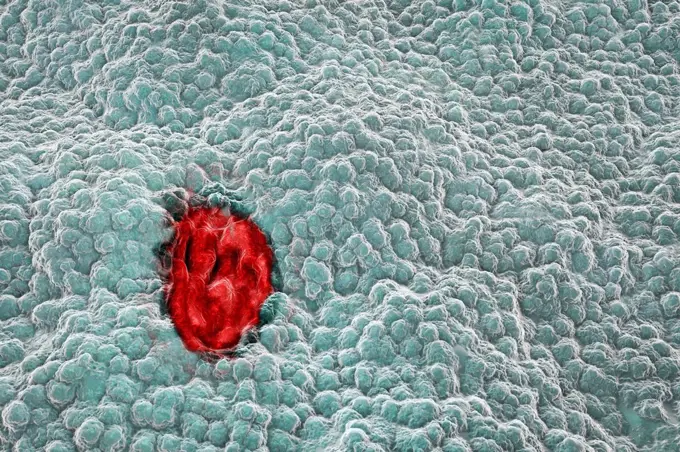
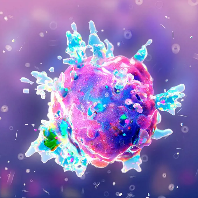
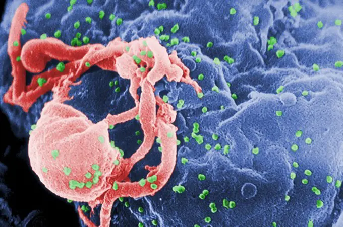
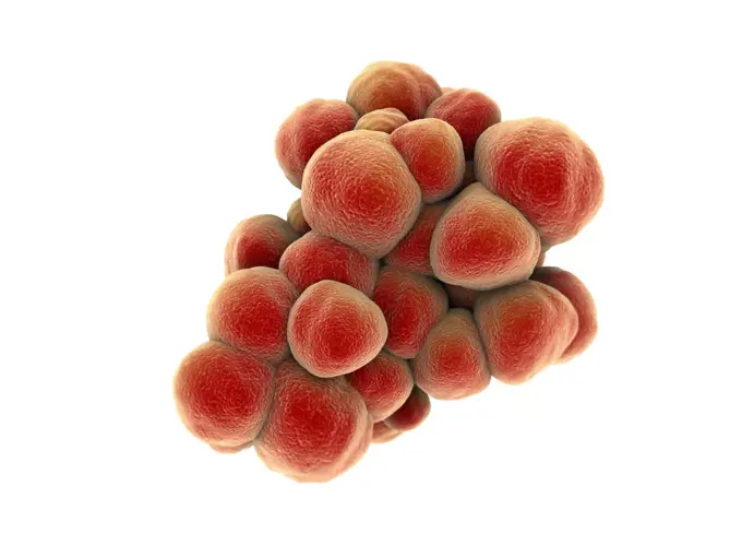
Close-up illustrations of various cells, including cancer cells and sperm, highlighting biological processes and disease mechanisms.

Embryonic Stem Cell
Embryonic Stem Cell
216 assets in this story
4128-30418603
Cancer cell, illustration
4128-16073760
SARS-CoV-2 viruses and immune cell, illustration
4128R-13575637
Stem cells, computer illustration.
4128R-11286455
Fertilisation. Computer artwork of a sperm fertilising an egg cell.
4128R-15516257
Illustration of white blood cells attacking a cancer cell
4128R-11310371
Superbug bacteria, computer artwork.
1815-18053609
3D Rendered Illustration coronavirus visualisation
4128R-15405934
T-lymphocytes attacking cancer cell, illustration
4378-4515
Giardia lamblia Parasites
4378-989
Microscopic styled view of a stem cell.
4128R-11312985
Swine vesicular disease virus, computer artwork.
4128-20043007
T cell attacking cancer cell, illustration
4239R-8386
Microscopic view of phagocytic macrophages, which are involved in the immune response within the body.
824-63194642
Scanning electron micrograph of a clump of Staphylococcus epidermidis bacteria in the extracellular matrix, which connects cells and tissue.
4378-3048
Structure of small virus-like particles assembled from the L1 protein of human papillomavirus 16 (PDB 1DZl) from the family Papillomaviridae.
4269-27699
Macrophage. Scanning electron micrograph (SEM) of a macrophage.
4128-30418602
Cancer cell, illustration
4128R-11543432
Virus, conceptual illustration.
4128-30418599
Cancer cell, illustration
4048-16067041
Novel Coronavirus SARS-CoV-2, causes 'coronavirus disease-2019', abbreviated as COVID-19. A transmission electron microscope image shows the virus isolated from a US patient, emerging from surface of cells cultured in the lab. When viewed by an electron microscope, the spikes on the outside of the virus capsid appear as a yellow corona around the virion. Colors are artificially assigned to a monotone image for clarity. Image by National Institute of Allergy and Infectious Diseases (NIAID)  (BSLOC_2020_2_247)
4292-76694
Leucocyte or white blood cells
4378-3021
Close up view of a single breast cancer cell.
4128-V58569148
4128R-13587466
Aspergillus fungus, computer illustration. This is a toxic fungus that causes diseases in humans. These include fungal ear, lung and skin infections (...
4378-2402
Close up view of a single breast cancer cell.
4128R-3299
Leukaemia blood cells, SEM
4128-15750967
Coronavirus particles, illustration
4128-111494849
Neisseria gonorrhoeae bacteria, illustration.
1899-65662491
Scanning electron micrograph of a cell (purple) infected with the Omicron strain of SARS-CoV-2 virus particles (orange), isolated from a patient sample.
4128R-14058084
T lymphocyte and cancer cell
4378-1795
Zygote development after 24-36 hours.
4128R-11544460
Cancer cells, computer illustration.
824-63219654
This transmission electron microscope image shows SARS-Cov-2 also known as 2019-nCoV, the virus that causes COVID-19 isolated from a patient in the United States. Virus particles emerge from the surface of cells grown in the laboratory. The spikes on the outer edge of the virus particles form a crown and give their name coronavirus.
4378-1871
Neisseria gonorrhoeae, the bacterium responsible for the sexually transmitted infection gonorrhea.
4128-19054231
Flu virus, illustration
4128-30418597
Cancer cell, illustration
4128-48286035
Immune response to cancer, illustration
4239R-20483457
Microscopic view of cancer virus.
4128R-11478358
Cancer cell, conceptual artwork.
1574R-018977
Cancer Cells
4128-111576783
Prostate cancer cell, Coloured scanning electron micrograph (SEM). of the surface of prostate cancer cells. The cells shows numerous processes and mic...
4128-V58557902
4128R-13282648
Chlamydia bacteria in a cell. Computer illustration showing an inclusion composed of a group of chlamydia (red) near the nucleus (violet) of a cell.
4128R-14181749
Dendritic cell, illustration
4128-18573566
Blood clot, illustration
824-63191253
Scanning electron micrograph of HIV particles infecting a human T cell.
4128-18800690
COVID-19 virus and activated platelets, illustration
4128R-20708
Cancer cells dividing, artwork
4128-24795780
Nematode worm eggs, illustration
4128R-13575543
Contraception, Conceptual illustration showing an ovum being protected from spermatozoans by a condom.
4128R-15516224
Illustration of a cancer cell
4128-20044104
Chimeric antigen receptor (CAR) therapy, illustration
4128-111584782
Osteoblast bone cell. Coloured scanning electron micrograph (SEM) of an osteoblast bone cell. Osteocytes are osteoblasts (bone-producing cells) that h...
4239R-20483454
Conceptual image of cancer virus.
4128-V58574838
4417-16039629
Illustration of the Corona Virus Variation Scene 02
4128-V58557944
4378-1880
Fertilization between egg and sperm.
4128R-13725888
Computer illustration of influenza viruses coated with antibodies (white). Antibodies bind to specific antigens, for instance viral proteins, marking ...
4128R-12577784
Embryonic stem cell, computer illustration.
4378-1017
Microscopic styled view of a stem cell.
4128R-14057765
Cancer cells, illustration
4128-30422496
Pancreatic cancer, illustration
4128R-11313466
Venezuelan haemorrhagic fever virus, computer artwork.
824-63194637
Scanning electron micrograph of S. aureus bacteria escaping destruction by human white blood cells.
4128-V58569346
4378-3811
A close up of a macrophage cell. Macrophages are phagocytes and their function is involved in both innate immunity as well as adaptive immunity.
4128R-15466814
Ovarian cancer cells, sem
824-57659317
This scanning electron microscope (SEM) image revealed two Acanthamoeba polyphaga protozoa, as they interacted through their pseudopodia, projecting from the surfaces of these organisms. These pseudopodia allow the amoebae to move, to grasp objects in their environment and, in this case, to communicate with each other. CDC/Catherine Armbruster Margaret Williams 2009
4128-V58562701
4128R-11473185
White blood cells attacking a cancer cell, computer artwork.
4128R-13587543
Stomach lining, computer illustration. The stomach is a muscular sac involved in storage and digestion of food. Here, the inner mucosa layer of the ce...
4128-15665308
Spleen tumour, illustration
4128R-11474108
Mimivirus, computer artwork.
4128-16073934
T-cell attaching to cancer cell, illustration
4128R-15465083
Ovarian cancer cells, sem
824-63225845
Colorized scanning electron micrograph of an apoptotic cell (green) infected with SARS-COV-2 virus particles (blue), isolated from a patient sample. Image captured at the NIAID Integrated Research Facility (IRF) in Fort Detrick, Maryland.
4128-28968446
Cancer cell, illustration
4128-28968419
Melanoma cells, illustration
4128R-13587545
Gastric (stomach) ulcer, illustration. Gastric, or peptic, ulcers are erosions of the mucosal lining of the stomach.
1525-19813502
cancer cell made in 3d software
4128-111495110
Cancer cell, illustration.
4128-20043225
Cell udergoing lysis process, illustration
4128R-15465072
Breast cancer cells, SEM
1525-56722635
The COVID-19 Inside Virus Structure 3D rendering. The COVID-19 Inside Virus Structure
4297-1229
Scanning electron micrograph of HIV-1 budding from cultured lymphocyte
4239R-20483592
Microscopic view of tumor.
4239R-8042
Cluster of blastomeres forming a developing morula (early stage of embryonic development).
4128R-15516230
Illustration of a cancer cell
4128R-14426
Cancer cells, computer artwork. The uneven surface and cytoplasmic projections are characteristic of cancer cells.
4128R-15557
Lymphocyte white blood cell, coloured scanning electron micrograph SEM. Its surface is covered in characteristic microvilli. Magnification: x6000 when...
4128R-11287093
Buckyballs, computer artwork.
1899-53513320
Ebola virus isolated in November 2014 from patient blood samples obtained in Mali. The virus was isolated on Vero cells.
4128R-13326006
Leucocyte attacking bacteria, illustration.
4128-V58566809
4128R-5448
Lymphoma lymph cell, SEM
4128R-5857
Dividing fibrosarcoma cells. Coloured scanning electron micrograph of fibrosarcoma fibroblastic sarcoma cells in the late telophase stage of mitosis. ...
4128R-13047368
Dendritic cell. Coloured scanning electron micrograph (SEM) of a protective cell of the human immune system known as a dendritic cell. Dendritic cells...
4128R-20546
Cancer cells dividing, artwork
4128-30420559
Marburg virus, illustration
PREVIOUS
of 3
NEXT
2401 S. Ervay, Suite 206
Dallas, Texas 75215
United States
Get Started
Free ResearchMy BoardsMy Cart
For Creators
How To License Your ContentContributor PortalFrame of Mind
Resources
API accessPricing
Contact
+1 866 236 0087help@viewpictures.co.uk Contact form
©2026 View Pictures. All Rights Reserved. -A
This site is protected by reCAPTCHA and Google Privacy Policy and Terms of Service apply.