My Boards
View Pictures
  • My Boards

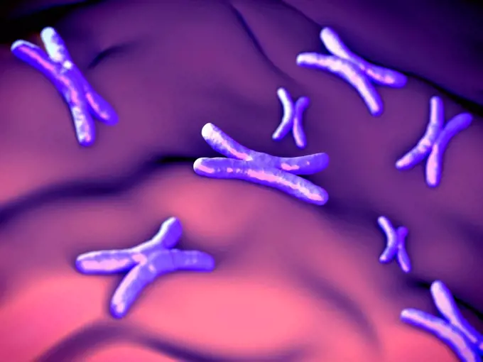
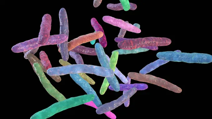
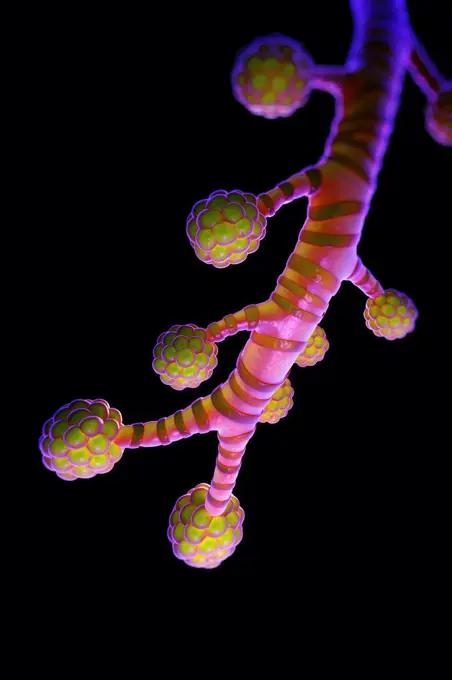
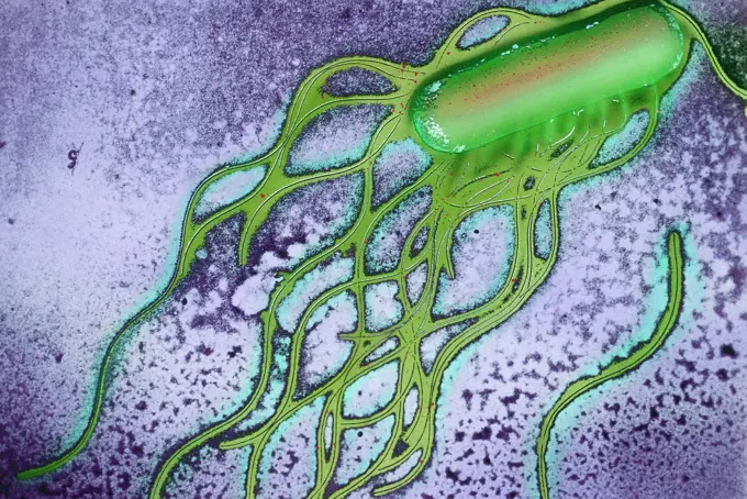
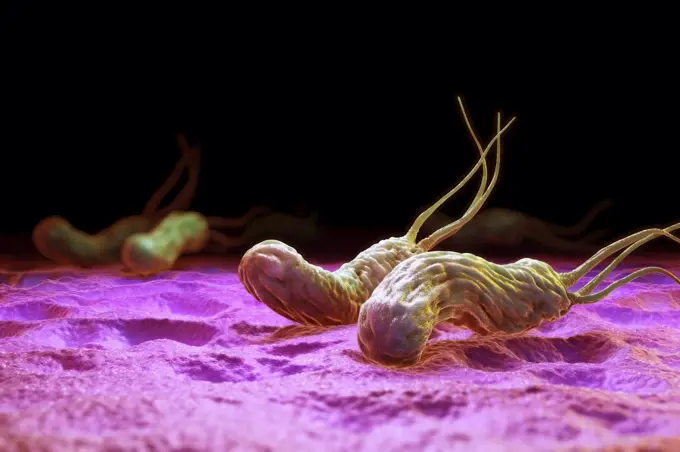
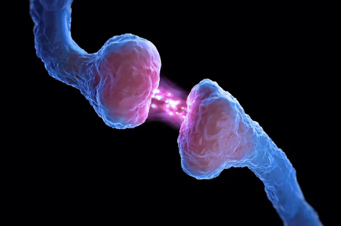
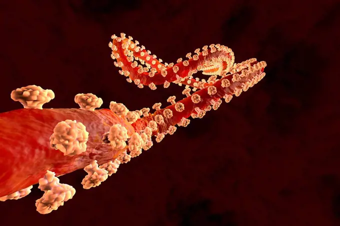
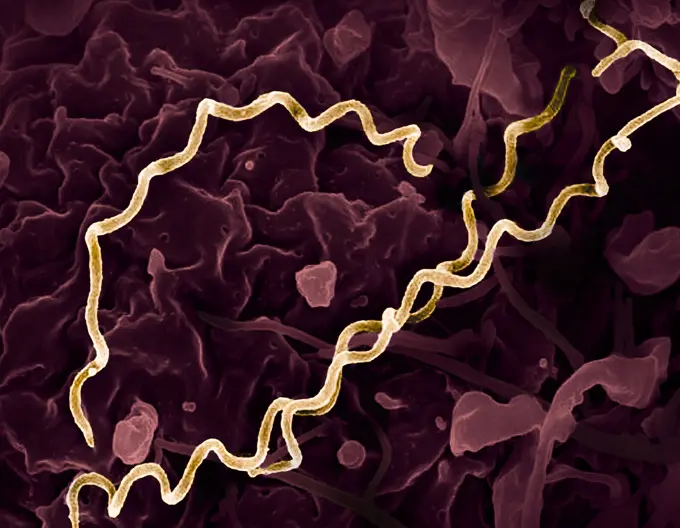
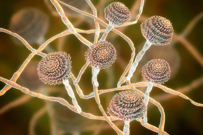
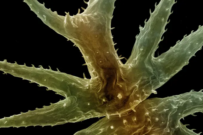
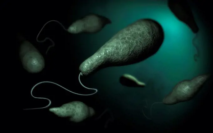
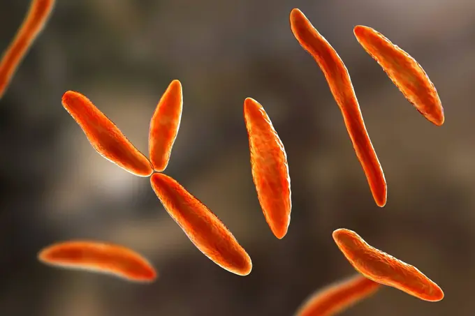
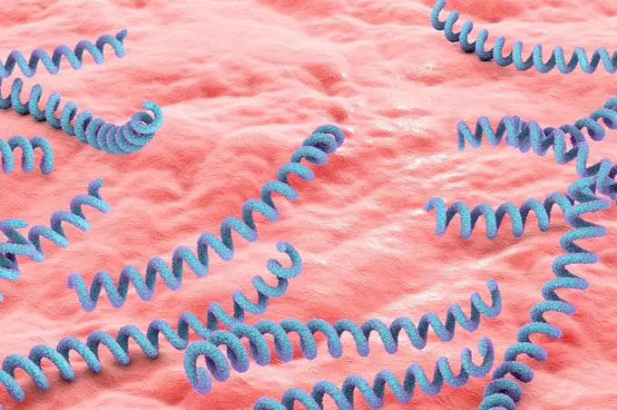
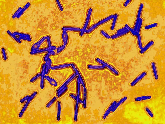
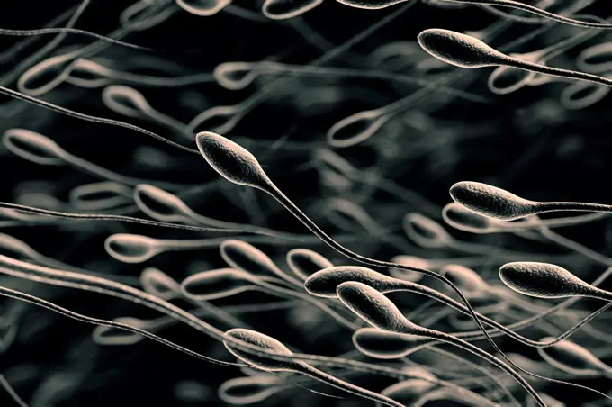
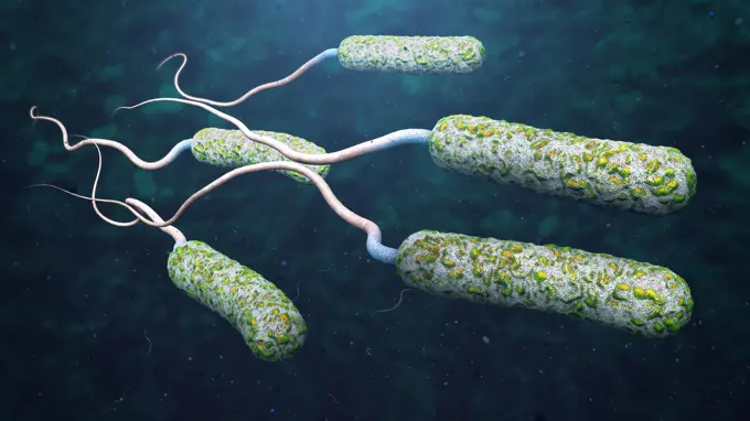
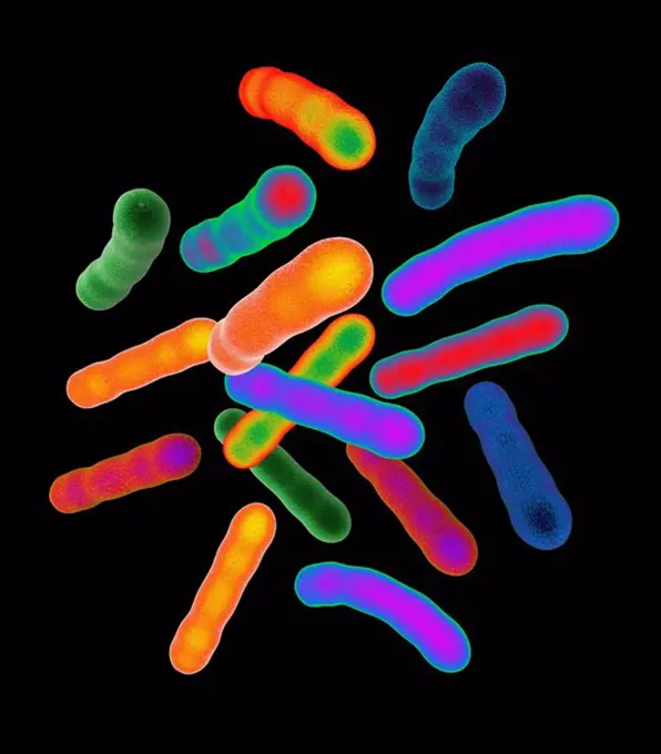
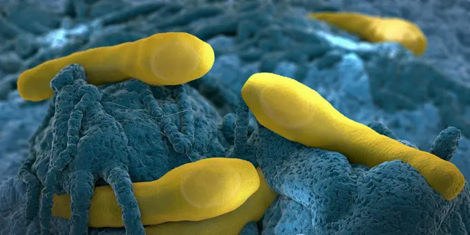
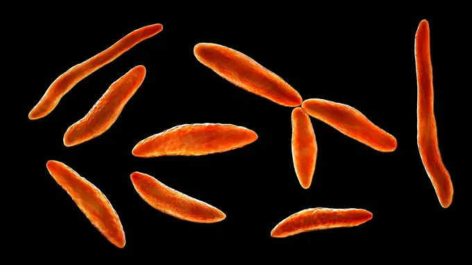
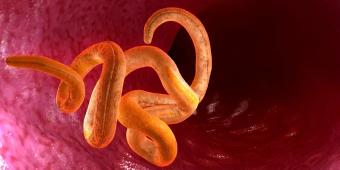
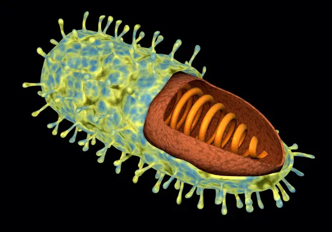
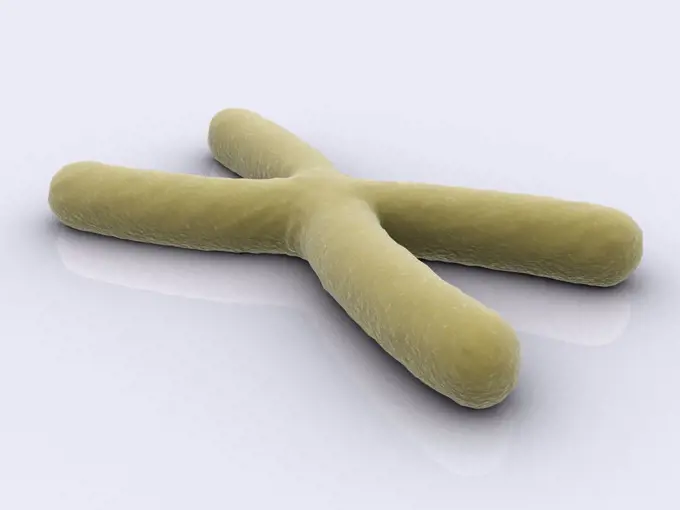
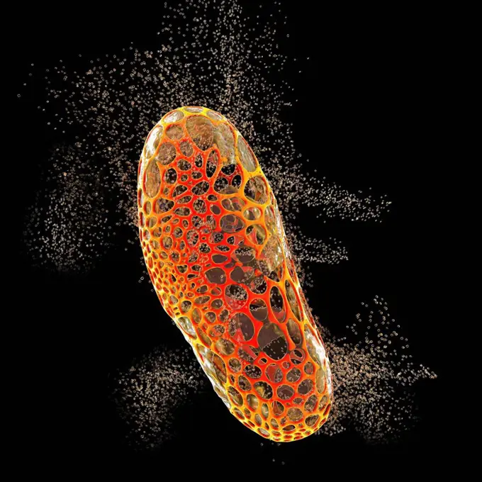
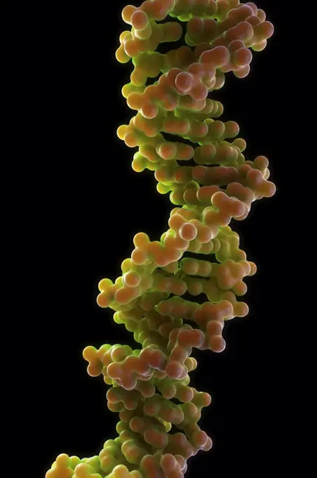
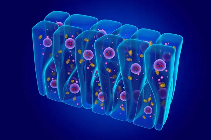
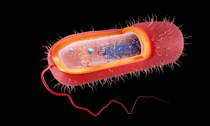
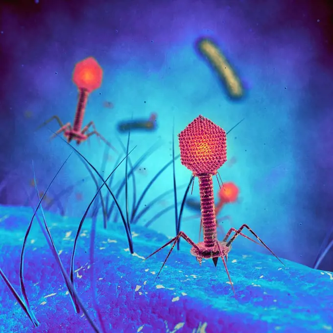
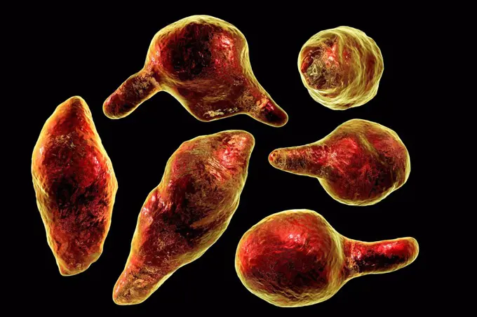
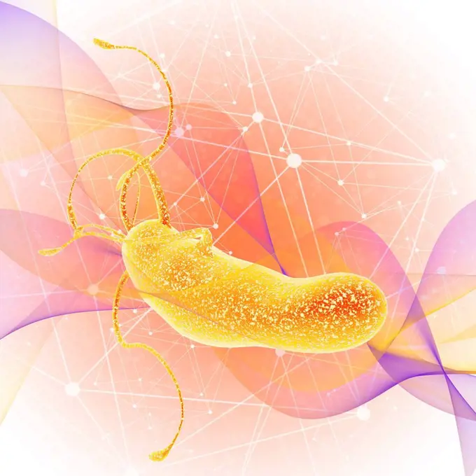
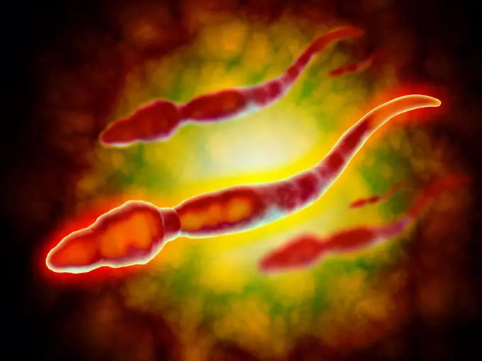
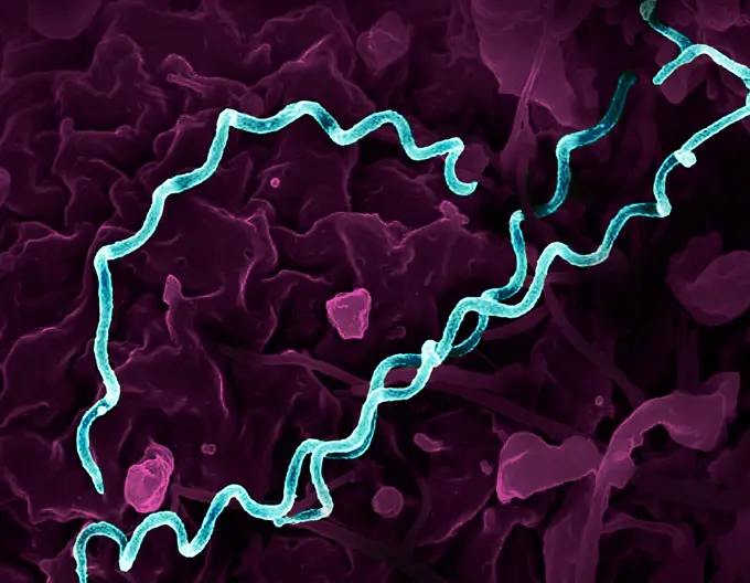
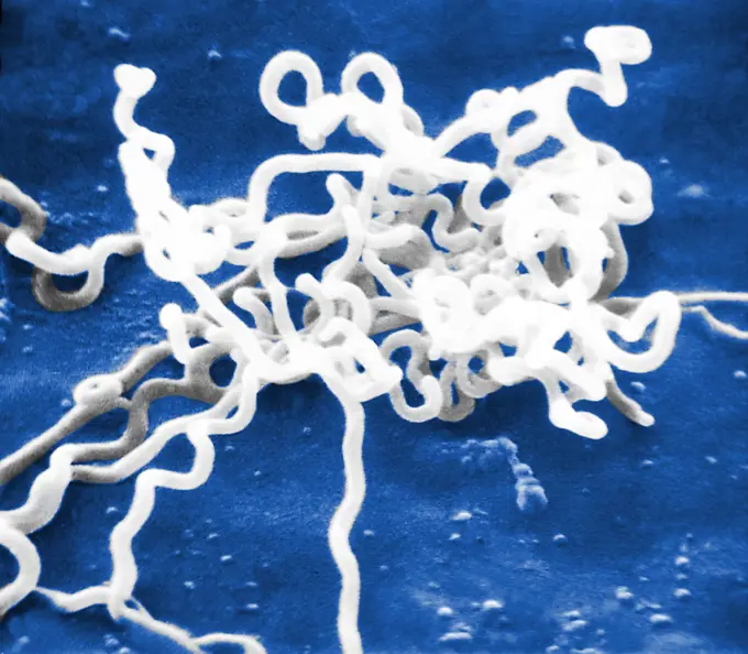
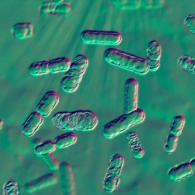
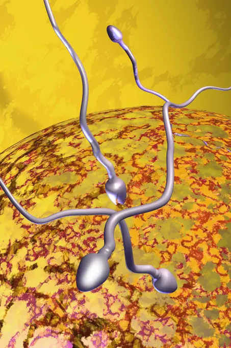
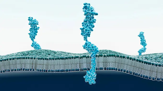
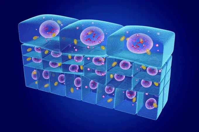
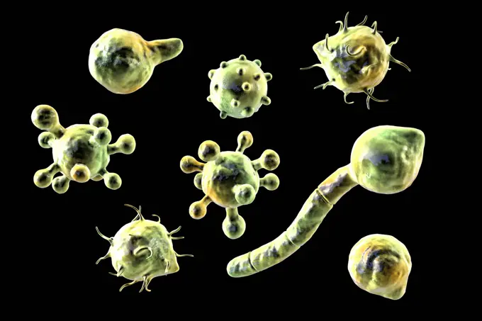
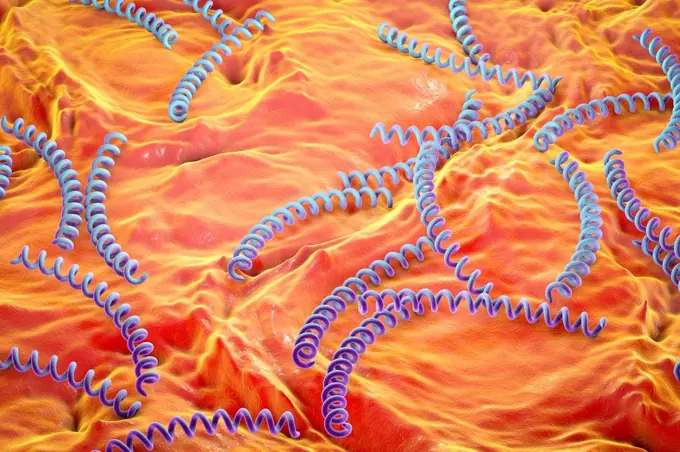
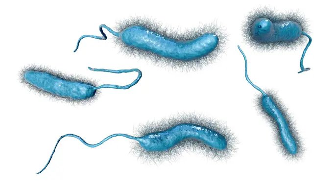
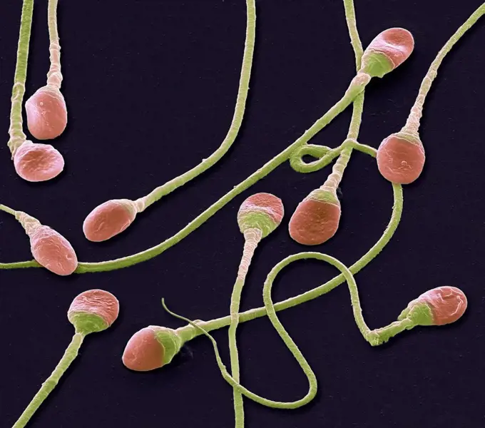
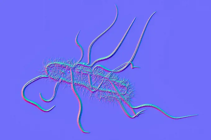
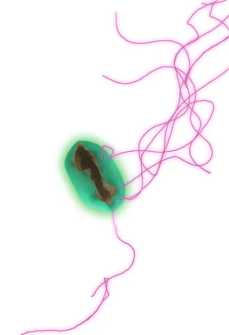
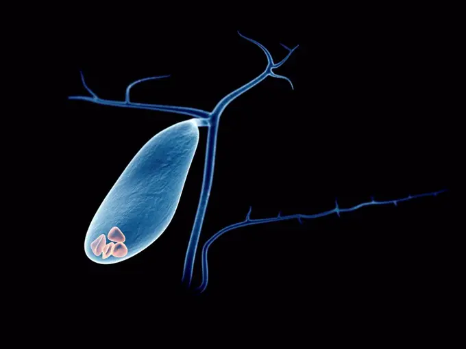
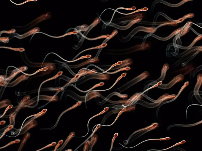
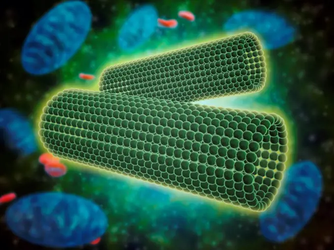
Fungi and Viruses Illustration

Detailed illustrations of various fungi and viruses, highlighting their structures and forms with vibrant, colorful backgrounds.

Chromosomes, illustration.
228 assets in this story
4128-20042948
Bacteria, illustration
4128-28575411
Mycobacterium bovis bacteria, illustration
4239-V53647210
4378-4606
Bronchioles
824-63127922
Marburg virus viewed under transmission electron micrograph.
4269-7071
Transmission electron micrograph (TEM) of Salmonella sp. When ingested with spoiled food, the Gram-negative bacteria of the genus Salmonella are responsible for salmonellosis, a small intestine infection (gastroenteritis).
4128R-5474
DNA molecule, artwork
824-57660013
Borrelia - pathogenic bacteria (Spirochaetota) responsible for Lyme disease, (Borrelia burgdorferi, Borrelia spielmanii, Borrelia garinii and Borrelia afzelii give other variants of Lyme disease).
4378-4596
Helicobacter pylori
4378-1167
Stylized view of strands of human DNA or deoxyribonucleic acid.
4378-5728
Synapse
824-65830869
Colorized transmission electron micrograph of HIV-1 virus particles (purple) budding and replicating from an H9 T cell (green). The virus particles are in various stages of maturity, which accounts for differences in shape.
824-63178988
Diphtheria bacilli (Corynebacterium diphtheriae or Klebs-Loeffler bacillus). Diphtheria is a contagious infectious disease. Localization of bacilli on the larynx can cause asphyxia (croup). This bacillus produces toxins that can lead to paralysis or even death of the patient by heart attack. Optical microscopy X 1850
4378-4438
Helicobacter pylori
1815-111459860
3D rendered illustration of an Ebola virus
4269-27243
Syphilis. Light micrograph of a Treponema pallidum. This spirochaete bacterium is the agent of syphilis.
1899-65662363
Colorized electron micrograph of Treponema pallidum, the bacteria that cause syphilis. Several spiral-shaped bacteria have been highlighted in gold.
4128-V58614790
4128R-15366400
Aspergillus fumigatus fungus, illustration
1899-65662482
Scanning electron micrograph of Mycobacterium tuberculosis particles (colorized red/yellow), the bacterium which causes TB.
824-65830987
Scanning electron micrograph of Mycobacterium tuberculosis particles (colorized red/yellow), the bacterium which causes TB.
4201-66365
SEM close-up view of sponge sclerites at 210x magnification
4220-21334539
Scanning Electron Micrograph (SEM): Freshwater Desmid, Micrasterias lux.     Date:
4128R-34251
Fallopian tube. Light micrograph of a cross-section through a fallopian tube. The lumen (white) is seen, along with mucosal folds lined with simple co...
4239R-8434
Conceptual image of Euglena.
4128-28767342
Corn smut fungus, illustration
4128R-13446643
Computer illustration of Treponema pallidum, the bacterium which causes syphilis.
4128-19053753
Penicillium, illustration
824-63178986
Clostridium botulinum bacterium (botulism bacillus). This bacterium secretes a very powerful toxin that inhibits the motor neurons of muscle contraction. This toxin (botox), in localized injection, reduces wrinkles, by paralysis of the muscles responsible for the wrinkle. Optical microscopy X 2000
4128-V58567875
4378-5456
Sperm
6188-66486162
3D illustration of cholera pathogens in dark, dirty water 3D illustration of cholera pathogens in dark dirty water
1760-4872
Canada, BC, Clown shrimp (Lebbeus grandimanus) on a crimson anemone C1966/nCribrinopsis fernaldi)
4128R-15290390
Lyme disease bacteria, illustration
4128R-10091
Tuberculosis bacteria, artwork
4128-48285282
Neuroblastoma cancer cells, illustration
6188-66486441
Clostridium botulism Erreger wachsen auf organischem Gewebe - 3d Illustration Clostridium botulism pathogens growing on organic tissue - 3d illustration Copyright: xZoonar.com/ChristophxBurgstedtx 22603191
824-63223121
Scanning electron micrograph of Borrelia hermsii, the causative agent of relapsing fever, interacting with red blood cells. Credit: NIAID.
4128R-12699858
Coloured scanning electron micrograph (SEM) of fungal cells. The round structures are sporangia, which house the fungus' reproductive cells (spores). ...
4128-28767340
Corn smut fungus, illustration
824-65831014
Colorized transmission electron micrograph of mature extracellular Nipah virus particles (red) near the periphery of an infected VERO cell (purple).
6188-65564752
Einzelne verdrehte Nematode innerhalb Darm auf einem schwarzen Hintergrund - 3D-Illustration Single twisted nematode inside intestine on a black background - 3d illustration Copyright: xZoonar.com/ChristophxBurgstedtx 22516006
4128-20040405
Fragile X chromosome, illustration
4239R-20483404
Conceptual image of rabies virus.
4128-V58615186
824-65830983
Scanning electron micrograph of Mycobacterium tuberculosis particles (colorized orange), the bacterium which causes TB.
4239R-20483563
Conceptual image of chromosome.
4201-66372
Spanish Moon Moth (Graellsia isabellae) SEM close-up view of cocoon silk fibers at 105x magnification
4128-19247841
Destruction of bacterium. Conceptual computer illustration showing a bacterium with holes in its cell wall as a result of action of antibiotics, nanop...
4378-3875
Structure of human DNA or deoxyribonucleic acid (PDB 1BNA).
4128R-13022304
Ebola virus, illustration.
4128R-12699234
Chromosome, illustration.
4128R-13844664
Pseudostratified epithelium, illustration.
4128-16171828
Sperm cells. Coloured scanning electron micrograph (SEM) of human sperm cells or spermatozoa. These tiny male sex cells are produced in the testes, an...
4128R-15366458
Human hair with dandruff, illustration
4128-V58569695
824-63227195
This detail of an artwork used on the 2013 cover of the Journal of the American Chemical Society shows functionalized fluorescent nanodiamonds for biological imaging applications.
4128-30418650
Microbiome, illustration
4128-38524197
Rod-shaped Gram-negative bacterium, illustration
4128R-13575564
Illustration of Helicobacter pylori bacteria. It is a curved, or spiral, Gram-negative bacilli with flagella at one end (seen here) that make it highl...
4128R-15366448
Human hair, illustration
4128-111612563
Illustration of bacteriophages infecting a bacterium.
1899-65662362
Colorized electron micrograph of Treponema pallidum, the bacteria that cause syphilis. Several spiral-shaped bacteria have been highlighted in gold.
4128R-15221444
Mycoplasma genitalium bacteria, illustration
4128R-15366664
Helicobacter pylori bacteria, illustration
4128-V58576134
1773R-101714
Illustration of colorful microbes
1525-56278031
Synapse and Neuron cells sending electrical chemical signals . 3D illustration. Synaptic transmission
4239R-8392
Microscopic view of a male sperm cell.
4128-V58615265
824-65830674
Colorized electron micrograph of Treponema pallidum, the bacteria that cause syphilis. Several spiral-shaped bacteria have been highlighted in teal.
4128R-13620209
Coloured scanning electron micrograph (SEM) Cat hair (Felis silvestris catus). Cats have four types of hair. There are short fluffy hairs called secon...
824-63127927
Bacterium responsible for syphilis. Electron micrograph of Treponema pallidum on cultures of cotton-tail rabbit epithelium cells (Sf1Ep).Treponema pallidum is the causative agent of syphilis. In the United States, over 35,600 cases of syphilis were repor
1525-25636382
Mitochondrion. Mitochondrion is a double membrane-bound organelle found in all eukaryotic organisms. 3D illustration
4128R-13620497
Molecular representation of stearic acid. Stearic acid is the saturated fatty acid with an 18 carbon chain and has the IUPAC name octadecanoic acid. I...
1848-53914862
Bacteria, illustration
4201-36576
Cayenne Slender-legged Tree Frog (Osteocephalus leprieurii) tadpoles, native to South America
4128-19055980
Bacteroides sp. bacteria, illustration
4128-20239378
Microsporum audouinii fungus, illustration
824-63126678
Fertilization, drawing
4128-30421539
RAGE receptors in membrane, illustration
4128-19249828
Tick, illustration.
4128R-13844700
Epithelium transitional, illustration.
4128-28767120
Conidiobolus coronatus microscopic fungi, illustration.
4128-V58574817
824-63123172
Cerebral arteriography
4128R-13446639
Computer illustration of Treponema pallidum, the bacterium which causes syphilis.
4128-28575430
Vibrio mimicus bacteria, illustration
4128-16171856
Sperm cells. Coloured scanning electron micrograph (SEM) of human sperm cells or spermatozoa. These tiny male sex cells are produced in the testes, an...
4128-15981020
Bacterium, computer illustration.
4128-19056147
Escherichia coli bacteria, illustration
4269-7077
Color enhanced transmission electron micrograph (TEM) of Escherichia coli 0157:H7 strain, magnification 6836x. E. coli are rod-shaped Gram-negative bacteria that normally colonize the digestive tract in humans and other animals. However, some strains can produce a toxin that leads to severe illness or even death.
4128-20041761
Ecoli bacteria, SEM
4128-15666328
Gallbladder stones, illustration
824-63123137
A view at the SEM: Scanning Electron Microscope.
4128R-15537120
Illustration of human sperm
4239R-8326
Microscopic view of barrel shaped structure of centrioles within a human cell. A centriole is a cylinder shaped cell structure found in most eukaryotic cells, family required for centrosome duplication in C. Elegans and in human cells.
824-65830681
Transmission electron micrograph of vesicular stomatitis virus (VSV) particles (green) found in extracellular space and budding from the plasma membrane of infected cells (blue).
4128-V58615095
4128R-31538
Avian flu virus, computer artwork. A virus is a tiny pathogenic particle comprising genetic material enclosed in a protein coat. The coat contains sur...
PREVIOUS
of 3
NEXT
2401 S. Ervay, Suite 206
Dallas, Texas 75215
United States
Get Started
Free ResearchMy BoardsMy Cart
For Creators
How To License Your ContentContributor PortalFrame of Mind
Resources
API accessPricing
Contact
+1 866 236 0087help@viewpictures.co.uk Contact form
©2026 View Pictures. All Rights Reserved. -A
This site is protected by reCAPTCHA and Google Privacy Policy and Terms of Service apply.