My Boards
View Pictures
  • My Boards

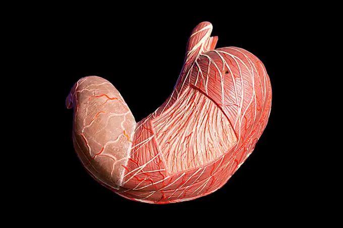
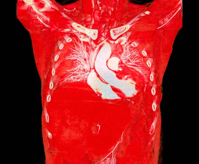
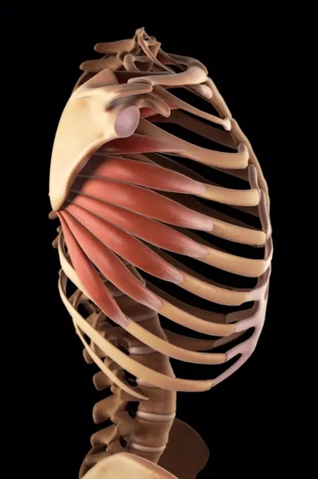
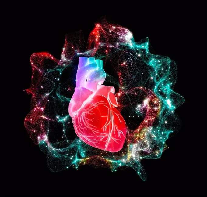
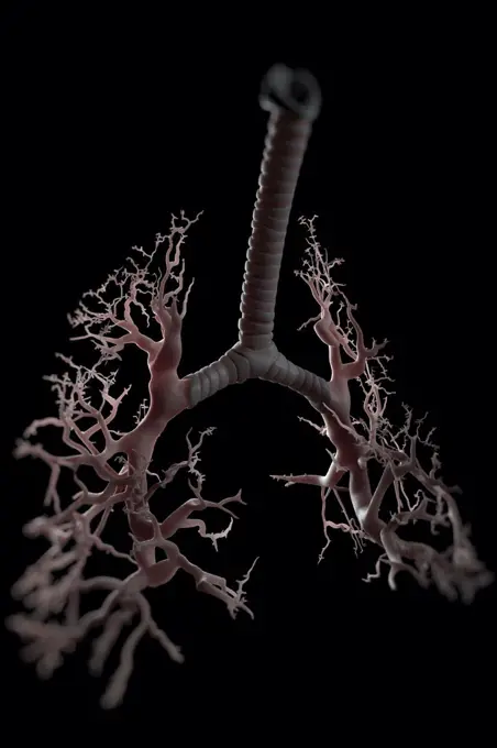
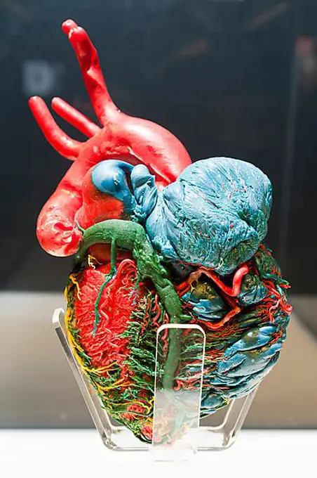
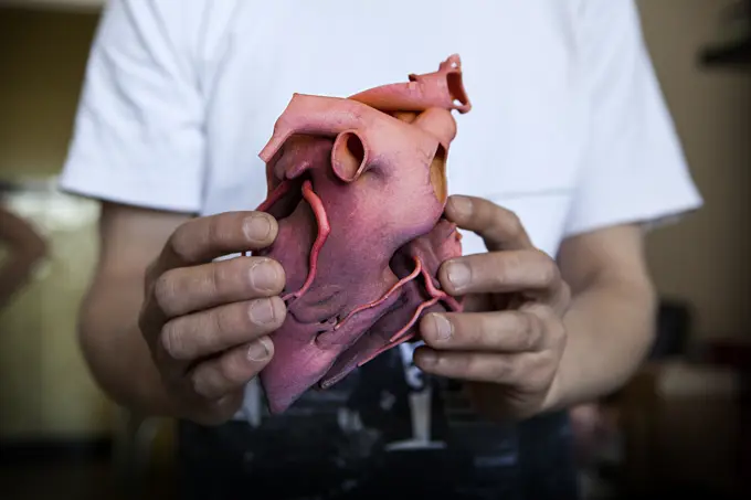
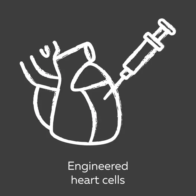
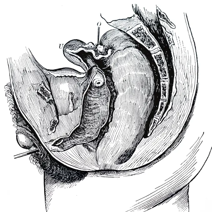
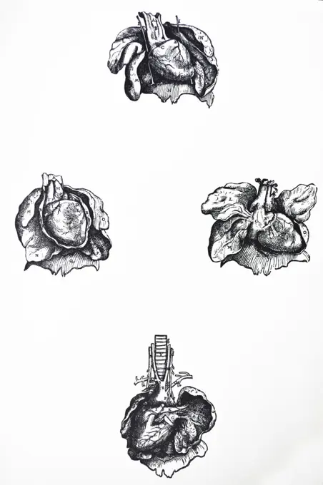
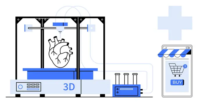
Heart Anatomy Illustrations

Detailed illustrations of the human heart, showcasing its anatomy, blood vessels, and educational models used in medical contexts.

Diagram of a heart.
Diagram of a heart.
201 assets in this story
1525-24087962
Anatomy heart, human organ illustration. Vintage tattoo. Vector drawn art
1525-20360067
Open artificial human heart model front view isolated on white background
4128R-26622
Heart valve. Computer artwork showing the tricuspid valve.
4128-V58614997
824-698
Human Heart
4128-111580870
Human heart, computer illustration.
1899-53508434
Complex congenital cardiopathy. The 3D CT scan allows three-dimensional visualization of the heart after surgery to treat a very complicated congenital cardiopathy.
4128-30416231
Human heart, illustration
4378-3470
Three-quarter view of the heart and its coronary blood vessels.
4378-3827
Three-quarter view of the heart and its coronary blood vessels.
4128R-11324443
Human shoulder joint, computer artwork.
1899-61459147
human anatomy, medicine, the circulatory system, blood circulation: section of the neck, shoulder and chest of a man, with aorta, arteries and blood vessels shown in red. Colored lithograph by J. Maclise, 1841.
4128R-14060453
Human heart, illustration
1848-53963542
Anatomical model, human stomach
4239R-20483378
Conceptual image of human lungs and rib cage.
4128-30419938
Pulmonary artery, CT scan
1899-54027750
Anterior view cross-section illustrating of the 4 cardiac valves. From left to right, the tricuspid valve, the pulmonary valve, the aortic valves and the mitral valve.
4128R-24161
Human heart, computer artwork.
4378-20393526
Serratus Muscle
1525-24588613
Heart sketch icon, cardiovascular system isolated vector. Blood circulation, human anatomy. Human heart sketch icon, cardiovascular system
4128-18929782
Human heart with energy field, illustration
6145-46025866
Prints of the Brain and the Heart. Around 1735 Jan lAdmiral printed several remarkable anatomical prints in colour, among others on commission for the celebrated physician Frederik Ruysch. For his illustrations of the cerebral membrane (the skull of an unborn child) and a human heart he experimented with ever different colours, and made various versions of them. The objective was to provide medical practitioners with the most precise and realistic images possible.
1525-26243946
Human heart, realistic anatomy 3d model of human heart on the monitor, visual heart beating. Human anatomy, cardiovascular system. 3D illustration. Human heart, realistic anatomy 3d model of human heart on the monitor,
4378-3313
Front view of the heart with coronary blood vessels visible set against a black background.
4128R-13586438
Illustration of human brain anatomy.
824-63126541
Heart, illustration
1525-28090091
The heart of man, vintage engraved illustration. Zoology Elements from Paul Gervais.
1525-26244050
Human Circulatory System Heart Beat Anatomy Concept. 3D illustration. Human Circulatory System Heart Beat Anatomy Concept. 3D
4128R-14637670
Human heart coronary blood vessels, illustration
824-63190789
Frontal illustration of the heart (on the left), and its coronary circulation, with the common trunk of the left coronary artery shown in orange with the circumflex artery leading off it. The right coronary artery leads to the auricular artery, then the sinus node artery. Rear illustration of the heart (on the right) and its circulation, with the left retro ventricular artery leading to the right marginal artery and the posterior inter ventricular artery.
824-63147973
Heart, illustration
1525-56726484
illustration 3D. Finding and preventing cardiac vital signs.
4128R-14181752
Heart with coronary vessels, illustration
4378-3992
Organs of the digestive system within the bones of the torso viewed from a front view.
6188-66528826
Thorax, anatomy Thorax, anatomy 016889_081
6188-58102989
Human Liver Model with all Organs and Bones medical science of human liver model with all organs and bones Copyright: xZoonar.com/StanislavxRishnyakx 12932574
1848-49387877
Bronchi lie in the lungs and are an organ, illustration, natural science, biology, illustration, natural science, biology, abstract art
4128R-20710
Bacterial lung infection, artwork
4128R-7039
Heart anatomy, artwork.
4378-3790
Front view of the heart with coronary blood vessels visible set against a black background.
824-63126600
Heart, illustration
1428-1336
Cross sections of human heart and major vessels on white background
824-63165986
Abdomen, 3D CT scan.
4378-2932
Diagram of showing the structure of the bronchi and bronchial tubes.
1525-57077559
Illustration human bones.The thoracic cavity vector.
824-63126628
Heart rate, illustration
1848-49200649
Human anatomy, medicine, the circulatory system, blood circulation: section of the neck, shoulder and chest of a man, with aorta, arteries and blood vessels shown in red. Coloured lithograph by J. Maclise, 1841, 1844, Historic, digitally restored reproduction from a 19th century original
1848-53940670
Plastination specimen of arteries and veins of human heart
1525-26235820
Human Circulatory System Heart Beat Anatomy Concept. 3D illustration. Human Circulatory System Heart Beat Anatomy Concept. 3D
824-63190800
Illustration of the Circle of Willis, for blood flow to the brain stem, the cerebellum and the occipital lobes.
6188-62268773
Human thorax flat isometric icon Human thorax flat isometric icon. Protextora bone rib cage or internal organs of thorax isometric flat illustration. Copyright: xZoonar.com/DanilxTrapeznikovx 8963869
4128-38524210
Junctional rhythm of the heartbeat, illustration
4269-25412
Calcifying pericarditis (armoured heart). 3D computed tomographic (CT) scan reconstruction of a heart affected by calcifying pericarditis (armoured heart) seen from above. Pericarditis is the inflammation of the pericardium, a thin layer of tissue that covers the outer surfaces of the heart. Here, the pericardium is covered by a fibrous calcarious rigid bag (white coat) and can not complete its natural mouvements.
1525-23339125
DNA, brain and heart. 3d illustration
4269-28017
Angioplasty. Treatment of carotid stenosis by angioplasty and setting stent prosthesis. Here, 3D reconstruction arteries of a patient after treatment. Limoges hospital, France.
4128R-1510
Heart anatomy, artwork.
4128-19249801
Tick, illustration.
1428-1335
Cross sections of human heart and major vessels on white background
4128R-11313849
Human respiratory system, computer artwork.
4269-24799
Rheumatoid polyarthritis. X-ray of a shoulder affected by a rheumatoid polyarthritis on the shoulder, evidenced by the narrowing of the joint space and the erosion of the cartilage and bones.
4128R-13373125
Human urinary system, illustration.
6188-66528813
Respiratory tract, drawing Respiratory tract, drawing 016889_072
5507-50351361
Set of Science cartoon doodle objects
5507-50749879
Set of Science cartoon doodle objects
1525-56593128
illustration 3D of cardiopulmonary and heart signal cabinet.
824-63186212
Reportage in Descartes University, Paris, France where Vizua 3D is based. Vizua provides real-time 3D visualisations for doctors using imagery from patients. Doctors can then print these visualisations in 3D. Heart printed with a 3D printer.
6145-29249024
Matthew Baillie, a series of engravings.
1525-22787329
medical science of human lungs model with all organs and bones. Human Lungs Model with all Organs and Bones
5507-31546231
Anatomical human heart. Strong and healthy cartoon character
6145-29246211
The heart, arteries of the neck and the aortic arch (): nine figures, with arteries indicated in red. Coloured lithograph by J. Roux, 1822.
4128R-26902
Female anatomy, computer artwork.
1525-27947776
Human kidneys icon
4378-2400
Multiple view of the skeletal structure of the rib cage.
1525-22197348
highlighted intestines
5507-48010015
Engineered heart cells red color icon. Human engineered cardiac tissues. HECTs. Heart injection. Study cardiac physiology. Bioengineering. Biotechnology. Isolated vector illustration
1660R-32481
Hanging carcass of large animal
4128R-15366990
Coffee beans making an electrocardiogram line
1525-56211131
The heart is the most important muscle in your body. There are two sides, the right and left, and four chambers. 3D illustration. The heart is the most important muscle in your body. There are two sides, the right and left, and four chambers.
4128-30415632
Left lung, illustration
4128-30419313
Female lung, illustration
4269-27198
Intestinal obstruction. 3D Computed Tomography (CT) reconstruction scan of the vascularization of the abdomen showing signs of whorls of the small intestine responsible for a bowel obstruction. It is evidenced by the winding of the blood vessels of the intestine's loops around their axis (bottom).
6145-52293601
Technical recordings Hr.Ms. Holland, (D 808) December 1976
5507-48010462
Engineered heart cells chalk icon. Human engineered cardiac tissues. HECTs. Heart injection. Study cardiac physiology. Bioengineering. Biotechnology. Isolated vector chalkboard illustration
1746-30002453
Hermaphroditism: On 20 August, 1864, a patient passing as a male entered the Hotel-Dieu, Paris, with pleurisy. They were identified as Marie-Madeline Lefort who had been examined at the Paris Faculty of Medicine in 1815. Passed first part of life as female and second part as male. From George M Gould and Walter L Pyle Anomolies and Curiosities of Medicine, Philadelphia, 1900. Mesial section showing the generative organs of Marie-Madeline Lefort.
1525-22348349
Close-up of human lungs
4128-V58558710
824-28706031
Anatomical mannequin for teaching human organs
1899-13015
Lungs and heart Anterolateral view
1746-19688388
The Plates from the Sixth Book of the De Humani Corporis Fabrica by Andreas Vesalius, (1514-1564) Plate 64 - since the two preceding figures indicate fully the position of the heart and lungs, we have drawn the present one as though evulsed from the thoracic cavity.
1525-64445601
Depicts a 3D printer creating a human heart alongside a digital shopping interface. Ideal for medical technology, innovation, healthcare, e-commerce, 3D printing. Modern flat style with blue. Advancements in Medical Technology 3D Printed Heart and E-commerce for Healthcare
4128R-14060409
Atomic structure of the body, illustration
4128R-11313376
Human respiratory system, computer artwork.
824-57655645
Electrocardiogram, human heart.
1570R-140071
Graphs of pulse traces
6188-68111506
Eine Kühlbox mit menschlichen Organen zur Transplantation. Illegaler Verkauf von menschlichen Organen. A cooling box with human organs for transplantation. Illegal sale of human organs. LicenseRF Copyright: xZoonar.com/MilanxVasicekx 12667031
5507-31494035
Thyroid structure and parts. Hand-drawn icon isolated on white background Human internal organs Engraving sketch style Design for medical project poster Pathology, disease, treatment
5507-31493865
Pancreas. Realistic hand-drawn icon of human internal organs. Line art. Sketch style. Design concept for your medical projects post viral rehabilitation posters, tattoos.
5507-31493779
Thyroid structure and parts. Hand-drawn icon isolated on black chalkboard background Human internal organs. Engraving art. Sketch style Design for medical project, poster Pathology, disease, treatment
4128-20040904
Thyroid cancer, illustration
5507-31493926
Liver. Realistic hand-drawn icon of human internal organs on chalkboard. Engraving art. Sketch style. Design concept for your medical projects post viral rehabilitation posters, tattoos.
PREVIOUS
of 3
NEXT
2401 S. Ervay, Suite 206
Dallas, Texas 75215
United States
Get Started
Free ResearchMy BoardsMy Cart
For Creators
How To License Your ContentContributor PortalFrame of Mind
Resources
API accessPricing
Contact
+1 866 236 0087help@viewpictures.co.uk Contact form
©2026 View Pictures. All Rights Reserved. -A
This site is protected by reCAPTCHA and Google Privacy Policy and Terms of Service apply.