My Boards
View Pictures
  • My Boards

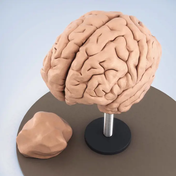
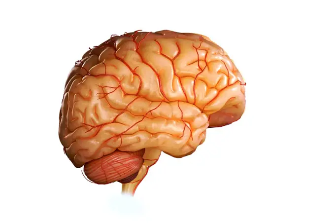
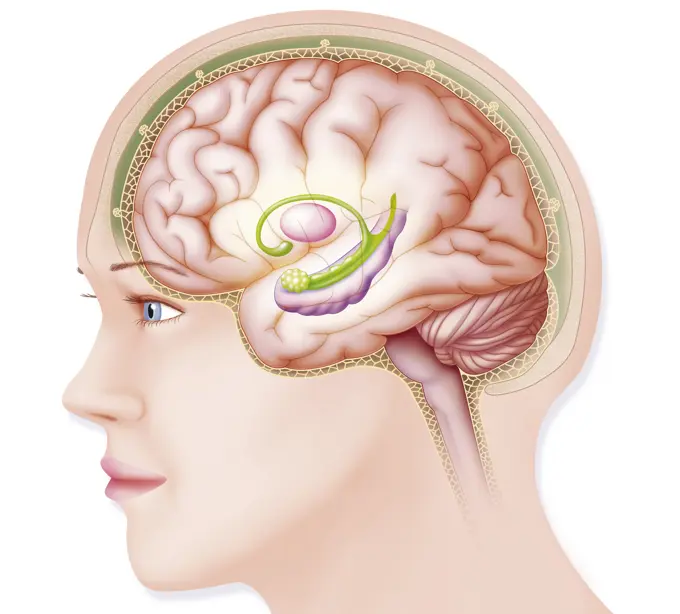
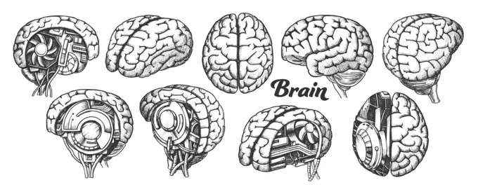
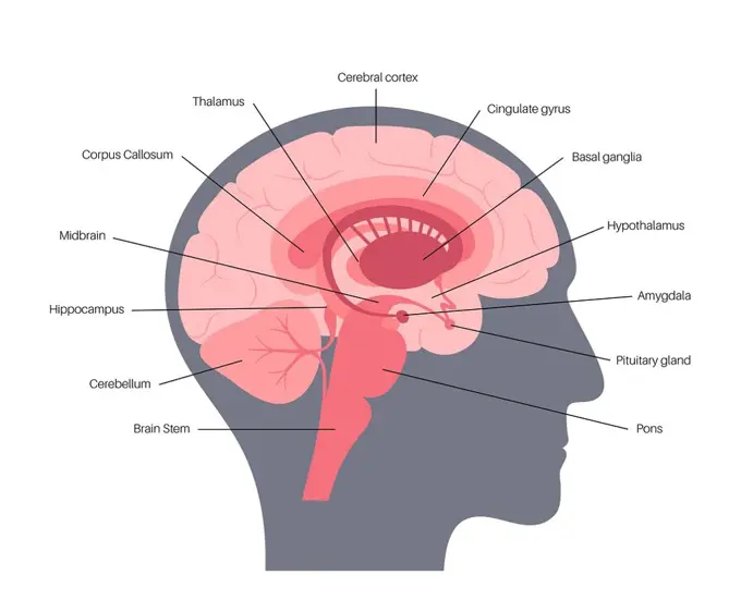
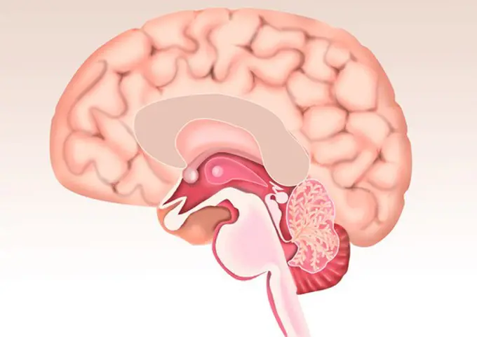
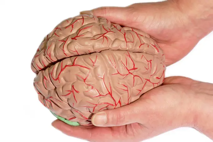
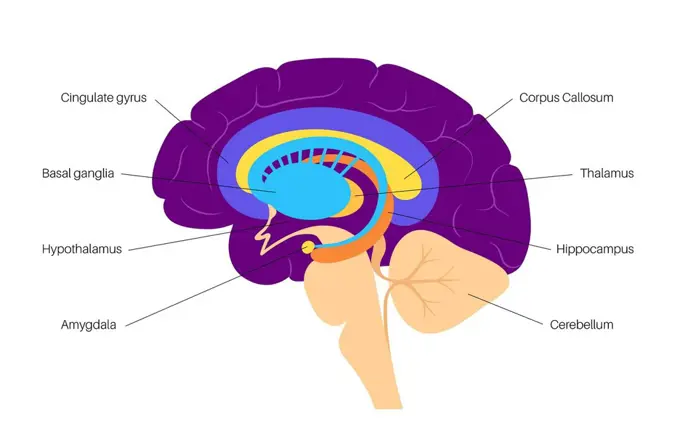
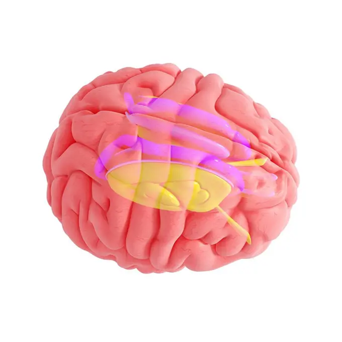
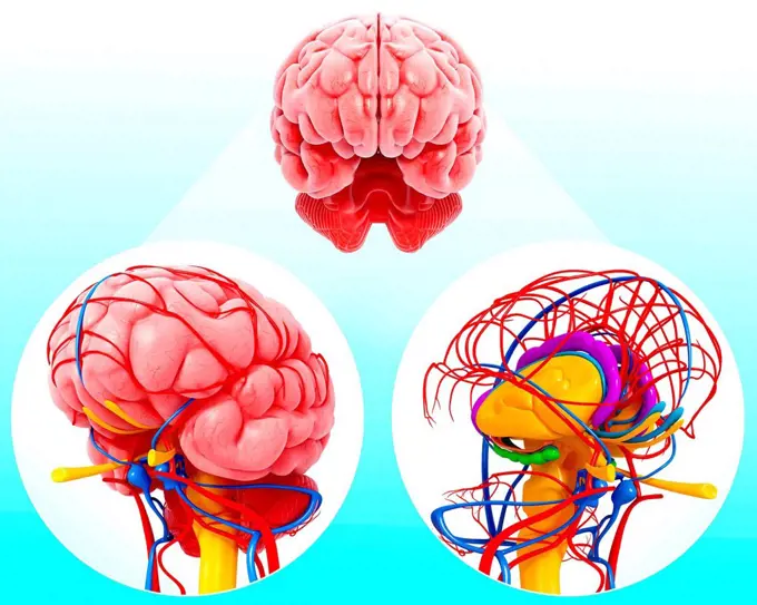
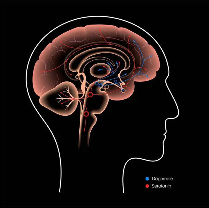
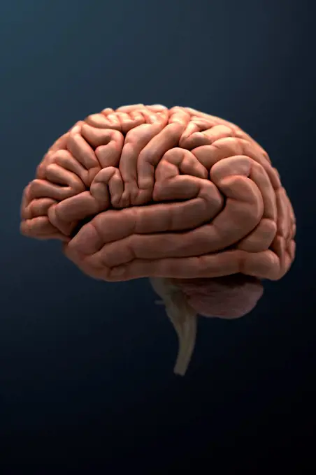
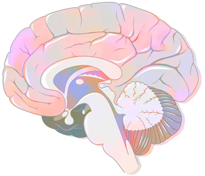
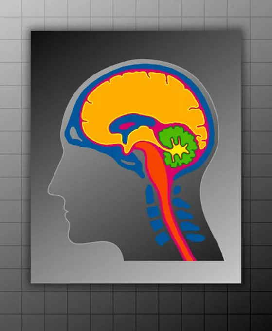
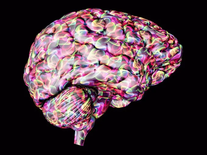
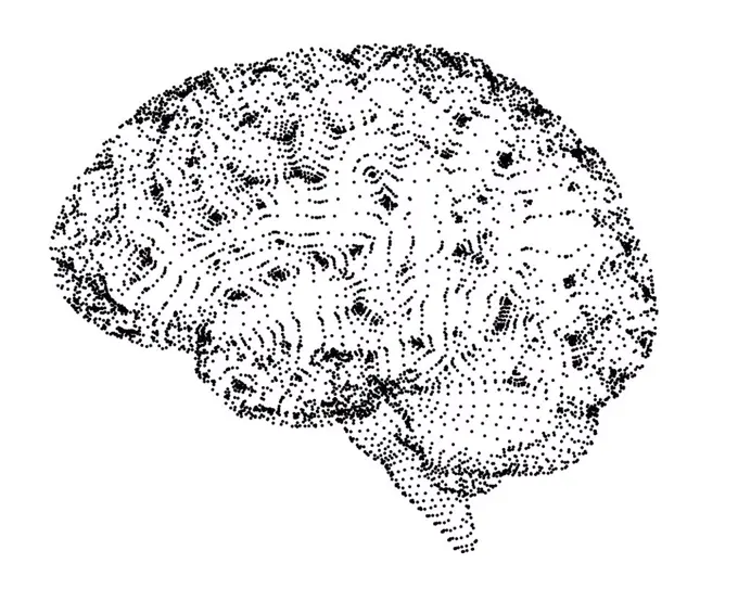
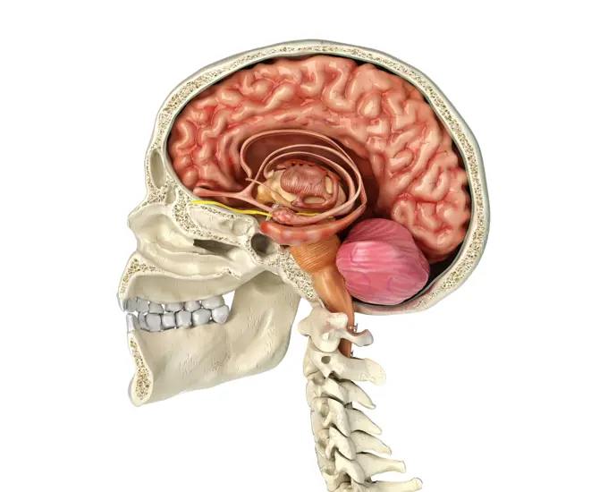
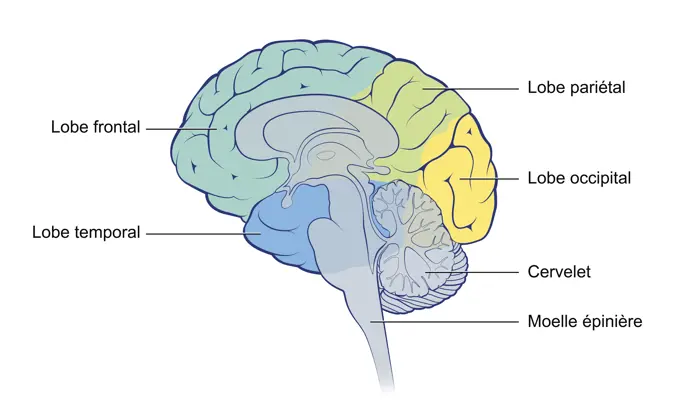
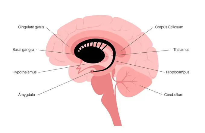
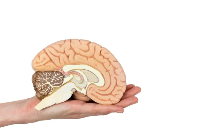
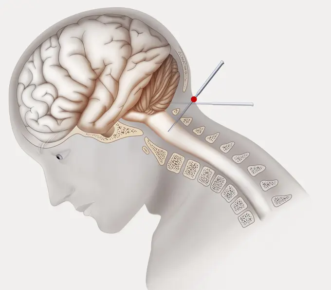
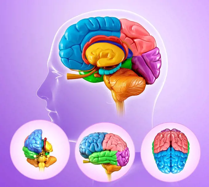
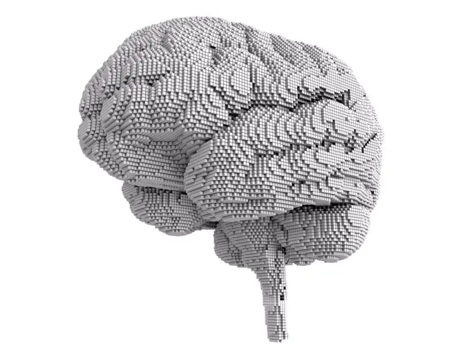
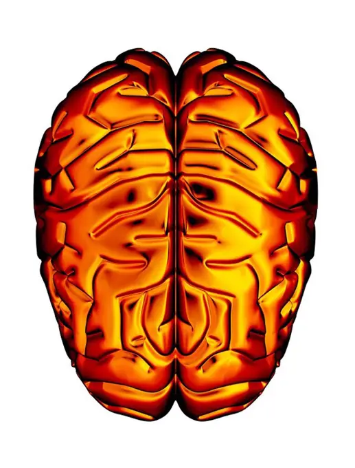
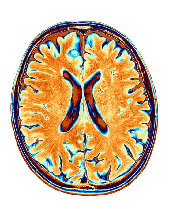
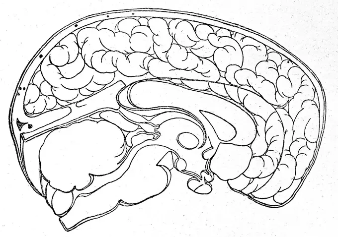
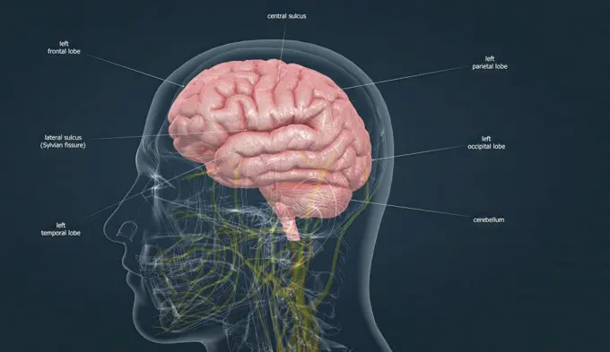
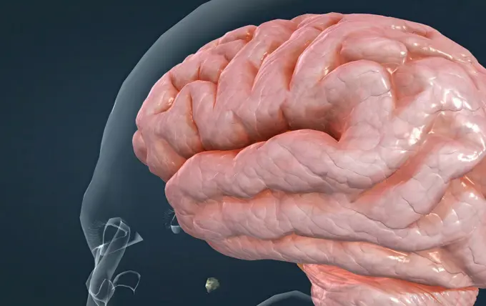
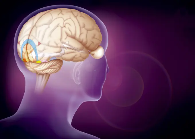
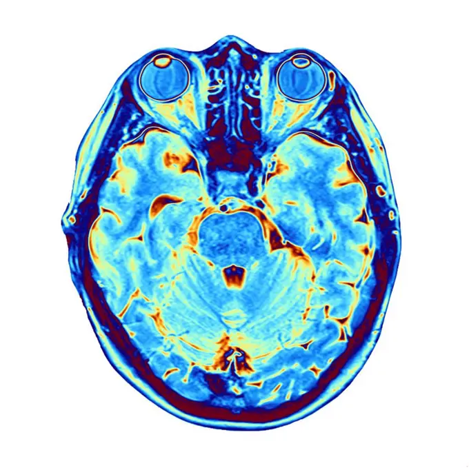
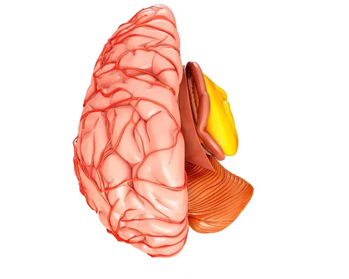
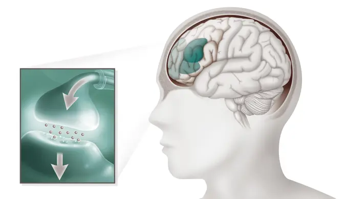
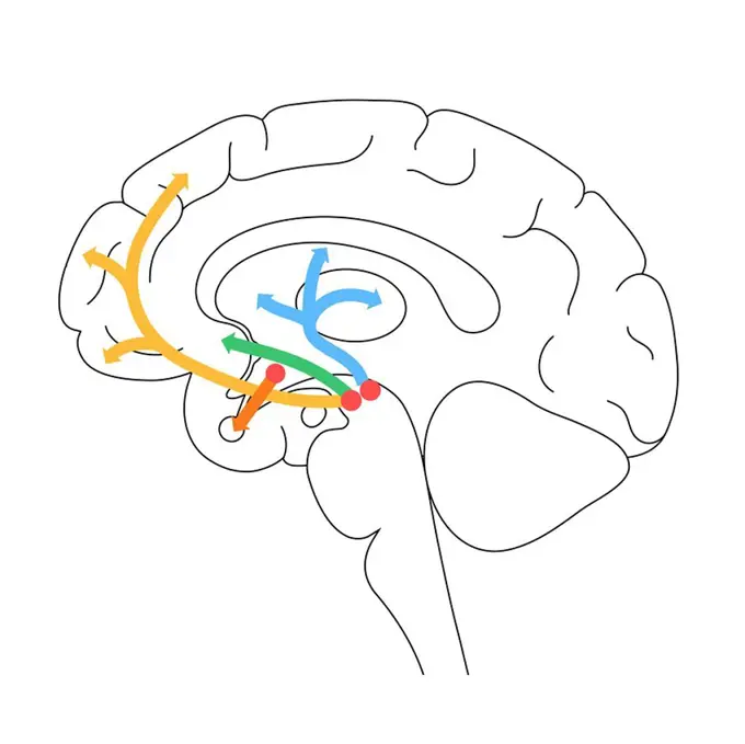
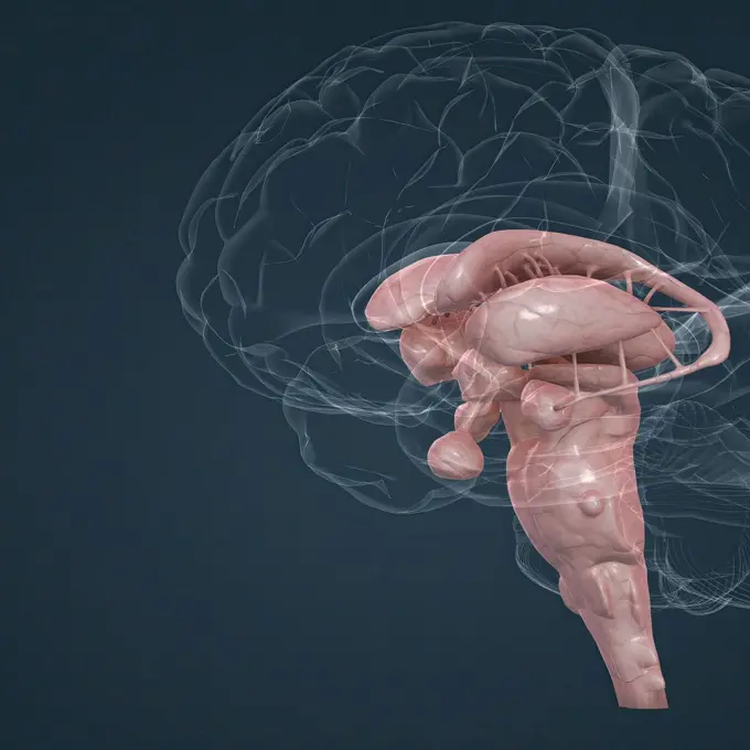
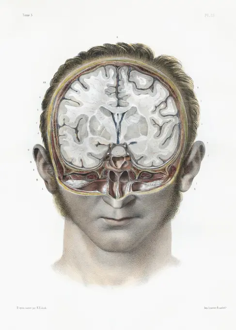
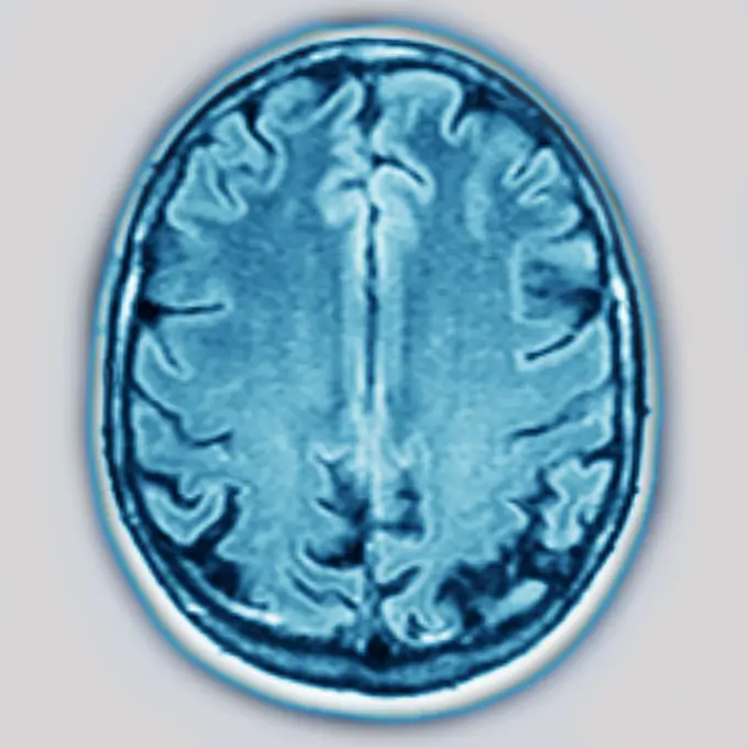
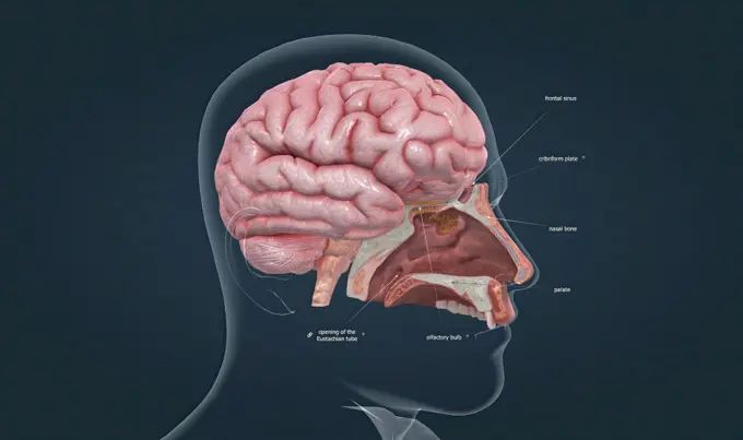
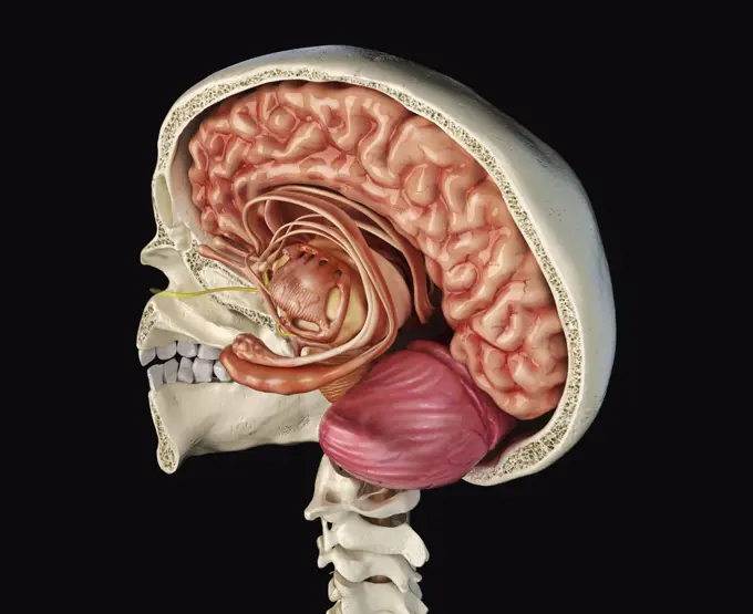
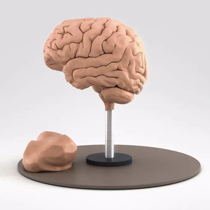
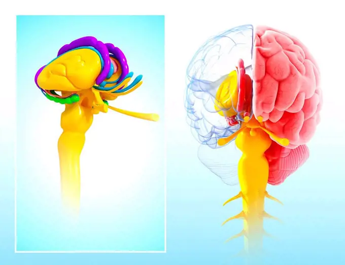
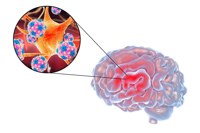
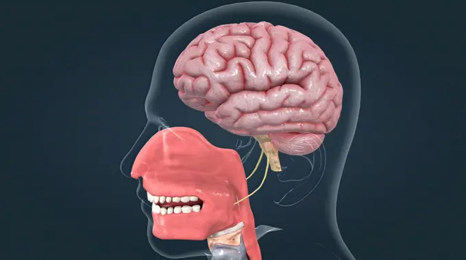
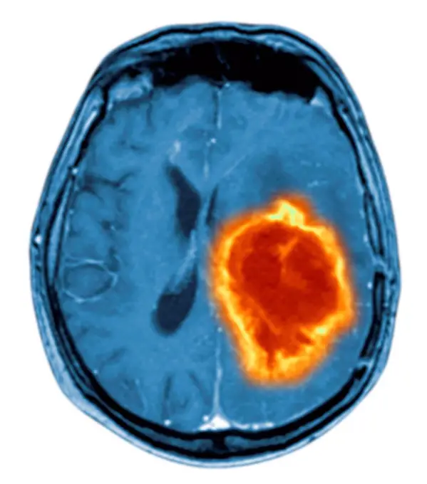
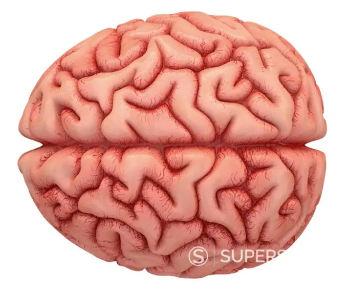
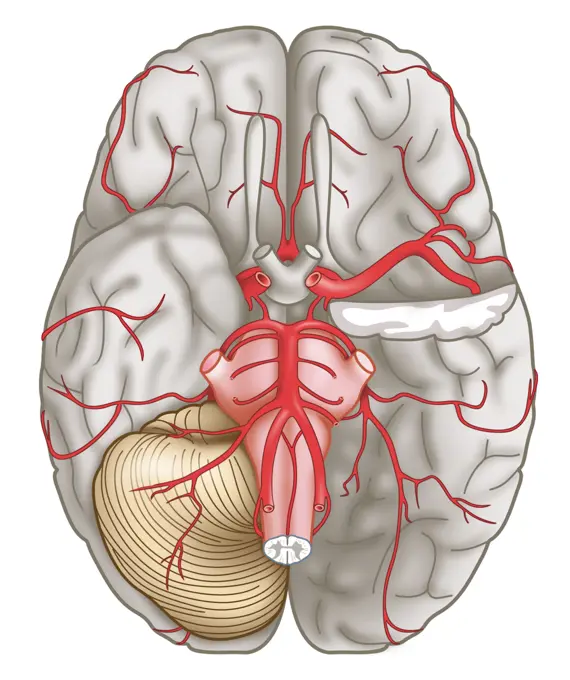
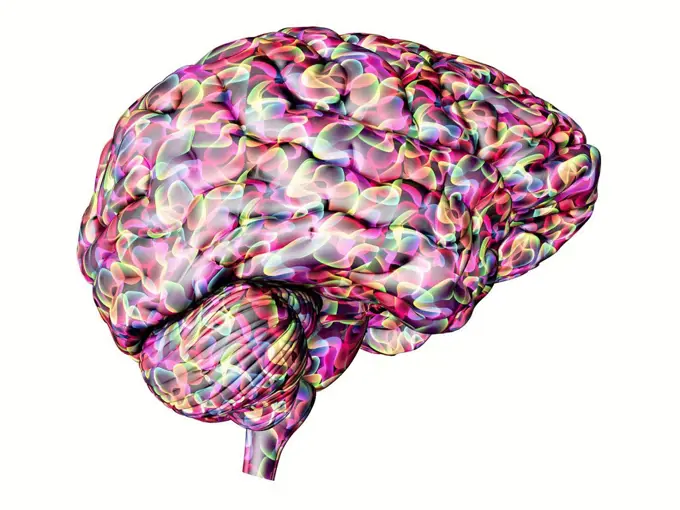
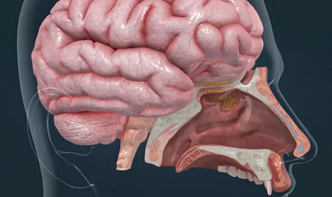
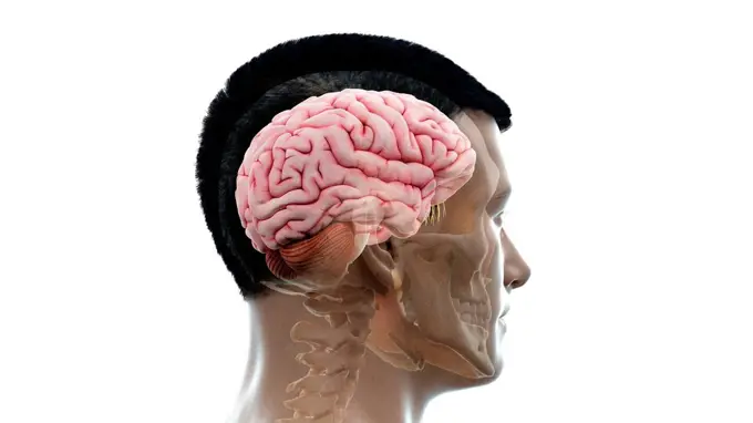
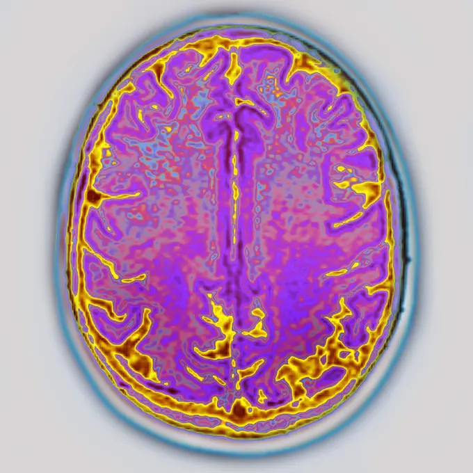
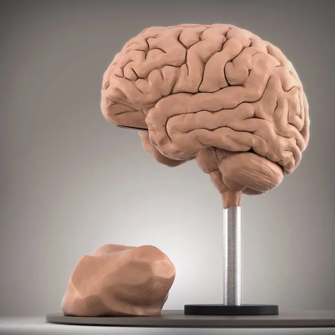
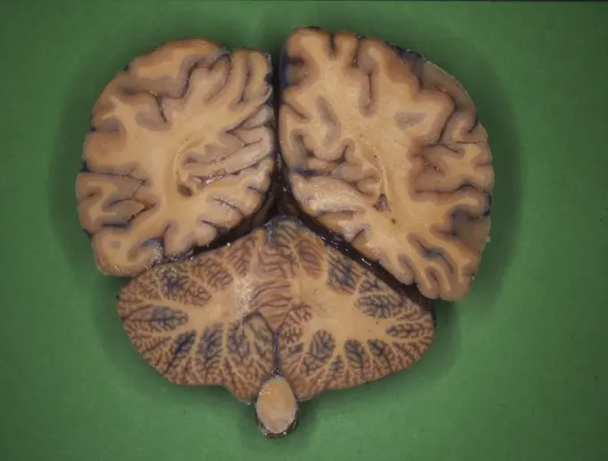
Human Brain Illustrations

Computer-generated and artistic representations of the human brain, displaying its anatomy and various regions in vibrant colors.

Human Brain
Human Brain
375 assets in this story
824-63180606
Anatomy of the brain from the exterior to the centre : the skull in cross-section (beige), the arachnoid (purple blue), the cortex (beige) followed at the bottom by the rachidian bulb and bone marrow (dark purple) and the cerebellum (pink).
5507-41644775
Vector illustration of human brain ( front view)
1525-27502886
Model kit set with pink brain parts isolated on white. 3D illustration. Model kit set with pink brain parts.
1525-28333427
Collection In Different Views Brain Set Vector. Many Kinds And Modification Of Cyber And Human Brain. Anatomy Medical Neurology Element Hand Drawn In Vintage Style Monochrome Illustrations. Collection In Different Views Brain Set Vector
4128-19246983
Brain anatomy, illustration.
1848-51343134
Human brain
4252-4446
Brain
6188-65132374
Gehaltenes Lehrmodell Human Brain Gehaltenes Lehrmodell eines Gehirnes Human Brain holded by two hands Copyright: xZoonar.com/Teamarbeitx 200290
4128-19358394
Brain limbic system, illustration.
824-63189394
A saggital median plane illustration of the location of the hypothalamus and the hypophyseal.
1788-39653
Medicine: Human anatomy, encephalon or human brain, inferior area. Drawing
4128R-13269870
Human brain, illustration.
4128R-29535
Brain anatomy, computer artwork.
1788-39652
Medicine: Human anatomy, encephalon or human brain. Drawing
1899-53513350
Prefrontal area: area of the brain activated by smell.
4128R-11296698
Human brain anatomy, computer artwork.
4128R-13387005
Illustration of child's brain anatomy.
824-63224028
Visual area: area of the brain activated by sight.
4128-19358411
Dopamine and serotonin hormones pathway in the human brain, illustration.
4128R-13681833
Human brain, illustration.
4378-678
Side view of the human brain (left side).
824-63164338
Sagittal section of the brain with meninges and cerebrospinal fluid.
4128-19248003
Brain in an open head, illustration.
4286-33611
Stylized view of head and brain
1525-23711701
3D illustration of human brain in different angles
4128R-12580884
Brain. Abstract computer artwork of a side view of a human brain.
4128R-14057815
Human brain, conceptual artwork
4128R-14637802
Human brain, illustration
6188-68089945
Menschlicher Schädel im Querschnitt mit Gehirn. Menschlicher Schädel im Querschnitt mit Gehirn. LicenseRF Copyright: xZoonar.com/Pixelchaosx 23158871
4128R-13386939
Illustration of human brain anatomy.
4128R-13446861
Illustration of a cross-section of the human brain.
824-63224059
Sagittal section of the brain with the lobes in color and captioned.
824-63224036
Prefrontal area: area active by smell and providing emotions.
4128-19358390
Basal ganglia anatomy, illustration.
1525-56184346
When light hits the retina , special cells called photoreceptors turn the light into electrical signals. These electrical signals travel from the retina through the optic nerve to the brain. Then the brain turns the signals into the images you see. 3d illustration. The mechanism of vision in the human eye
1525-23433885
Hand holding left brain hemisphere isolated on white background
824-63205871
Illustration of the location of the Yamen acupuncture point, one of the points of the governing vessel. The needle at the top indicates the correct position, which is between the C1 and C2 cervical vertebrae, but does not penetrate the spinal cord. The lower needle shows the wrong angle and position.
4128R-13446965
Illustration of human brain regions and anatomy.
1525-26182465
Central Organ of Human Nervous System Brain Anatomy 3D illustration. Central Organ of Human Nervous System Brain Anatomy
4128R-14058273
Child's brain anatomy, illustration
4239-18641649
Dog brain, multiple angles.
4128-30415842
Skull and brain, illustration
1525-56211151
When light hits the retina , special cells called photoreceptors turn the light into electrical signals. These electrical signals travel from the retina through the optic nerve to the brain. Then the brain turns the signals into the images you see. 3d illustration. The mechanism of vision in the human eye
4128R-28141
Conceptual computer artwork of a pixellated human brain.
824-63168353
Frontal section of the brain showing the ventricles.
4128-111582578
Male brain, computer illustration.
4128R-3477
Human brain, artwork
4128R-13682187
Normal brain. Coloured magnetic resonance imaging (MRI) scan of an axial section through a healthy brain, The images shows the cortex and lateral vent...
6145-29255500
Saggital section of brain
5507-51256278
Sticker of human brain. Vintage anatomy engraving sketch organ isolated on white background. Good idea for design retro medicine poster in hand drawn style. Anatomical body part vector illustration
1525-56183629
Brain lobes , Frontal Lobe, Temporal Lobe, Parietal Lobe, Occipital Lobe, Cerebellum and Brainstem. Each has a separate function. 3D illustration. Human Anatomy of the Brain
1525-56211154
Thyroxine plays a crucial role in heart and digestive function, metabolism, brain development, bone health and muscle control. 3D illustration. Thyroxine is the main hormone secreted into the bloodstream by the thyroid gland.
4128-18631399
Brain, illustration
4128R-13711080
Illustration of the anatomy of the human brain and its arteries.
824-63214049
Illustration of the migraine process, or migrainous attack. External factors trigger the stimulation of the hypothalamus (green area), which triggers the stimulation of neurons innervating the arteries in the meninges (green area round the brain). These neurons release neuropeptides in the lining of these arteries, causing inflammation and dilatation, causing the migrainous pain.
824-63195040
Illustration of the brains visual cortex and the path taken by visual information from the eye (sensorial receptor) to the visual cortex (centre for visual elaboration), via the optic chiasma and the lateral geniculate body. When the information finally arrives at the end of its journey, the brain deciphers it.
4128R-28109
Brain scan. False-colour magnetic resonance imaging (MRI) scan of a human head containing a healthy brain, seen in horizontal view. At upper frame, th...
4128R-13711079
Illustration of the anatomy of the human brain and its arteries.
1525-24377200
man showing poster of hand drawn brain as medical concept
1525-24113757
Illustration with brain internal organ. Human body anatomy. Health care and medical education image.. Illustration with brain internal organ. Human body anatomy. Health care and medical image.
1899-54028641
Illustration of the ADHD process, attention deficit disorder with or without hyperactivity. The cerebral areas located in the frontal lobe, responsible for attention, organizational skills (dark green area) and emotional responses (light green area) are abnormally active or have a specific anatomy. There is also an imbalance in levels of some chemical messengers (neurotransmitters) in the brain, such as dopamine and noradrenaline. (Photo by: BSIP/UIG via Getty Images)
1660R-41552
brain with a straw in it
4239-18642044
Human skull transversal cross section with brain. Front view on white background.
1848-77189234
3D Illustration Concept of Central Organ of Human Nervous System Brain Lobes Parietal Lobe Described with Labels Anatomy
824-63214051
Illustration of the ADHD process, attention deficit disorder with or without hyperactivity. The cerebral areas located in the frontal lobe, responsible for attention, organizational skills (dark green area) and emotional responses (light green area) are abnormally active or have a specific anatomy. There is also an imbalance in levels of some chemical messengers (neurotransmitters) in the brain, such as dopamine and noradrenaline.
4128-19358404
Dopamine pathway in the human brain, illustration.
1525-56183595
The brainstem is the posterior stalk-like part of the brain that connects the cerebrum with the spinal cord. 3D illustration. The brainstem is the posterior stalk-like part of the brain that connects the cerebrum with the spinal cord.
6188-62240822
Human brain Human brain Copyright: xZoonar.com/SergiixSvyerchkovx 5957428
1889-60419466
Vertical section of the human brain.  Plate from volume 3 of Traite Complet de l'Anatomie de l'Homme by Jean-Baptiste Marc Bourgery.  Illustration by Nicolas-Henri Jacob.  The picture comes from volum
1899-53511264
Cerebral atrophy - Posterior cortical atrophy (posterior cortical degeneration responsible for agnosia, apraxia, aphasia, Gerstmann syndrome, acalculia, indistinction, agraphia, Balint syndrome, optic ataxia, psychic paralysis. Radial section of the brain, visualized by a cross-sectional MRI radial.
4128R-11478493
Human brain, computer artwork.
1525-56198053
Olfactory organ there are two olfactory bulbs on the bottom side of the brain, one above each nasal cavity 3d illustration. Olfactory organ there are two olfactory bulbs on the bottom side of the brain, one above each nasal cavity.
4239-18641151
Brain surface anatomy, with labels.
824-63224368
Brain ventricles transparent in a brain in external profile view. The ventricles are a collection of cavities. They are located inside the brain. They are continuous with the ependymal duct of the spinal cord. They participate in the secretion and circulation of cerebrospinal fluid (or cerebrospinal fluid). The cerebral ventricles are four in number. They are represented in transparency in bluish gray in this profile view of the brain. The two lateral ventricles are connected to the third ventricle in the center of the brain through the interventricular foramen. Then the structure descends downwards through the midbrain aqueduct. The fourth ventricle is located slightly above and in front of the cerebellum. Then finally, the structure descends into the brainstem. It forms the canal of the spinal cord.
6188-68116752
Menschliches Gehirn mit weißrussischer Flagge. Wissenschaftliche Forschung und Bildung in Belarus Konzept, 3D-Rendering Human brain with Belarusian flag. Scientific research and education in Belarus concept, 3D rendering isolated on white background LicenseRF Copyright: xZoonar.com/alexlmxx 23212494
4239-18642026
Human skull mid sagittal cross-section with brain. Perspective view on black background.
1525R-243118
3d rendered medical illustration of a human brain
4128R-13410000
Human brain anatomy, illustration
4378-2565
Clay model showing the anatomical structure of a brain.
4128R-11296208
Human brain anatomy, computer artwork.
4128R-14181696
Viral encephalitis, illustration
1525-56184257
Olfactory organ there are two olfactory bulbs on the bottom side of the brain, one above each nasal cavity 3d illustration. Olfactory organ there are two olfactory bulbs on the bottom side of the brain, one above each nasal cavity.
4128R-13844781
Brain tumour. Coloured Magnetic Resonance Imaging (MRI) scan of an axial section through the brain showing a metastatic tumour. At bottom left is the ...
4128R-11478495
Human brain, computer artwork.
824-63168368
Inferior view of the brain showing the arteries that supply it, anterior cerebral arteries, anterior and posterior communicating arteries, basilar artery, vertebral arteries and posterior cerebral arteries.
1788-39819
Medicine - Anatomy - Nervous system - Brain. Drawing.
4128R-12580904
Brain. Abstract computer artwork of a side view of a human brain.
4128-18929762
Spinal cord, LM
4128R-14323976
Brain anatomy, illustration
4128R-15303116
Human appendix, illustration
1525-56184330
Olfactory organ there are two olfactory bulbs on the bottom side of the brain, one above each nasal cavity 3d illustration. Olfactory organ there are two olfactory bulbs on the bottom side of the brain, one above each nasal cavity.
4128-18183276
Human brain, illustration
4239-18641126
Human brain anatomy, lateral view.
4128-19055743
Illustration of a human brain
1899-53511267
Cerebral atrophy - Posterior cortical atrophy (posterior cortical degeneration responsible for agnosia, apraxia, aphasia, Gerstmann syndrome, acalculia, indistinction, agraphia, Balint syndrome, optic ataxia, psychic paralysis. Radial section of the brain, visualized by a cross-sectional MRI radial.
1746-30002693
Vertical transverse section of the human brain.
4378-2766
Clay model showing the anatomical structure of a brain.
6188-55645511
Human brain in the longitudinal and cross -section of human brain in longitudinal and cross -section
1428-139
Close-up of two human brains
4378-2623
Clay model showing the anatomical structure of a brain.
PREVIOUS
of 4
NEXT
2401 S. Ervay, Suite 206
Dallas, Texas 75215
United States
Get Started
Free ResearchMy BoardsMy Cart
For Creators
How To License Your ContentContributor PortalFrame of Mind
Resources
API accessPricing
Contact
+1 866 236 0087help@viewpictures.co.uk Contact form
©2026 View Pictures. All Rights Reserved. -A
This site is protected by reCAPTCHA and Google Privacy Policy and Terms of Service apply.