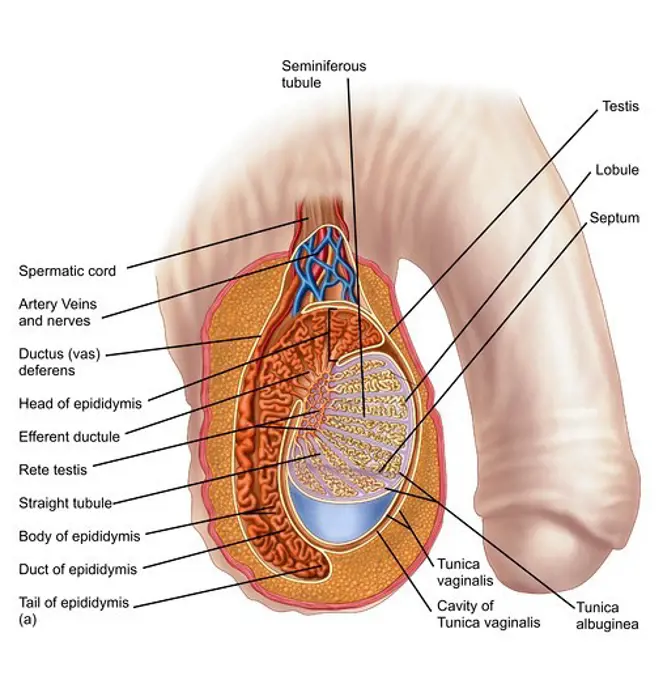
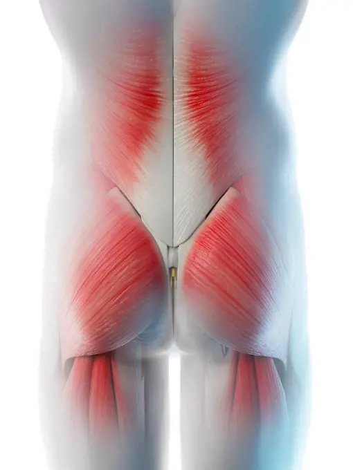
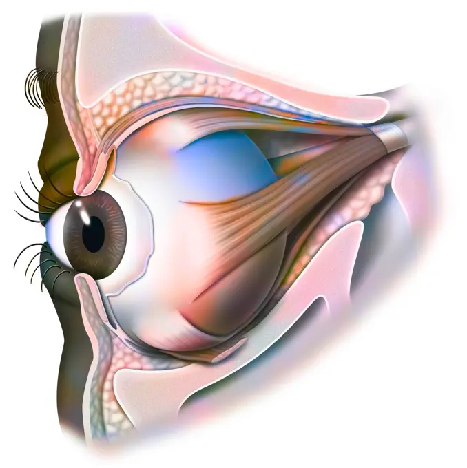
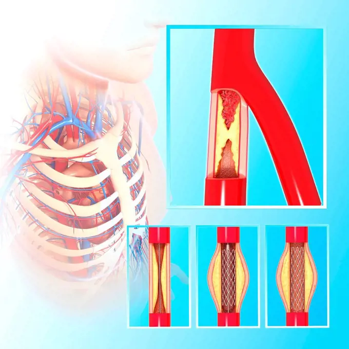
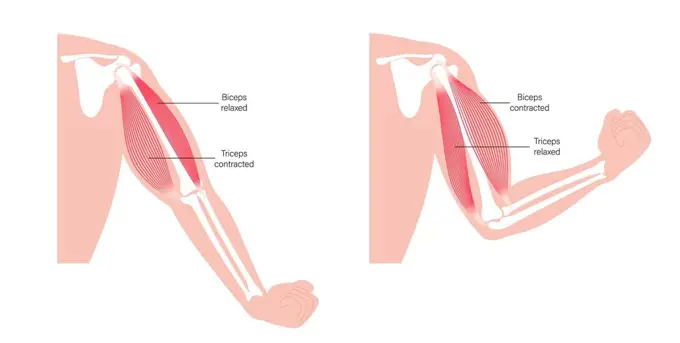
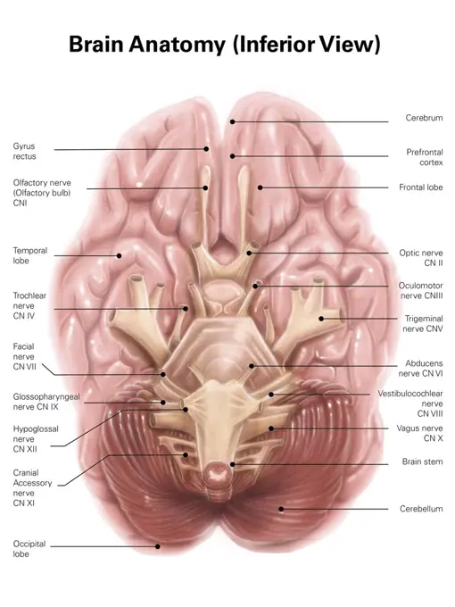
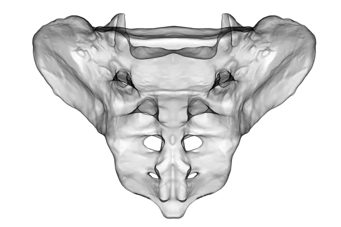
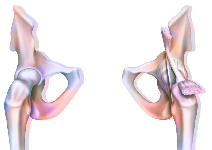
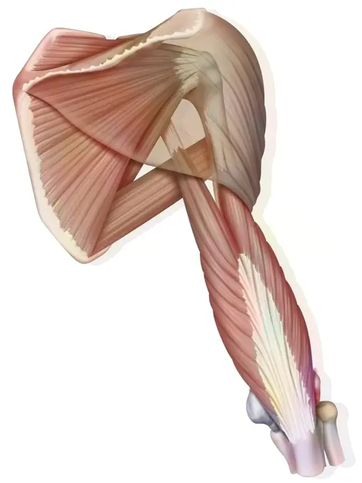
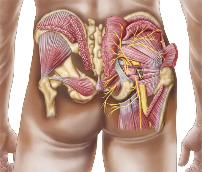
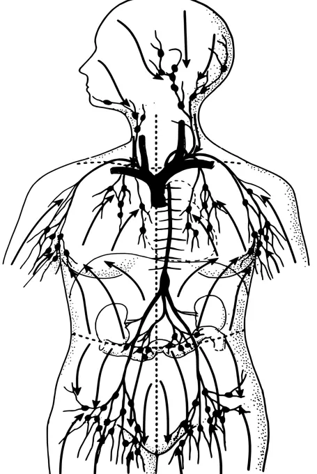
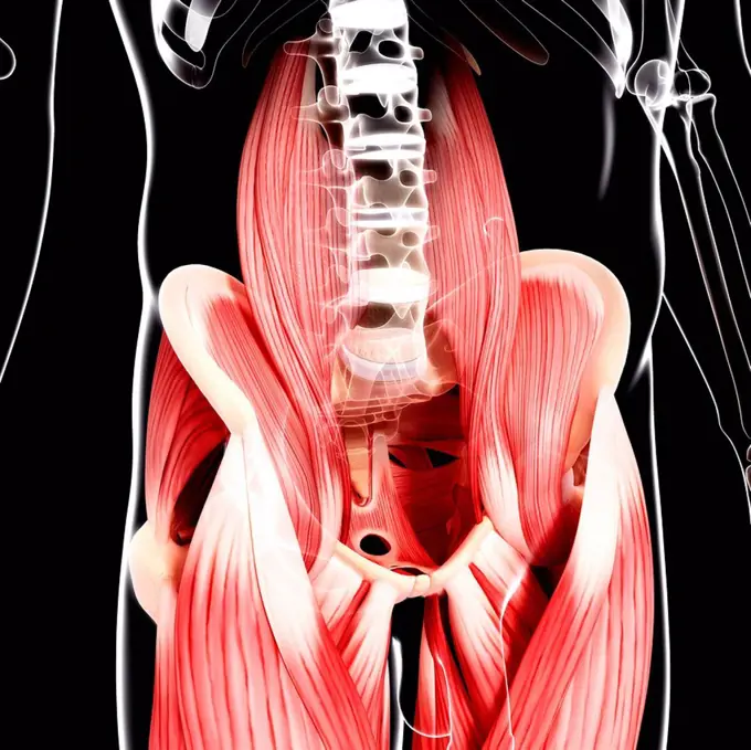
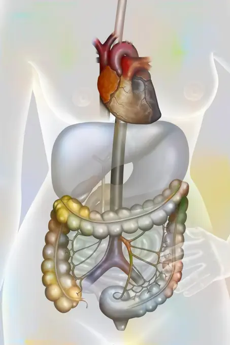
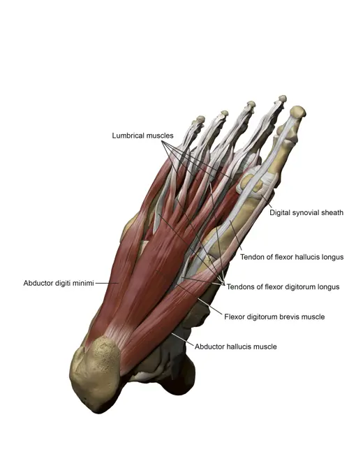
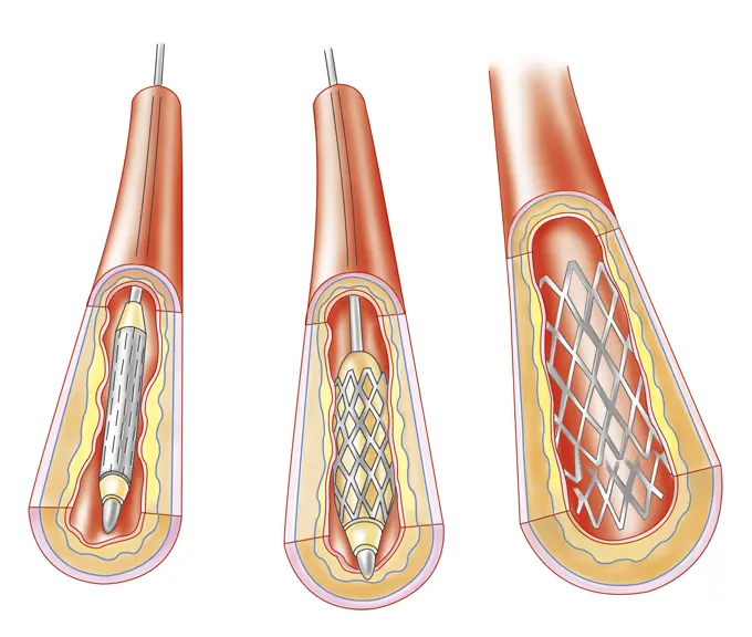
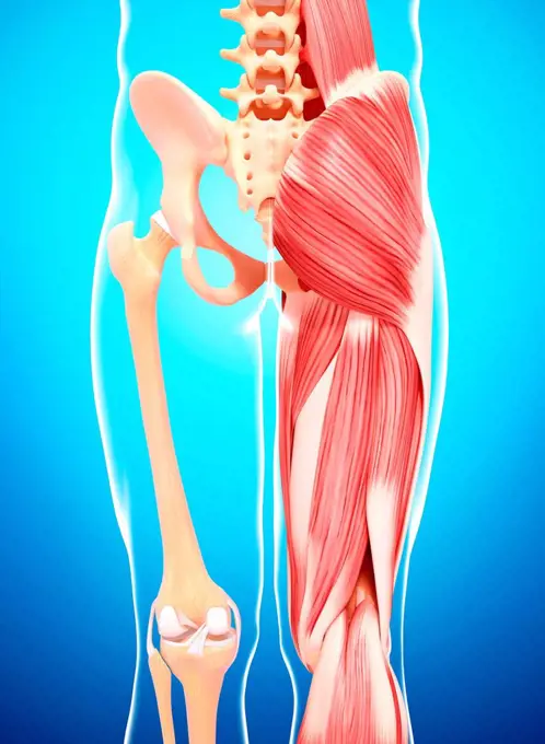
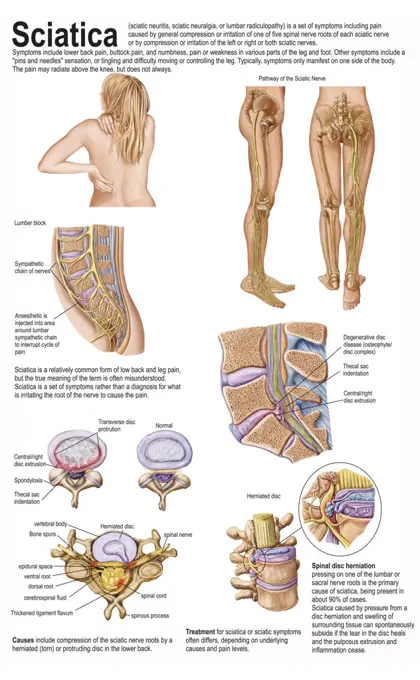
My Boards
View Pictures
  • My Boards

Internal Human Anatomy

Illustrations depicting various internal body structures including the spine, ribs, and musculature, emphasizing human anatomy in detail.

Human hip musculature, computer artwork.
206 assets in this story
824-63187030
Illustration of a herniated disc which compresses and swells the nerve root of the sciatic nerve.
1899-53509135
Muscular system of the muscles of the right shoulder in posterior view.
1788-20761546
Thigh muscles seen from the front, anatomical plate.
4239R-20483626
Anatomy of the gluteal muscles in the human buttocks.
1788-39756
Medicine: Human anatomy, abdominal organs. Drawing
4128R-5914
Intestines, artwork.
6188-55565466
Die menschliche lymphe Die menschliche lymphe Copyright: xZoonar.com/curryx 2414036
4128R-31607
Human hip musculature, computer artwork.
4252-4319
Thorax muscles
4462-21420133
Cross section of uterus
6145-29256078
Respiratory organs from F.-Achille Longet's Anatomie et physiologie du systeme nerveux
4128-19247542
Human foetus anatomy at week 42, illustration.
4128-19052730
Hip muscles, illustration
6243-72452461
Medical - Victorian Anatomical Illustrations - isolated on a white background for cut out.
824-63195443
Illustration of an owls skull. The auditory canals are not symmetrical.
824-63217367
Illustration of spinal stenosis in L4 and L5. Spinal stenosis is a pathology located in the lumbar spinal column where the cauda equina nerves pass. It is caused by an intrinsically narrow vertebral canal or a degenerative narrow canal related to a herniated disc, an increase in the volume of rear joints and ligaments that join the vertebrae together, or an instability in the vertebrae with vertebral displacement (or degenerative spondylolisthesis), which decreases the diameter of the canal, compressing the nerves, causing lumbar pain in the lower limbs.
4128-20042472
Leg anatomy, illustration
4128R-28327
Atherosclerosis. Computer artwork of a narrowed artery, due to a cholesterol plaque.
4239-18641476
Abdominal surface of diaphragm through abdomen (organs removed).
1848-77187618
3D Illustration Concept of Female Reproductive System Anatomy
824-63198639
3/4 inner side illustration of the anatomy of muscles in the right eye.
4128-20042278
Arm anatomy, illustration
824-63189809
Illustration of leukemia. This pathology is a cancer of the bone marrow cells. Leukemia is characterised by an abnormal and excessive proliferation of immature white blood cells, from stem cells in the red bone marrow, stuck at a differentiation stage (leukoblasts, purple cells), that end up completely invading the bone marrow then blood.
4128R-11312448
Human vertebra, computer artwork.
6188-55565428
Ribs of the human chest ribs of the human breast body
4128R-13586547
Illustration of the structure of a vertebra with the spinal cord.
824-63191334
Illustration of Couinaud liver segmentation. The liver is divided into sectors, which are in turn divided into segments. The hepatic veins divide the liver into sectors and the branches of the portal vein divide the liver in segments that are numbered from I to VIII.
824-72488481
Vascularization of the colon. The colon is supplied by the superior and inferior mesenteric arteries, derived from the abdominal aorta, and by their branches.
4239-18642004
Side view of pelvic positions affected by posture.
4128-30416839
Prostate, illustration
4239-69152004
3D model of the foot depicting the plantar superficial muscles and bone structures.
4239R-8521
Anatomy of human thigh muscles, anterior view.
824-63185328
Stages of an arterial stent placement to treat an atheromatous plaque.
824-63188528
Illustration of how a movement is carried out, from the brain to the muscle, thanks to motor neurons.
4128R-21874
Arm muscle. Computer artwork of the biceps brachii muscle of the arm.
4239-18641120
Colored base of human skull, inferior view.
4128R-32416
Human leg musculature, computer artwork.
4128-20042485
Psoas muscles, illustration
824-63217366
Illustration of the spinal chord, in the vertebral canal, protected by the spinal column.
4128-20042350
Biceps injury, illustration
6188-66528721
Hip osteoarthritis femoral head and acetabulum. The cartilage at the femoral head and acetabulum is worn, osteophytes bony growths have formed. Bone tissue remodeling also occurs: at the friction regions, bone condensation occurs osteocondensation while geodes cavities form in the contiguous areas. See images 1531507 for the coxofemoral joint without pathology, 1531707 for osteoporosis and femoral neck fracture. Hip osteoarthritis drawing 016238_051
1848-72927270
Medical illustration, The human digestive system, school picture, authentic reproduction of a school mural, around 1900, exact date unknown, historical
4239R-8172
Anatomy of human pelvic bone.
824-63194728
Cell junctions : tight junction (or occluding).
4239-18641123
Base of human skull, inferior view, with labels.
4239-18641421
Medical chart showing the signs and symptoms of sciatica.
4128R-23956
Knee anatomy, computer artwork.
824-63219896
Immune system organs, tonsils, ganglia, spleen, red bone marrow. The immune system consists of organs connected by blood and lymph vessels. These organs are meeting places between lymphocytes and microbes circulating in the blood and lymph. Microbes are pathogens such as bacteria and viruses. These immune cell storage organs are the tonsils, lymph nodes and spleen shown in green. The thymus and the bone red marrow represented in orange is the organ of formation of immune cells. Thymus plays a very important role in the establishment of the immune system in children by ensuring the maturation of T lymphocytes (thymocytes)
6188-55565429
Ribs of the human chest ribs of the human breast body
6188-55611175
[Translation failed]
4239R-8161
Anatomy of male testis.
4128-111583006
Male hip muscles, computer illustration.
824-63165341
Anatomy of the eye and eyelid (viewed from 3/4) with iris, pupil.
4128R-14060106
Thyroid gland and cartilage anatomy, illustration
4128R-24813
Balloon angioplasty. Computer artwork of a stent being placed in a narrowed blood vessel.
4128-20042372
Biceps and triceps muscles, illustration
4239-18641125
Anatomy of human brain, inferior view.
4128-28968834
Sacrum bone, illustration
4252-4240
The ovaries
824-63163848
Bone joint of the hip without and with the coxofemoral joint capsule.
4239R-8165
Anatomy of thyroid gland with larynx & cartilage.
4128R-7264
Musculature of the neck, artwork.
824-63184534
Illustration of a herniated disc which compresses and inflames the nerve roots.
1788-20761863
Medicine - Human body - Circulatory system - Aortic arch, illustration
4269-5423
Artwork of a goblet cell. These cells produce mucus to protect gastric or intestine mucosa.
4128R-11542643
Human spinal cord, computer illustration.
4128-20042324
Arm anatomy, illustration
5507-41161010
nose anatomy
6243-71428067
Medical - Victorian Anatomical Illustrations - isolated on a white background for cut out.
4128R-13326257
Human lumbar spine, illustration.
4409-17348426
Rear view of the pharynx.
4128-19052760
Lower arm and hand muscles, illustration
824-63208871
Illustration of hearing, journey of the sound wave in the ear. The sound wave is captured by the auricle, penetrates in the auditory canal, vibrates the eardrum membrane, which in turn vibrates the ossicles (malleus, incus, stapes). The vibration of the stapes make the fenestra vestibuli membrane vibrate which causes hydraulic waves in the cochlea perilympha (blue), appliying pressure on the endolymph inside the cochlea channel, moving the basilar membrane (pink), causing the hair cells to move against the tectorial membrane, producing a nerve impulse in the cochlea nerve fibres (yellow). This nerve impulse moves to the auditory nerve and carries the message to the auditory area of the brain, which analyses the data.
5507-34946804
Digestive System Chart
4239-18641168
Digital illustration of the anterior muscles of the leg.
1848-77392600
3D Illustration Concept of Human Skeleton System Thoracic Skeleton Described with Labels Anatomy
824-63190455
Illustration of the sensory innervation of the upper limb, front view on the left and posterior view on the right.
1848-77392601
3D Illustration Concept of Human Skeleton System Rib Cage Described with Labels Anatomy
1525-21777020
spinal column islolated on a white background
1525-26034195
Hand drawn illustration of the leg muscles isolated on white, artistic anatomy graphic study
4239-18641148
Anterior view of human pelvis, with labels.
5507-47564827
Bodybuilder torso. Color
1848-77405020
3D Illustration Concept of Human Skeleton System Thoracic Skeleton Described with Labels Anatomy
4430-10694
zoology/animals monkeys chimpanzees left: musculature of head & neck right: median cut through head drawing after Professor G. Ruge to Hans Kraemer ""Weltall und Menschheit"" Berlin 1900,
4128-20042347
Arm anatomy, illustration
1848-77397956
3D Illustration Concept of Human Skeleton System Pelvis Bone Joints Described with Labels Anatomy
1525-23843012
Human Spine In Details: Vertebra, Bone Marrow, Disc And Nerves 3d illustration. Human spine in details: Vertebra, bone marrow, disc and nerves
4128-20042333
Arm anatomy, illustration
824-63175525
Representation of the patellar reflex. On the left representation of the path of the nerve message with in blue the path of the afferent influx from the neuromuscular spindle at the level of the muscle towards the spinal cord, counterbalanced by the response of the efferent influx in red which induces a contraction of the muscle which resists and s opposes stretch and efferent influx in purple which inhibits contraction of the antagonist muscle. The leg remains static. On the right, representation of the actors of the patellar reflex. The afferent impulse (in blue) goes from the muscle to the marrow with the sensory neuron which responds to the stimulation sent by the contact of the malleus on the patellar ligament. It activates the efferent influx (in red) which induces muscle contraction and instantaneous extension of the leg. The efferent influx (in purple) which induces the contraction of the antagonist muscle is inhibited.
4391-330
A line drawing showing the colon, rectum, stomach, cecum, appendix, small intestine, and anus.
4409-28579129
Infographic about the spinal cord, about how and why it is protected. Adobe InDesign (.indd).
824-63190813
Illustration of the Bundle of His in a cross-section of the heart.
4128-19490354
Intervertebral hernia, illustration
1848-77405019
3D Illustration Concept of Human Skeleton System Rib Cage Described with Labels Anatomy
5507-31699027
Arm muscle anatomical illustration ( biceps and triceps )
1848-77397806
3D Illustration Concept of Vertebral Column of Human Skeleton System Described with Labels Anatomy
1848-77417831
3D Illustration Concept of Vertebral Column of Human Skeleton System Described with Labels Anatomy
824-63175494
Production of energy during physical activity. Comparison at rest and during physical activity, between the blood supply of the muscle, oxygen (red arrow) and glucose (green arrow) leading to a chemical reaction releasing energy (orange explosion) and waste in the form of carbon dioxide (purple arrow).
1848-77397959
3D Illustration Concept of Human Skeleton System Hip Bone Joints Described with Labels Anatomy
5507-31699028
Arm muscle anatomical illustration ( biceps )
PREVIOUS
of 3
NEXT
2401 S. Ervay, Suite 206
Dallas, Texas 75215
United States
Get Started
Free ResearchMy BoardsMy Cart
For Creators
How To License Your ContentContributor PortalFrame of Mind
Resources
API accessPricing
Contact
+1 866 236 0087help@viewpictures.co.uk Contact form
©2026 View Pictures. All Rights Reserved. -A
This site is protected by reCAPTCHA and Google Privacy Policy and Terms of Service apply.