My Boards
View Pictures
  • My Boards

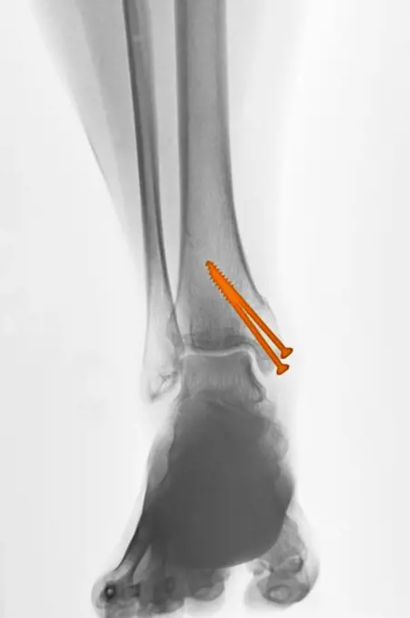
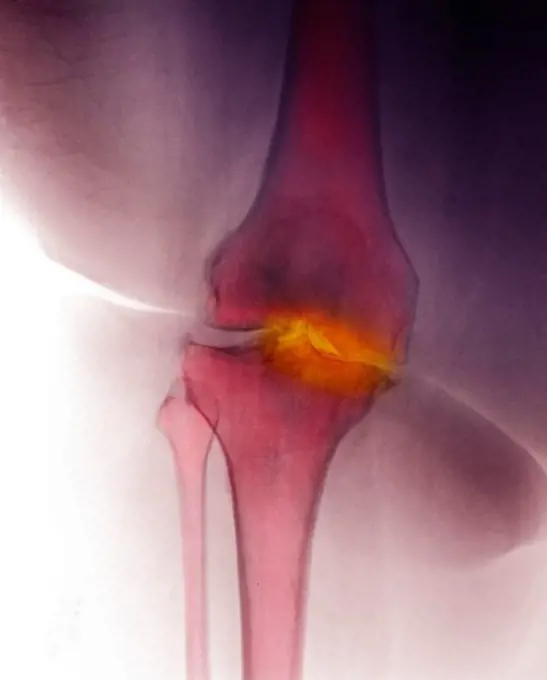
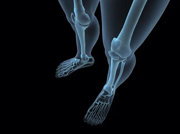
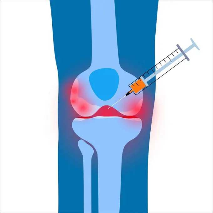
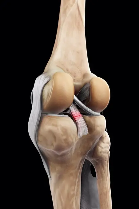
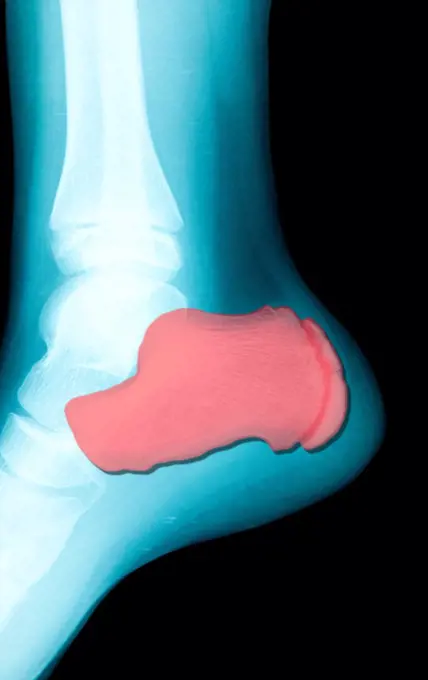
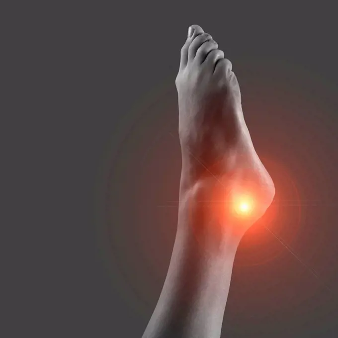
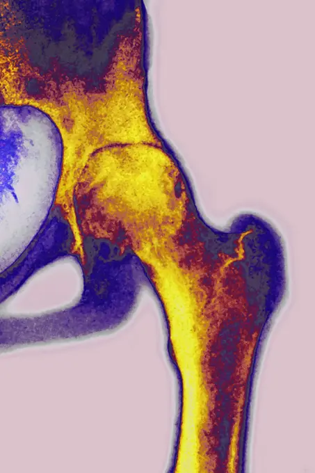
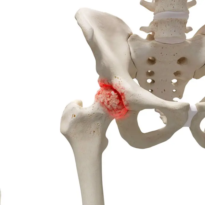
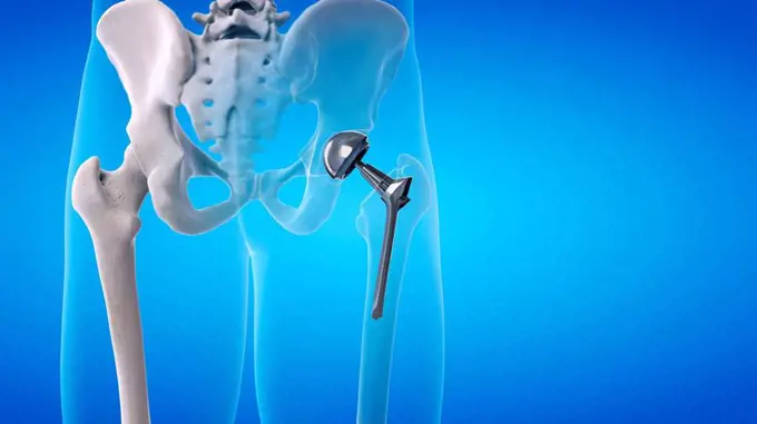
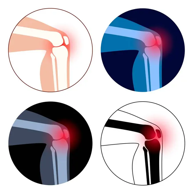
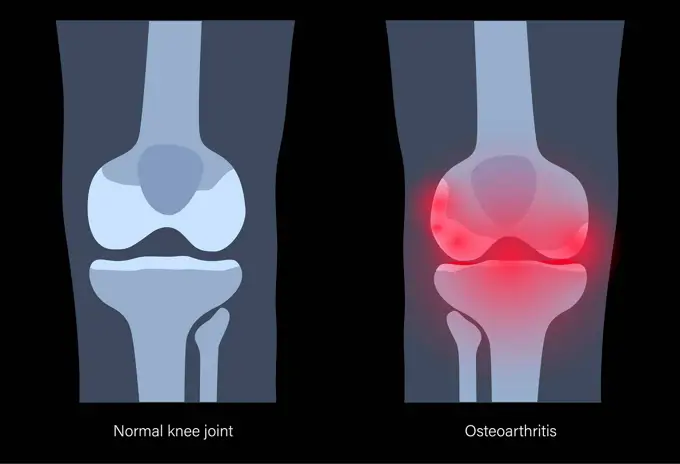
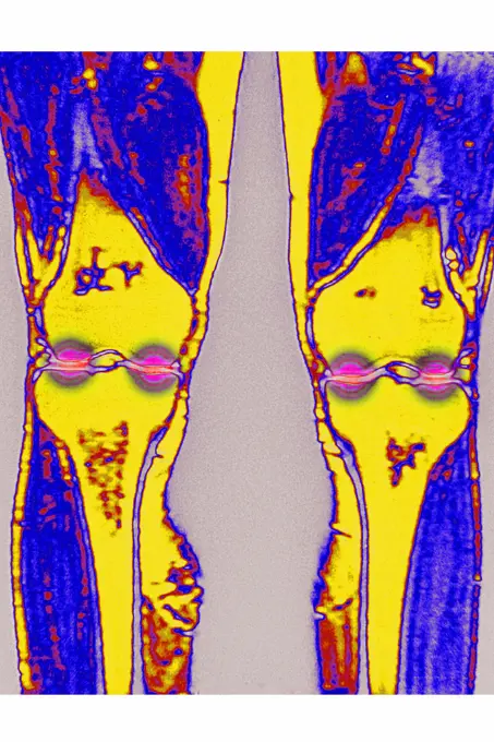
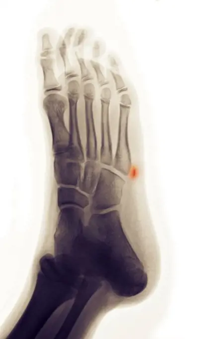
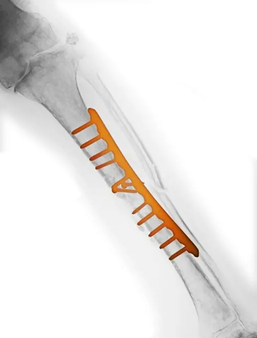
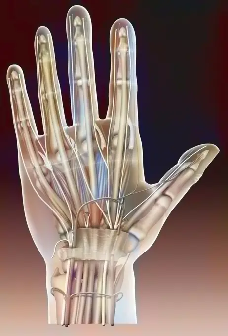
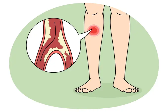
Knee and Ankle Pain Illustrations

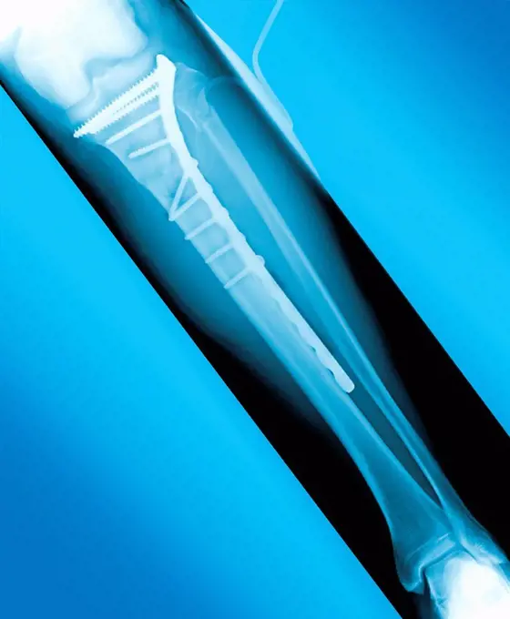
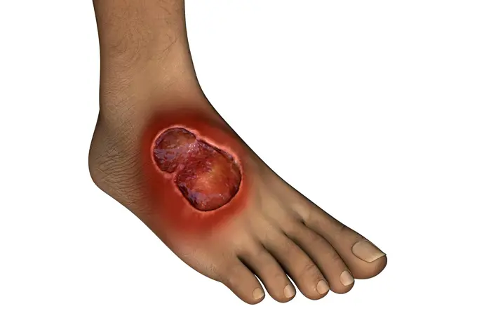
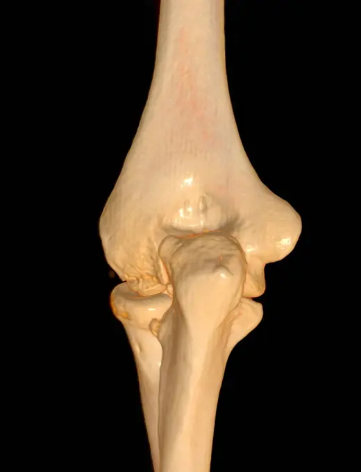
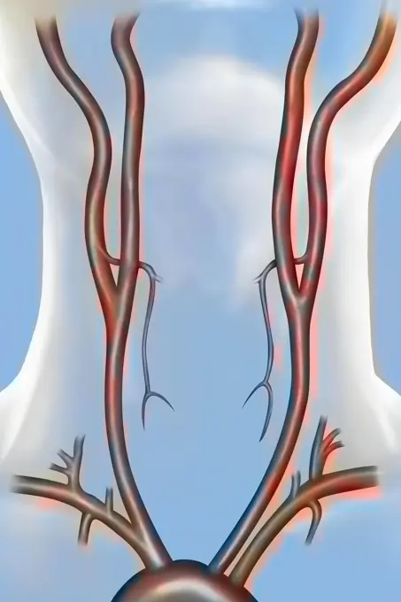
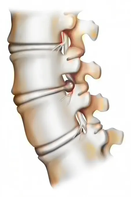
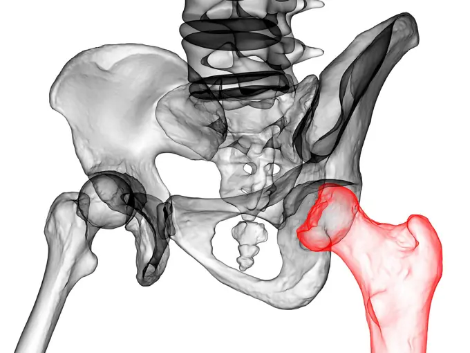
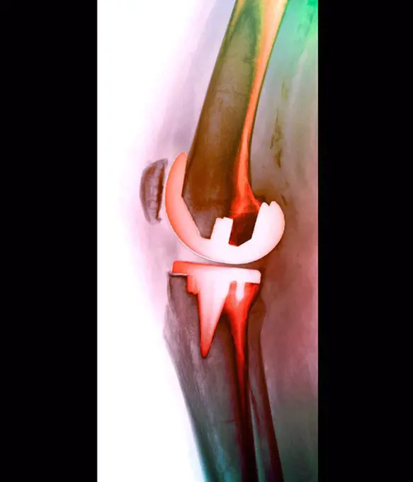
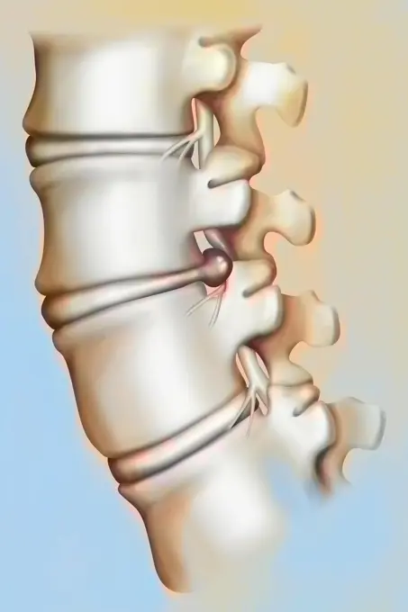
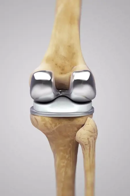
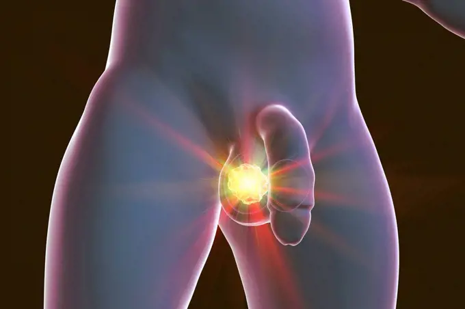
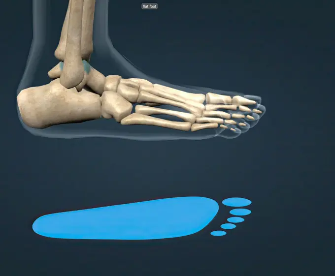
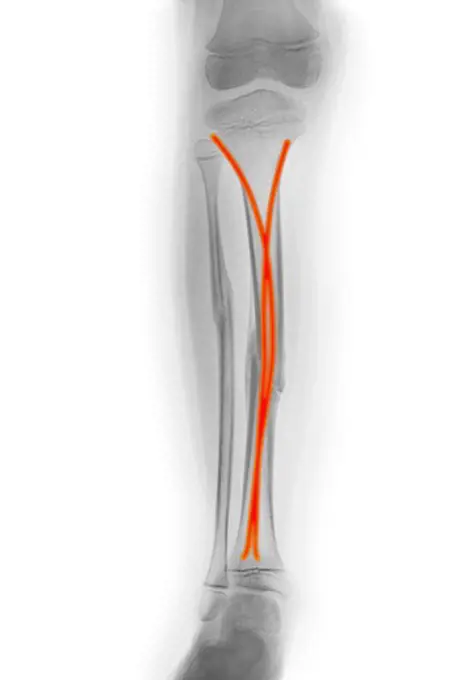
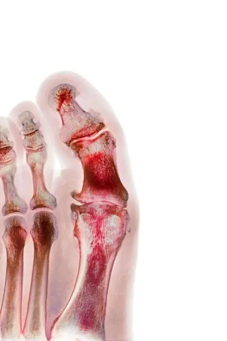
Anatomical images of painful knees and ankles, focusing on inflammation and joint health in a clear, illustrative style.

Human knee pain
Human knee pain
261 assets in this story
4297-1396
Colorized x-ray of an ankle fracture in a 19 year old woman surgically repaired with two large screws
824-63178455
X-ray of the left knee of a 30-year-old man. No anomaly
4297-1188
Colorized x-ray of the knee in a massively obese 57 year old woman showing degenerative caused the extreme stress on the knee from being overweight
4128-V58575846
4128-30420309
Arthritis of the foot, conceptual illustration
4128-30420349
Hip pain, conceptual illustration
1428R-463
X-ray view of knees
4128-19248939
Painful knee joints, illustration.
4128R-11288578
Painful knee, computer artwork.
4128-20045113
Knee injection, illustration
4128R-13586909
Human calf pain, illustration.
4128-30420324
Arthritis of the foot, conceptual illustration
824-63193842
Illustration of a right side laparotomy-thoracotomy. This operation involves the partial removal of the oesophagus, carrying out a thoracic lymph node excision and reconstruction of the continuity between the remaining oesophagus and gastroplasty (transformation of part of the stomach to replace the oesophagus). This operation is practised in the case of cancers in the lower and upper third of the oesophagus.
4128-19247912
Arthritic foot, illustration.
4269-20408361
Frontal X-ray of a total hip replacement , the replacement hip consists of a ball and shaft, embedded in the femur (thigh bone, running up from bottom centre), which moves freely in the prosthetic socket implanted in the pelvis .
4128R-14637712
Arthritis in the hip, illustration
1525-56857791
Hip Replacement Surgery concept as an artificial joint or prosthesis with orthopedic surgery inserting a metal ball and socket to replace a damaged by disease Femure joint in a 3D illustration style.
4128-30420407
Arthritis of the knee, conceptual illustration
4378-5277
Right Knee Ligaments
5507-45886354
Swelling of the feet and ankles from infected or injury
4378-20393184
Broken Metatarsal
4128R-14012
Pinned broken leg. Coloured X_ray of a metal plate and screws in a fractured tibia shin bone.
4378-20393344
Tennis Elbow
1525-57102544
X-ray shots. Human skeleton and body parts, medical diagnosis and healthcare concept, anatomy and science vector set. Radiology for medicine, chest, knee, teeth and foot trauma or fracture. X-ray shots. Human skeleton and body parts, medical diagnosis and healthcare concept, anatomy and science vector set
824-63178481
Severe osteonecrosis of the knee of a 78 years old patient. RMN
4128R-11477896
Human shoulder muscle pain, computer artwork.
4128-19052631
Painful Sacroiliac joint, illustration
4378-20014039
Foot Bones
824-63163791
Bone system: hip prosthesis.
4128R-11472889
Human hip replacement, computer artwork.
4128-30418886
Painful knee, illustration
4378-20393435
Tennis Elbow
4128-28769242
Buruli ulcer on a foot, illustration
4128-30419963
Healthy elbow joint, CT scan
824-63163804
Modification of the silhouette linked to osteoporosis (vertebral compression).
6188-66528719
Arterial vascularization of the neck. The arteries of the neck are essentially composed of the carotids, anteriorly, and the vertebral arteries not visible here, posteriorly. They irrigate the structures of the neck, the head and the brain. The carotid arteries arise from the aortic arch. The common carotid arteries, right and left, divide at the level of the thyroid into internal and external carotids. The external carotids give rise to numerous collaterals, among which the superior thyroid arteries, represented here. The internal carotids give rise to only one branch: the ophthalmic artery. The course of the carotids is similar on the right and on the left, the only difference being that the right common carotid arises from the brachioc 016238_048
4128R-15405182
Man with foot pain, illustration
4128-19358339
Achilles tendon injury, illustration.
1525-23407918
Pain in legs. knee pain, heel pain. Vector illustration.
5507-45870598
Swelling of the knee  from infected or injury
4378-5751
Posterior Cruciate Ligament Injury
6188-66528613
Herniated disc. A herniated disc is a protrusion of an intervertebral disc, which can then compress the spinal cord. It is generally located at the level of the lumbar vertebrae. Herniated disc 016238_024
6188-56009409
Kompressionsverband bei Venenschwäche *** Compression bandage at Venous insufficiency 1026902177
1899-86029
Sever's Disease
4128-28968669
Legg-Calve-Perthes disease, illustration
1525-56849494
Watercolor Illustration of firs aid person with injured hand with wrist, bandaged arm. Physiotherapy clipart. Watercolor Illustration of firs aid person with injured hand, bandaged arm. Physiotherapy clipart.
1525-75819423
Patient with arm problem, x ray scan of elbow joint. Anatomical structure, care and diagnosis for pain in arm. Rheumatism and surgery, clinics and hospital treatment and cure. Vector in flat style. X ray of human elbow, screen or scan in clinics
4128-111579522
Hip replacement, computer illustration.
824-63164257
Healthy ankle and ankle with hemophilic arthropathy in frontal cut.
1525-56234392
A tear in the anterior cruciate ligament is one of the most common knee injuries. 3D rendering. A tear in the anterior cruciate ligament is one of the most common knee injuries.
4128-V58575815
4128R-14939488
Ankle pain, conceptual image
1525-76004826
Knee pain icon cartoon vector. Arthritis joint. Treatment articulation. Knee pain icon cartoon vector. Arthritis joint
4128R-5600
Prosthetic knee joint, X_ray
824-63186559
Representation of a varicose vein. On the left of the drawing, a healthy vein, with its normally functioning valves ensuring normal venous return to the heart without reverse blood return. On the right, zoom on the varicose vein caused by a malfunction of the valves. The varicose vein is between the muscle and the skin which blisters under the swelling of the vein.
6188-66528612
Herniated disc. A herniated disc is a protrusion of an intervertebral disc into the spinal canal, which then compresses the spinal cord. It is usually located at the level of the lumbar vertebrae. Herniated disc 016238_023
4378-20393178
Knee Replacement
4128R-14939786
Testicular cancer treatment, illustration
1525-76004831
Patient spine pain icon cartoon vector. Arthritis joint. Medical treatment. Patient spine pain icon cartoon vector. Arthritis joint
824-63211559
Osteoarthritis of the hip seen on a frontal x-ray.
4128-19247927
Hip joint, illustration.
4269-25683
Knee arthrosis. X-ray of knees affected by arthosis (green), also called osteoarthritis or gonarthrosis. It is a joint disease that can cause cartilage destruction (seen as a reduction in the joint space), bone erosions, and tendon inflammation.
4128R-14637710
Arthritis in the hip, illustration
4128-15980501
Hip replacement, computer illustration.
4128-30420355
Arthritis of the knee, conceptual illustration
4128-19247914
Arthritic foot, illustration.
4128-30420412
Arthritis of the knee, conceptual illustration
824-63164609
Angioplasty (stenting) helps restore blood flow to an artery blocked by an atheroma.
824-63164235
Healthy ankle with hemophilic arthropathy in frontal cut.
4128-30419980
Fractured knee, CT scans
824-63164259
Healthy ankle and ankle with hemophilic arthropathy in frontal cut.
824-63218200
Osteoarthritis of the knee. Wear then destruction of cartilage in various areas of the knee joint. Frontal MRI scan of the knees.
4297-1337
Colorized x-ray of an avulsion fracture of the 5th metatarsal of the foot
4128-V58576261
824-72488699
Illustration of leg pain, and more particularly venous insufficiency (here, at the level of the popliteal vein and the external saphenous vein).
4128R-15545616
Illustration of a wrist replacement
4128-30420312
Arthritis of the foot, conceptual illustration
4297-1514
Colorized x-ray of a fracture of the tibia and fibula repaired with a plate and screws
824-63164238
Healthy ankle with hemophilic arthropathy in frontal cut.
4128-30418901
Obese man's painful knee, illustration
824-63164959
Anatomy of the palmar face of the hand with the median nerve, tendons, radial artery.
5507-33599687
Person suffer from PAD disease in legs
1525-56184097
Flat feet is a common condition in which the arches on the inside of the feet flatten when pressure is applied to them. 3D illustration. Flat feet is a common condition in which the arches on the inside of the feet flatten when pressure is applied to them.
1525-27452422
Foot Bone Anatomy Vector Illustration
1525-57010227
3D illustration folding knife camping, compact expedition knife. Penknife multi-tool.
4297-1392
Lower leg x-ray of a 7 year old boy who fractured his tibia and fibula. This is the post-operative x-ray with two rods inserted into the marrow space of the tibia to stabilize the fracture
4128-19247902
Arthritic lumbar spine, illustration.
4128R-13374580
Foot bones
1525-75819540
Aches above knees, isolated personage showing problematic part of body. Symptom of hamstring tendonitis, arthritis or bursitis. Healthcare minimalist drawing with area of pain, vector in flat style. Pain above knee, aches and health issues vector
4128R-12922489
Degenerative foot deformation, X-ray
4269-1059
Side viewed three-dimensional computed tomographic reconstruction of the lombar vertebras and the intervetebral spaces. The sacrum is at the bottom
4128R-5275
Total hip replacement, X_ray
4269-25677
Knee arthrosis. X-ray of knees affected by arthosis (green), also called osteoarthritis or gonarthrosis. It is a joint disease that can cause cartilage destruction (seen as a reduction in the joint space), bone erosions, and tendon inflammation.
1525-56234216
Dialysis is a procedure that filters blood outside the body and returns the filtered blood to the bloodstream. 3D rendering. Dialysis is a procedure that filters blood outside the body and returns the filtered blood to the bloodstream.
4128R-13411879
Foot bones
1525-56184099
Flat feet is a common condition in which the arches on the inside of the feet flatten when pressure is applied to them. 3D illustration. Flat feet is a common condition in which the arches on the inside of the feet flatten when pressure is applied to them.
4128R-28750
Knee bones, computer artwork.
824-63178465
Left knee prosthesis in a 45-year-old man.
1525-27393989
Disease of  head and neck, the joints and bones,human joints, skeletal spinal bone structure of Human Spine, medical health care flat vector illustration.
824-63165072
Childbirth, stage 2: the baby's head is out.
PREVIOUS
of 3
NEXT
2401 S. Ervay, Suite 206
Dallas, Texas 75215
United States
Get Started
Free ResearchMy BoardsMy Cart
For Creators
How To License Your ContentContributor PortalFrame of Mind
Resources
API accessPricing
Contact
+1 866 236 0087help@viewpictures.co.uk Contact form
©2026 View Pictures. All Rights Reserved. -A
This site is protected by reCAPTCHA and Google Privacy Policy and Terms of Service apply.