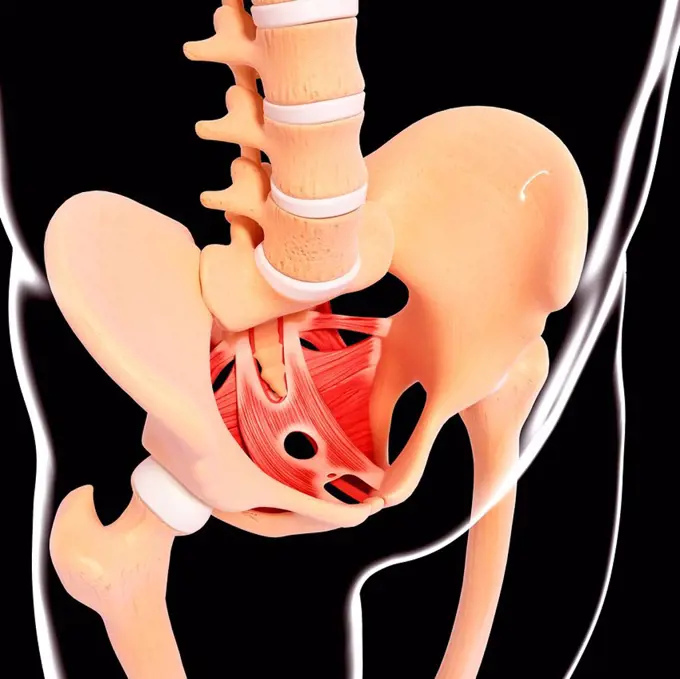
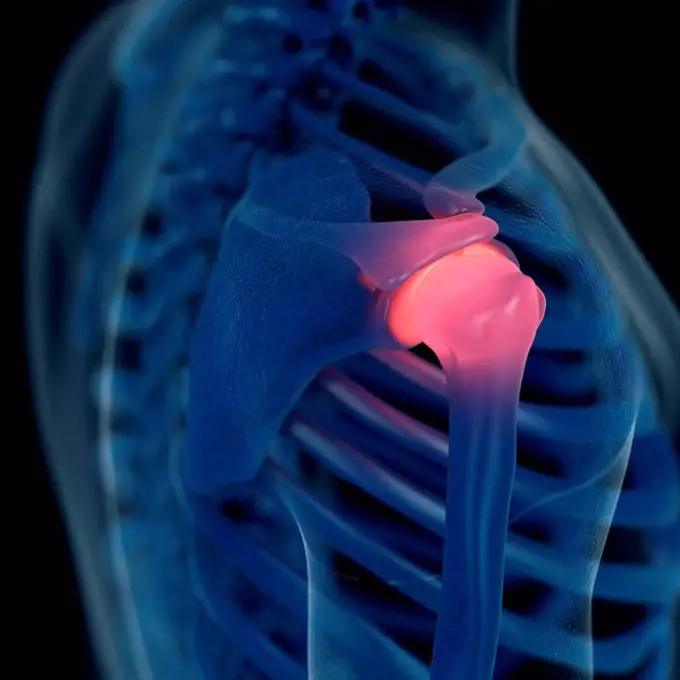
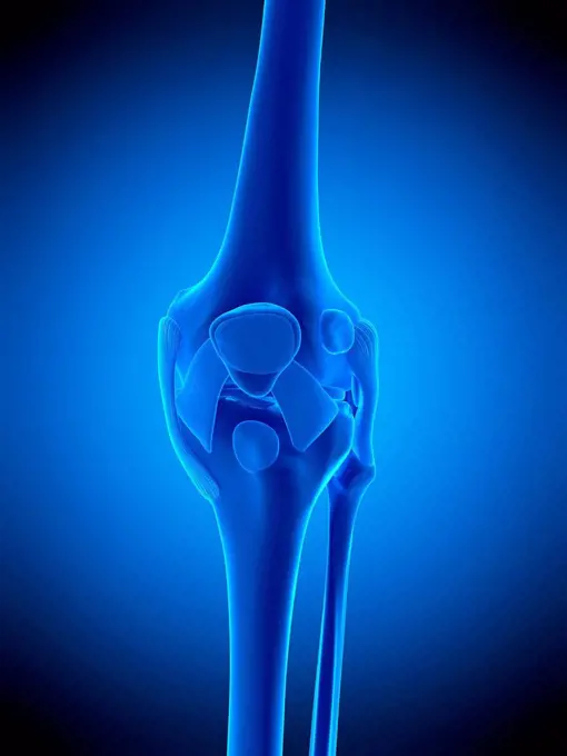
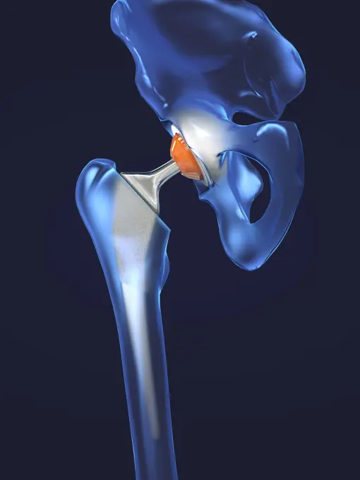
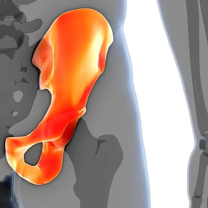
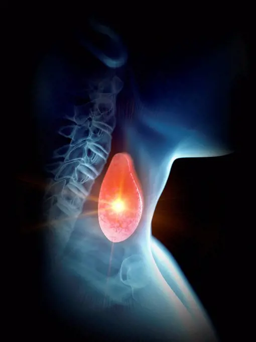
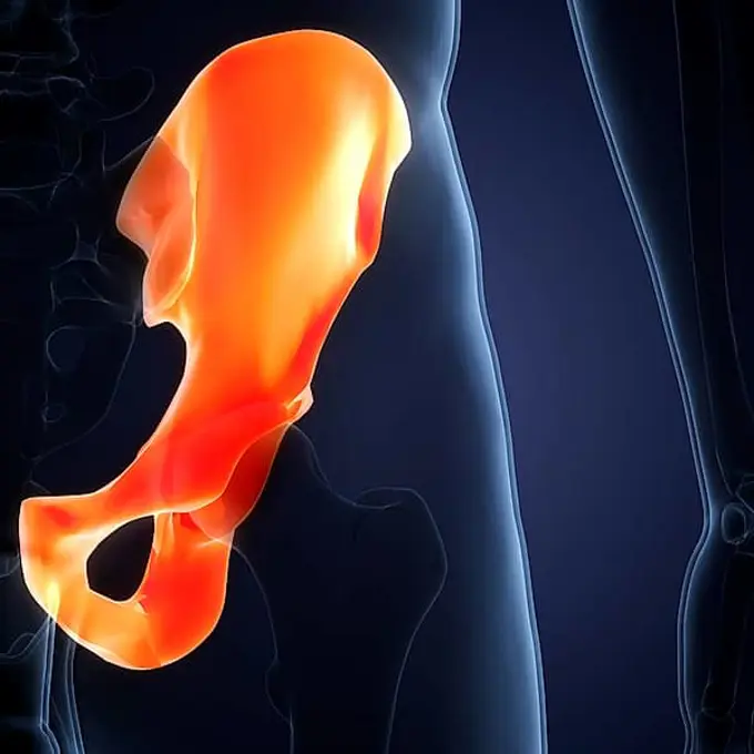
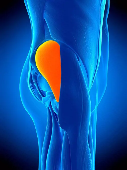
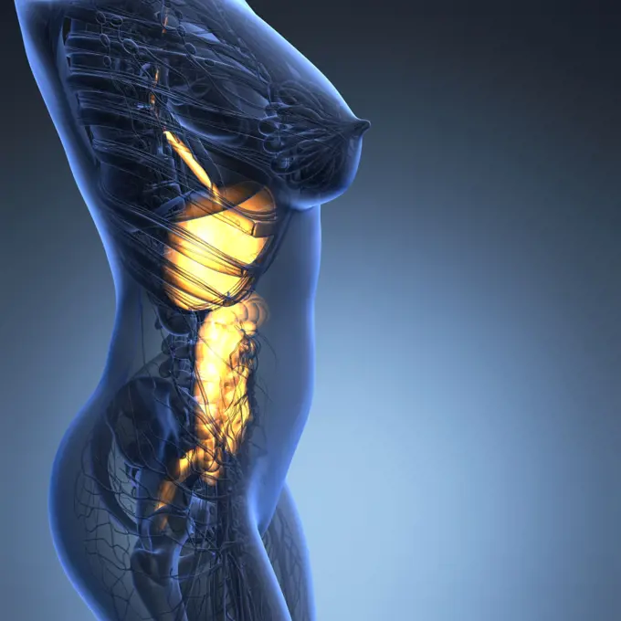
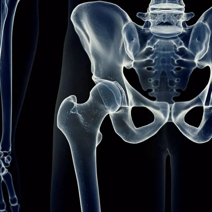
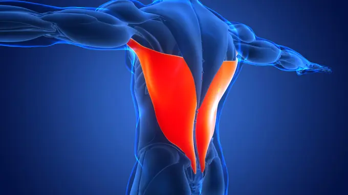
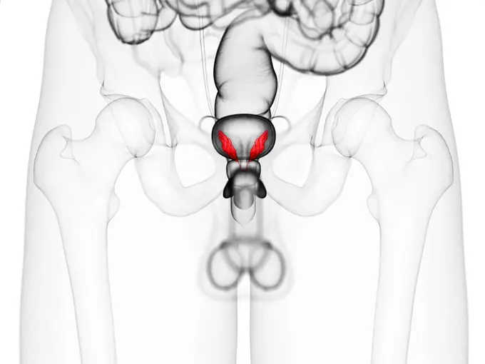
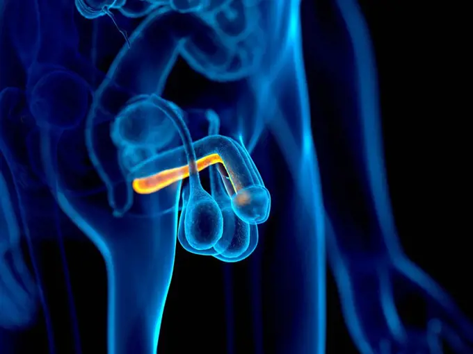
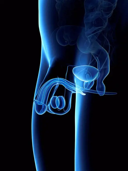
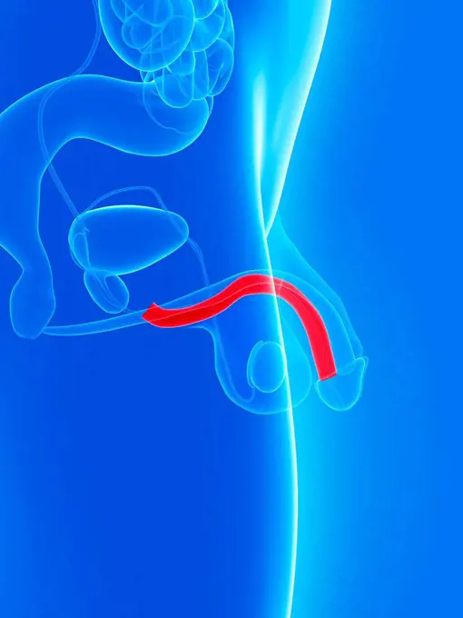
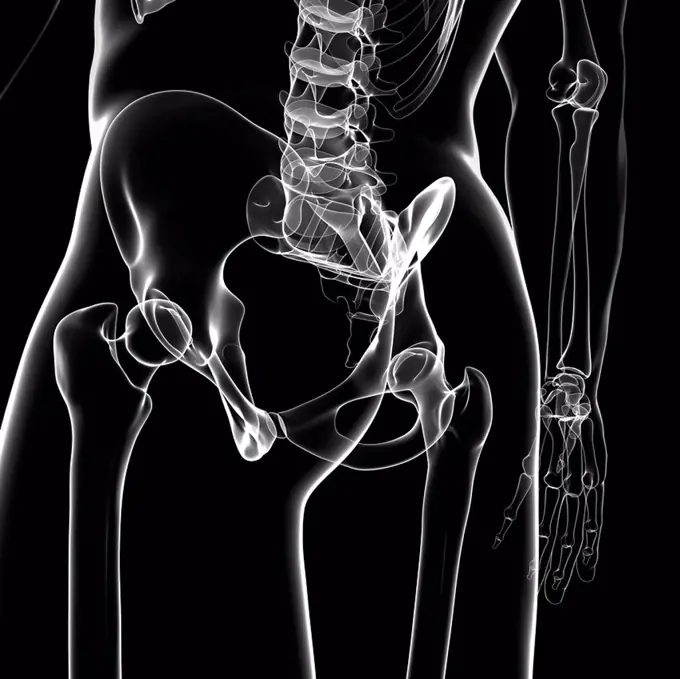
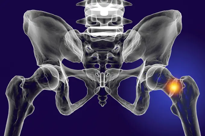
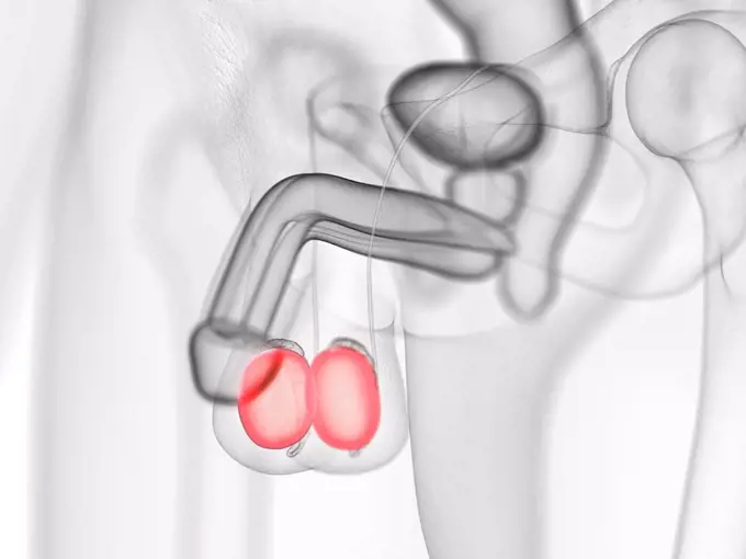
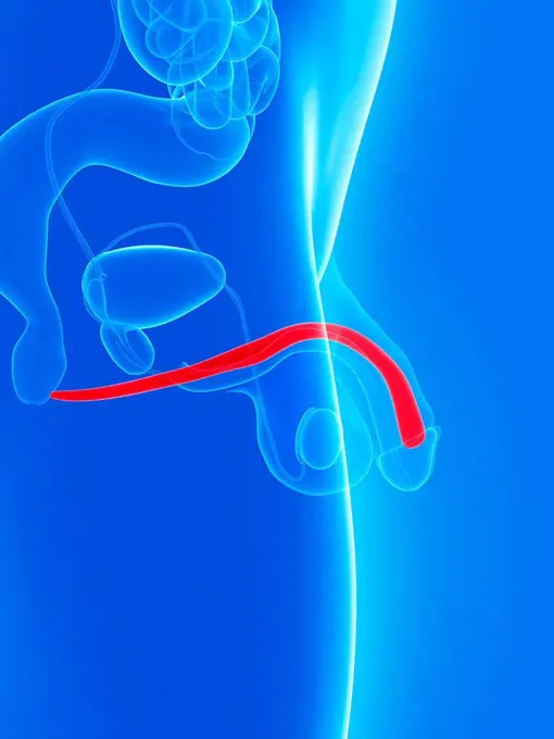
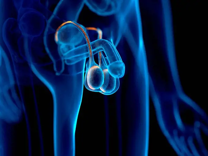
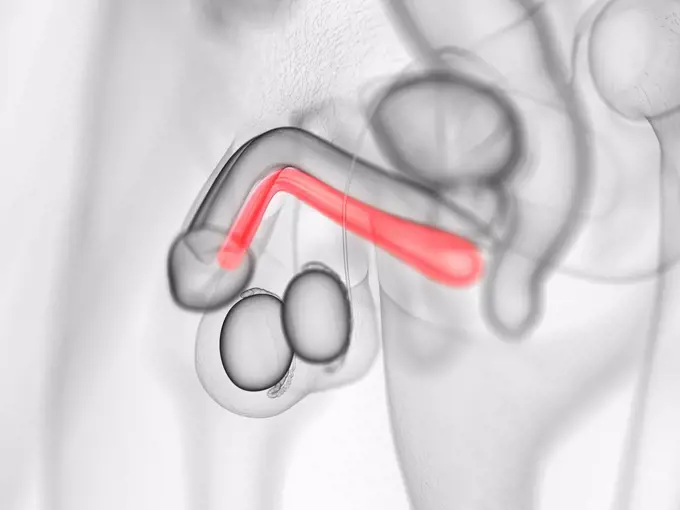
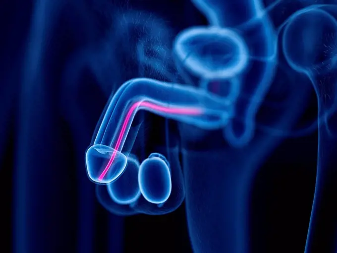
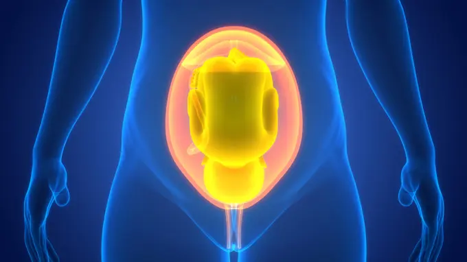
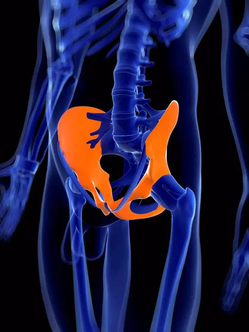
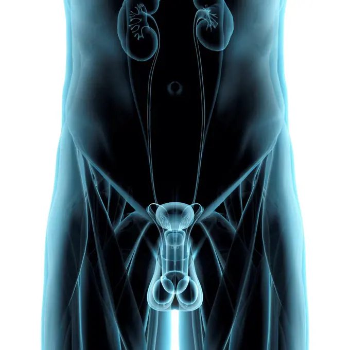
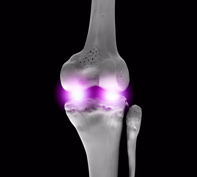
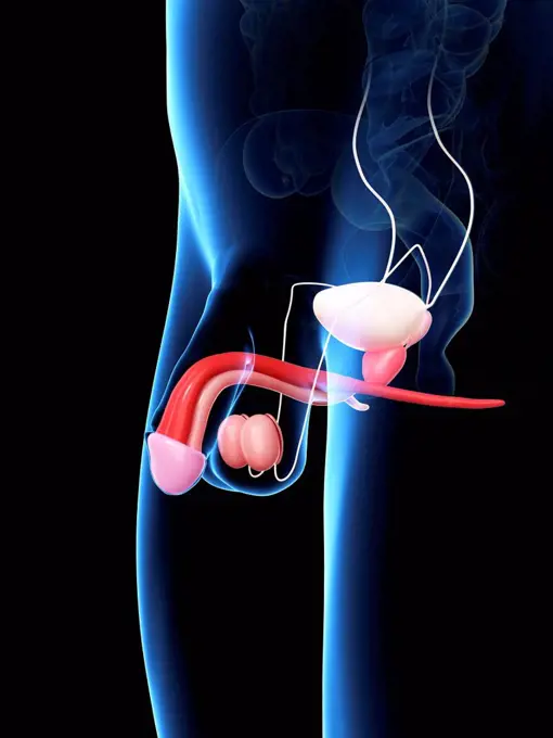
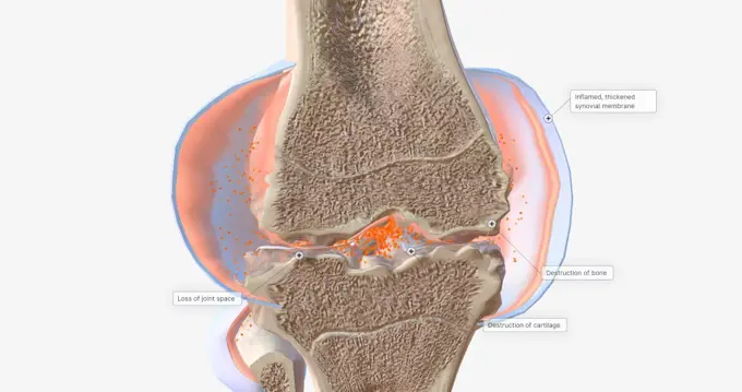
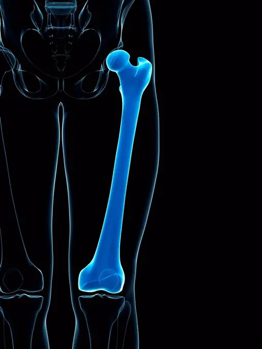
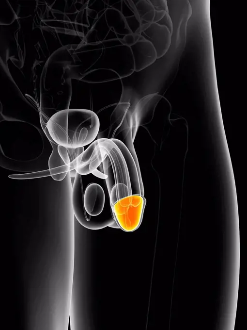
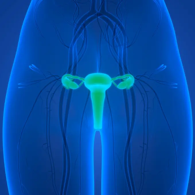
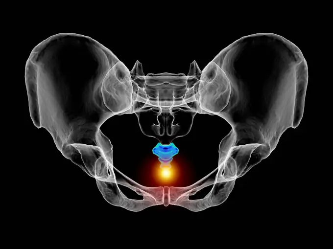
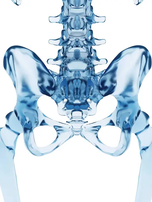
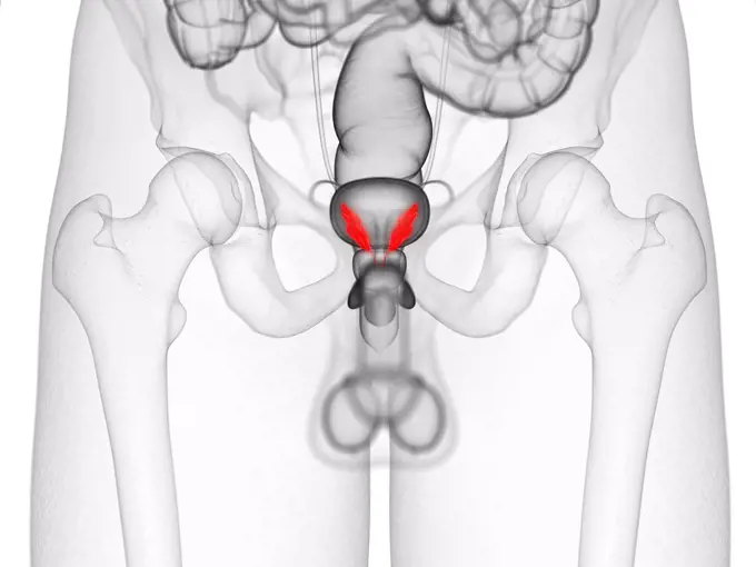
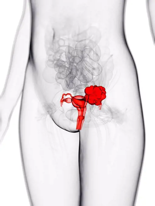
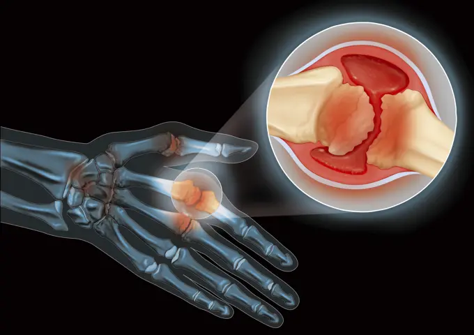
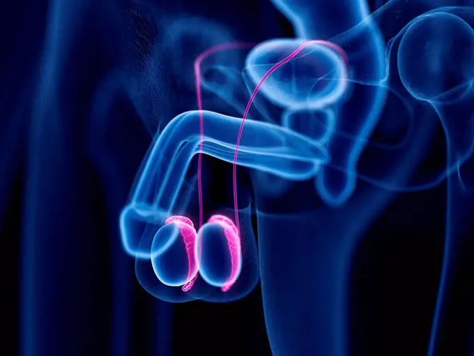
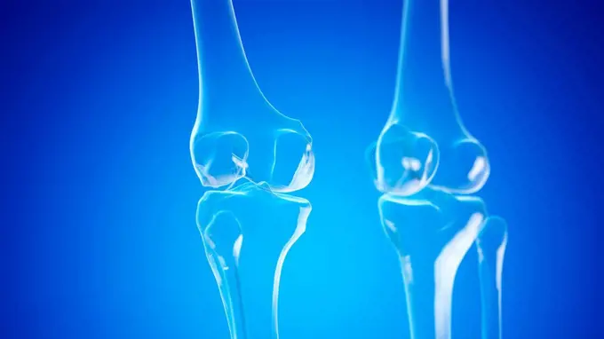
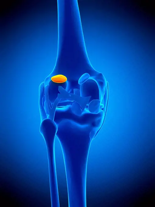
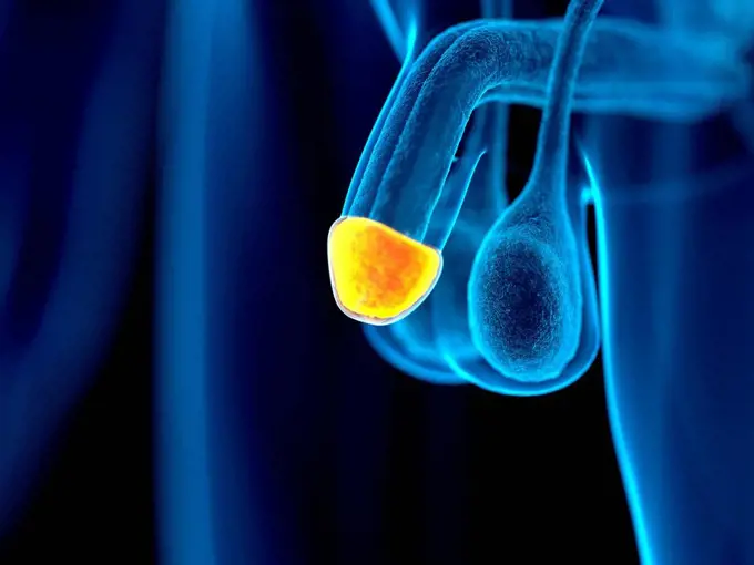
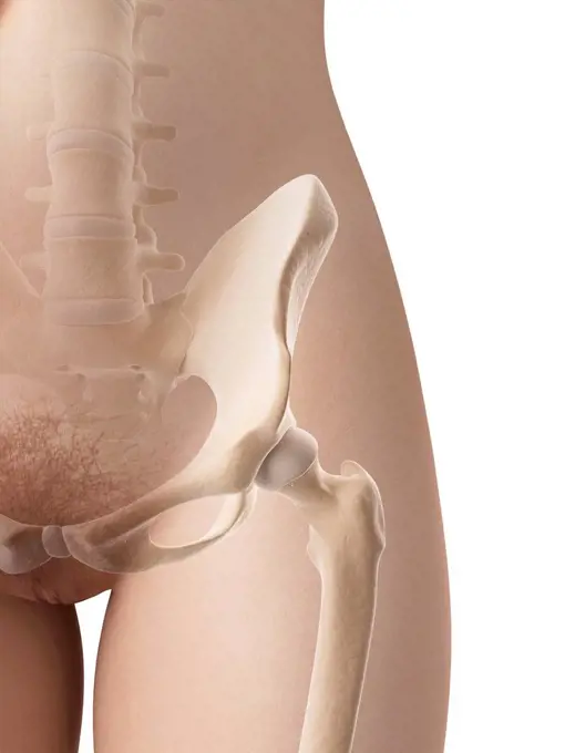
Male Skeleton And Pelvic Anatomy

Computer-generated images of the male skeleton focusing on pelvic and urinary system anatomy, highlighting joints and organs with a blue glow.

Human knee joint, computer artwork.
Human hip musculature, computer artwork.Splenic cancer, illustrationIllustration of a painful shoulderIllustration of the knee ligaments.Symphysis pubis dysfunction, illustrationKnee bones, illustrationSeminal vesicles, illustrationFunktion eines Hüftgelenksimplantats oder einer Hüftprothese in Frontalansicht - 3d-Illustration Function of a hip joint implant or hip prosthesis in frontal view - 3d illustration Copyright: xZoonar.com/ChristophxBurgstedtx 22602631Human back pain, computer illustration.Arteriography or angiography (radiological examination of the arteries of a lower limb seen in profile).Male pelvis bones, computer artwork.3D Illustration Concept of Human Muscular System Leg Muscles Gluteus Medius Muscle AnatomyIllustration of the gluteus minimus muscle.Human spine and back pain, illustration.Illustration of the obturator internus muscle.3D Illustration Concept of Human Skeleton System Hip Bone Joints AnatomyHuman hip bone, computer artwork.Knee bones, illustrationIllustration of a painful thyroid glandHip bone, illustrationFemale anatomy, computer artwork.3D Illustration Concept of Human Skeleton System Hip Bone Joints AnatomyIllustration of the gluteus medius muscle.science anatomy of woman body with glow digestive systemIllustration of the hip jointHuman Muscular System Torso Muscles Latissimus Dorsi Muscle Anatomy 3D Illustration Concept of Human Muscular System Torso Muscles Latissimus Dorsi Muscle Anatomy Copyright: xZoonar.com/magicminex 217478273D Illustration Concept of Human Fetus Baby in Womb AnatomySacrum bones, illustrationFemale anatomy, computer artwork.Seminal vesicles, illustrationCorpus cavernosum, illustrationPenis anatomy, computer artwork.Penis anatomy, computer artwork.Human upper leg muscles, computer artwork.Human fetus age 15 weeks, illustration.Human knee jointHuman pelvic bones, computer artwork.3D Illustration Concept of Human Fetus Baby in Womb AnatomyFracture of the femur neck, illustrationAnatomy of the pelvis bones, illustration3D medical man with skeleton spine highlightedTestes, illustrationIllustration of the navicular bone.Three-quarter view of the male pelvis and hip joints.Human elbow joint, computer artwork.Penis anatomy, computer artwork.Human diaphragm, illustration.Human appendix, illustrationFront view of knee joint opened to show pannus.Epididymis, illustrationSkeletal system of someone jogging, computer illustration.Corpus spongiosum, illustrationHuman knee bonesUrethra, illustrationBladder cancer, computer artwork.Menschlicher Fötus Baby im Mutterleib Anatomie 3D Illustration Concept of Human Fetus Baby in Womb Anatomy Copyright: xZoonar.com/magicminex 22683670Hip bones, computer artwork.Hip joint, illustrationMale reproductive system, frontal xray blue on white backgroundHuman knee joint, computer artwork.Penis anatomy, computer artwork.RA is caused by immune cells attacking the body’s own synovial membranes, leading to joint swelling and the formation of auto-antibodies. 3D rendering. RA is caused by immune cells attacking the body’s own synovial membranes, leading to joint swelling and the formation of auto-antibodies.Illustration of a liver tumourCorpus cavernosum, illustrationHuman hip pain, computer artwork.Thigh bone. Computer artwork showing the femur bone.Painful atlas bone, illustration.Penis anatomy, computer artwork.Female Reproductive System Anatomy 3D Illustration Concept of Female Reproductive System Anatomy Copyright: xZoonar.com/magicminex 21262342Coccyx pain, conceptual illustrationFemoral Head Disease and osteonecrosis or avascular necrosis and aseptic necrosis with a healthy hip compared to an osteoarthritis damaged pelvic joint in a 3D illustration style.Human pelvis, IllustrationSeminal vesicles, illustrationHuman fetus age 9 weeks, illustration.Human larynx, computer illustration.Male prostate gland, computer illustration.Uterine cancer, computer artwork.human bones radiography scan image human bones radiography scan. x-ray image Copyright: xZoonar.com/OxanaxRishnyakx 6223857Illustration of a hand suffering from rheumatoid arthritis, with a close-up of the joint between the second metacarp and the proximal phalanx of the index finger. The illustration shows the deterioration of the bone surface and cartilage, the thickening of the synovial membrane, the effusion of the synovial fluid and swelling.Epididymis, illustration3D Illustration Concept of Human Skeleton System Clavicle Bone Joints AnatomyButtock muscles, computer artwork.Breast cancer, conceptual computer illustration.Knee joints, illustrationIllustration of the lateral gastrocnemius bursa.Illustration of testicle cancerGlans penis, illustrationHuman shin bone and ligament, computer artwork.Anatomy of the female breast, computer illustration.Illustration of pancreas cancerFemale reproductive system, artworkProstate caverns and diverticulum in the bladder. Frontal pelvic x-ray. (Photo by: BSIP/UIG via Getty Images)Calcaneus bone, illustrationColoured computed tomography (CT) scan of the healthy full bladder (green) of a 45 year old patient.Human fetus age 11 weeks, illustration.Corpus spongiosum, illustrationFemale hip bone, computer illustration.