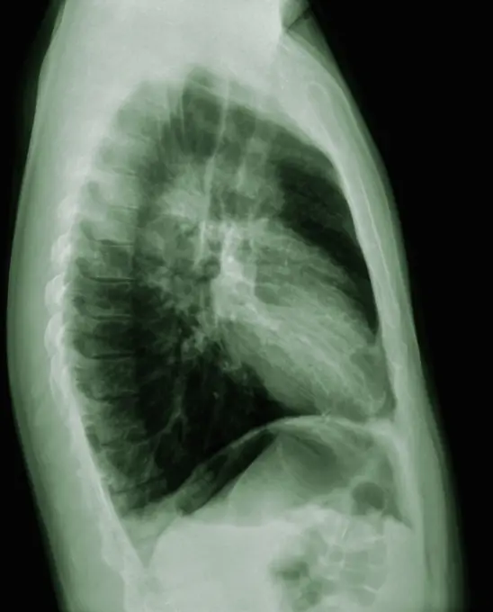
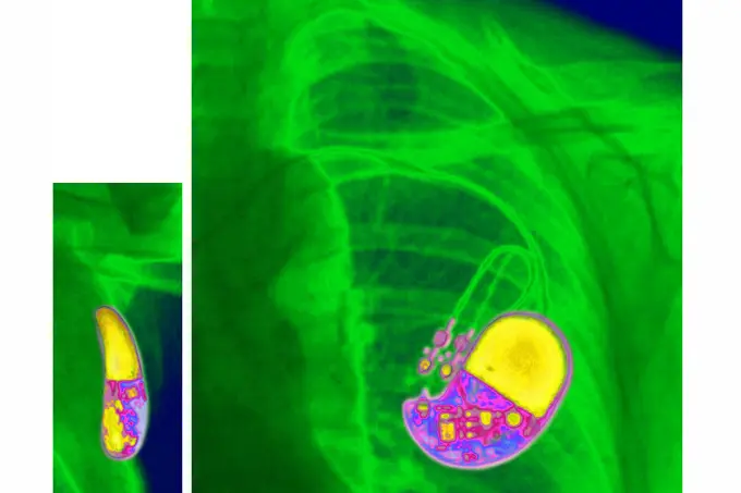
My Boards
View Pictures
  • My Boards

Medical Imaging and Diagnostics

Enhanced CT and MRI scans highlighting medical conditions such as stents, lung cancer, and vertebral issues, with color-coded identifiers for clarity.

Lung cancer located in the upper right lobe. Frontal chest x-ray.
207 assets in this story
1899-53511469
MRI of pulmonary cystic fibrosis (viscous mucus disease, cystic fibrosis affecting glandular epithelia). Radial MRI of the lungs.
824-63224702
MRI of pulmonary cystic fibrosis (viscous mucus disease, cystic fibrosis affecting glandular epithelia). Radial MRI of the lungs.
6145-44783470
This 2001 Mars Odyssey image shows a region of Aonia Terra criss-crossed with a multitude of dust devil tracks. The tracks show up well on the dusty plains.
4128R-11287909
Secondary lung cancer. Coloured X-ray of the chest of a 52 year old female patient with metastatic (secondary) lung cancer (red).
824-63214250
X-ray of urinary schistosomiasis. Frontal abdominal-pelvic x-ray.
824-63192443
Horizontal cross-section scan of the abdomen.
4413-28972469
Ichthyosaur. Themnodontosaurus sp. Toarcian (180 million years). Germany. Cross-section of a rostrum, showing the different stages of tooth growth (large tooth with root, small sprouting tooth without root), diagnostic of the mechanism of continuous tooth growth in ichthyhyosaurs. - Blouet brothers collection
4128R-15545768
Spine in ankylosing spondylitis, X-ray
824-63211582
Posterior cortical atrophy (Bensons syndrome) seen on a radial CT-scan.
6188-55645086
Abdomen man, man abdomen man, man, ct recording copyright: xzoonar.com/dr.xnorbertxlangeex 13790157
824-57656855
Arteriography or angiography (radiological examination of the arteries of a lower limb seen in profile).
6188-55645083
Abdomen man, man abdomen man, man, ct recording copyright: xzoonar.com/dr.xnorberbertxlangeex 13790140
824-63217270
Pulmonary sarcoidosis seen on a frontal chest x-ray.
4269-24988
Lungs malformation. 3D computed tomographic (CT) scan reconstruction of the left lung affected intra-lobal pulmonary sequestration, a congenital malformation of the lung caracterized by the separation of a part of bronchopulmonary tissue from its normal connexions.
1525-28067503
Transverse segment through the lower part of the femur of a giraffe, vintage engraved illustration. From the Universe and Humanity, 1910.
824-63170800
This axial MRI image of the lumbar spine reveals a spondylodiscitis at the L5 level.
824-63224708
MRI of pulmonary cystic fibrosis (viscous mucus disease, cystic fibrosis affecting glandular epithelia). Radial MRI of the lungs.
4269-27279
Cerebral aneurysm. 3D Computed Tomography (CT) scan reconstruction of an aneurysm of the right vertebral artery. An aneurysm is a bulge of a vessel due to the dilatation of its wall. A ruptured aneurysm can be fatal.
4128R-30937
Narrowed arteries. Coloured 3D computer tomography (CT) angiogram of the pelvis of a 66 year old male patient with narrowing (stenosis) of the iliac a...
824-63207408
Hip algodystrophy, seen in a frontal section MRI scan.
1439-57940616
Intricate patterns of human anatomy revealed in a chest Xray
6188-55645089
Abdomen man, man abdomen man, man, ct recording copyright: xzoonar.com/dr.xnorbertxlangeex 13790162
4269-24914
Uterine fibroma. Ultrasound scan of a uterus with a benign tumour of the connective tissue, a fibroma, seen as the irregular white mass.
824-63217245
Pacemaker (cardiac pacemaker) implanted in the thorax. Lateral and frontal chest X-ray.
824-63208116
Osteoporosis with compression of the dorsal and lumbar vertebrae, seen on a sagittal plane MRI scan.
824-63207432
Heart and lungs seen in a radial cross-section MRI scan.
6188-55645098
Abdomen man, man abdomen man, man, ct recording copyright: xzoonar.com/dr.xnorberbertxlangeex 13790172
1899-54026860
Intercranial neurinoma, seen on a radial section MRI scan.
4128R-11287630
Diverticulitis. Coloured X-ray of the colon of a patient with diverticulitis. A diverticulum is a small pouch-like structure that protrudes out of wea...
1899-53511914
Pulmonary metastases (secondary to kidney cancer), visualized by a frontal chest X-ray. (Photo by: CAVALLINI JAMES/BSIP/UIG)
824-63211058
Fracture of a dorsal vertebra, with damage to the spinal cord with a bony fragment in the neuroforamen. Cross-section scanner.
6188-68090444
Human Digestive System Anatomy 3D Illustration Concept of Human Digestive System Anatomy LicenseRF Copyright: xZoonar.com/magicminex 23022630
4269-27488
Shoulder chondrosarcoma. 3D Computed Tomography (CT) reconstruction scan of the shoulder showing a chondrosarcoma of the shoulder blade. The tumor is evidenced by the presence of a vascularized heterogenous lytic area with bone calcifications. The sub-clavian artery is seen in yellow.
824-63224576
Prostate cancer (adenocarcinoma of the prostate), visualized by ultrasound.
4269-6990
Ultrasound scan of a 3 months old fetus sucking his thumb.
4269-27850
Intestine tumor. Three-dimensional computed tomographic reconstruction scan of the small intestine showing a Gastrointestinal Stromal Tumor (GIST), seen as a red spot in the jejunum. The intestinal probe is seen in the duodenum (brown tube).
1746-21106104
Photographic cross-section of the abdomen from the Visible Human Male
824-63165972
Aortic coarctation. Here, a considerable narrowing of the aorta is visible, just past the point where the aorta and the subclavian artery meet, causing arterial hypertension. 3D CT scan.
824-63175228
Axial CT scan of the brain showing a subdural hematoma, a collection of blood between the dura and the arachnoid.
4128R-13817880
Kidney, CT scan. False colour 3-D computed tomography (CT) scan of a kidney in frontal view.
824-63218209
Fracture of the skull, seen on a radial section scanner.
4297-1602
Radionucleotide full body scan showing cancer Radiolabeled monoclonal antibodies confirm that this patient's cutaneous t-cell lymph cancer involves the lymph nodes and skin
4128-16224852
Pomegranates, X-ray
1899-53511757
Lung cancer located in the upper right lobe. Frontal chest x-ray. (Photo by: CAVALLINI JAMES/BSIP/UIG)
824-63204737
Multiple hemorrhagic foci, in contact with the meninges. Cross-section brain MRI.
824-63224761
Uterine cancer visualized by frontal MRI.
824-65830281
Ischemic mitral insufficiency (MI) - Pathology of the left ventricle (mitral ring and subvalvular apparatus). transthoracic echocardiography (TTE).
1525-24902451
Water
4269-24997
Slipped disc. 3D computed tomographic (CT) scan reconstruction of the lower rachis showing a slipped disc. The vertebral disk (green) is crusched between the lumbar vertebras L5 and L4, and cause a lombosciatica.
6145-29296965
Reperfusion, blood flow through brain
1848-53667140
X-ray of a turtle
824-63173382
Lumbar vertebra, infiltration
4128-20041792
Fluorescein angiogram of a healthy retina, illustration
824-63173334
Time Of Flight MRA (TOF).
4269-24944
Iliac artery aneurysm. Three-dimensional (left) and plane (right) computed tomographic (CT) scan reconstruction of an internal iliac artery aneurysm. It appears as swollen bulge (red, at right). An aneurysm is a bulge of a vessel due to the dilatation of its wall. A ruptured aneurysm can be fatal.
4269-25364
Abdominal aorta. 3D computed tomographic (CT) scan reconstruction of the abdominal aorta, showing the renal arteries going to the kidneys (in orange) and the division of the aorta into the common iliac arteries (in the pelvis).
824-63178598
Sinus X-ray of a 35-year-old woman. No anomaly noted
6188-55565410
digestive system/ Verdauungsapparat digestive system/ Verdauungsapparat Copyright: xZoonar.com/curryx 2215373
4128-16224227
Cycling helmet, X-ray
824-63204709
Frontal x-ray exmaining gastrointentinal transit.
824-63222961
Stenosis on the left popliteal artery (arteriopathy obliterating of the lower limbs). Angiography of the lower limbs.
824-63170775
This axial MRI image of a left hip shows osteoid osteoma of the femoral head.
4128-20041373
Retina damage from diabetes, illustration
824-63123127
Aorta-coronary bypass (ACB), viewed under angiography.
824-63217829
Screw and plate osteosynthesis on a fracture of the fibula. Frontal x-ray: tibia, fibula, malleolus and astragalus.
824-41259
RECTAL CARCINOMA. RECTAL CARCINOMA Institute of Nuclear Medicine, University Hospital of Lille, France.   PET Scan (Positron emission tomography).  Colorectal carcinoma. Several liver and lung abnormalities which contra-indicate surgical intervention.
1899-53511897
Dislocation of the cervical vertebrae, by a spinal trauma, between the sixth and the seventh cervical vertebra visualized by MRI in sagittal section. (Photo by: CAVALLINI JAMES/BSIP/UIG)
4128-20041450
Retina damage from diabetes, illustration
824-63206287
Evolutionary metastases of kidney cancer, seen on a dorsal view scintiscan.
6145-46703647
X-ray photography of heels taken by Oscar Karlowitch Larsson at Åminne, Mount or Sockholm.
4128-30419943
Human heart, CT scan
4128-20041370
Retina damage from diabetes, illustration
824-63178574
Normal X-ray of the lungs in a 26-year-old patient.
6145-44625090
North Polar Cap
4128R-11287652
Prosthetic heart valves. Coloured X-ray of the chest of a patient that has had their mitral and aortic heart valves replaced with prosthetic valves (b...
4269-24985
Lung cancer. Three-dimensional computed tomography (CT) scan of upper thorax showing a Pancoast's syndrome associated with a lung cancer.
4128-20041335
Retina damage from diabetes, illustration
4128-20041508
Retina damage from diabetes, illustration
824-63128430
X-ray showing a pacemaker. See image n0261305 on the site.
824-47216
ADENOCARCINOMA OF COLON. ADENOCARCINOMA OF COLON Institute of Nuclear Medicine, University Hospital of Lille, France.   PET Scan (Positron emission tomography).  Adenocarcinoma right side of colon, due to Metastasis of lung tumor (right middle lobe).
4269-25939
Lung scintigraphy. Gamma scans of healthy lungs.
4128-20041517
Retina damage from diabetes, illustration
824-63204738
Homogenous hepatomegaly revealed by an abdomino-pelvic scan.
824-63178737
Ultrasound of a 3 month old foetus.
824-94994
MENINGIOMA. MENINGIOMA Institute of Nuclear Medicine, University Hospital of Lille, France.   PET Scan (Positron emission tomography).  Meningioma. Upper left lobectomy: bronchial Adenocarcinoma. Hemorrhagic cerebral metastasis.
1525R-8959
824-63170761
This sagital MRI image of the thoracic spine shows a malignant tumor of a intervertebral disc with medullar compression.
6145-29294005
Lung: nocardiosis
4269-24811
Carpal tunnel. Three-dimensional computed tomographic (CT) scan reconstruction of the bones of the carpe (wrist), showing the carpal tunnel (red arrow), where the ligaments of the hand and wrist pass. The compression of a nerve by the ligaments causes the carpal tunnel syndrome (CTS).
6145-29295479
Lung: Kaposi's sarcoma
824-63204730
Breast fibroadenoma seen on a scan.
6145-54033808
Montana - Custer National Momument, Aerial Photograph.
4201-66330
Orchid (Dendrobium sp) SEM close-up view of stamen at 28x magnification
4128R-7821
Noses, artwork.
4269-24945
Intracranial aneurysm. Three-dimensional computed tomographic view of the base of a skull showing a 9 mm diameter intracranial aneurysm (red ballon shaped swelling) located on the left internal carotid artery termination,
824-65830271
Retinal detachment occurs when the tissue at the back of the eye becomes detached from the layer of blood vessels that supply the eye with oxygen. Sagittal MRI of the eye.
824-35279
LUNG TUMOR. LUNG TUMOR Institute of Nuclear Medicine, University Hospital of Lille, France.   PET Scan (Positron emission tomography). Tumor in left, upper lobe. Evolution of disease until recovery.  29 Aug 2002 : upper left lobectomy, malignant tumor if bronchi, recurrent, ganglionic micrometastasis.  18 Oct 2002 : thyroidectomy, thyroid metastasis of tumor  5 Aug 2003 : mormal PET scan.
824-63218214
Fracture of the skull, seen on a radial section scanner.
1899-54027124
Prostatic adenocarcinoma seen in a transverse section ultrasound scan.
1899-54027144
Breast cancer (adenocarcinoma), seen in two mammograms.
PREVIOUS
of 3
NEXT
2401 S. Ervay, Suite 206
Dallas, Texas 75215
United States
Get Started
Free ResearchMy BoardsMy Cart
For Creators
How To License Your ContentContributor PortalFrame of Mind
Resources
API accessPricing
Contact
+1 866 236 0087help@viewpictures.co.uk Contact form
©2026 View Pictures. All Rights Reserved. -B
This site is protected by reCAPTCHA and Google Privacy Policy and Terms of Service apply.