My Boards
View Pictures
  • My Boards

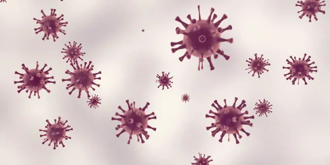
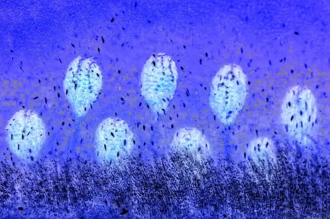
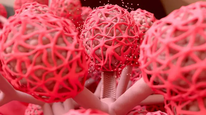
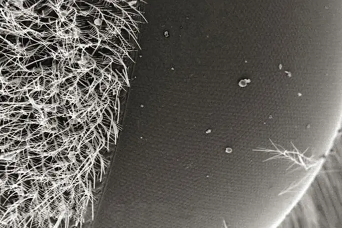
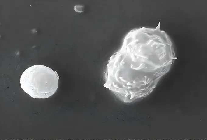
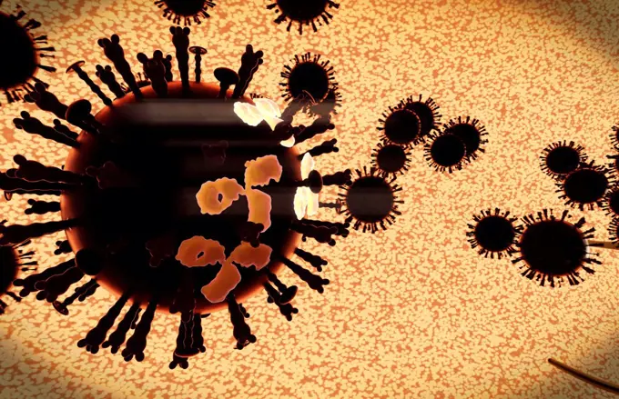
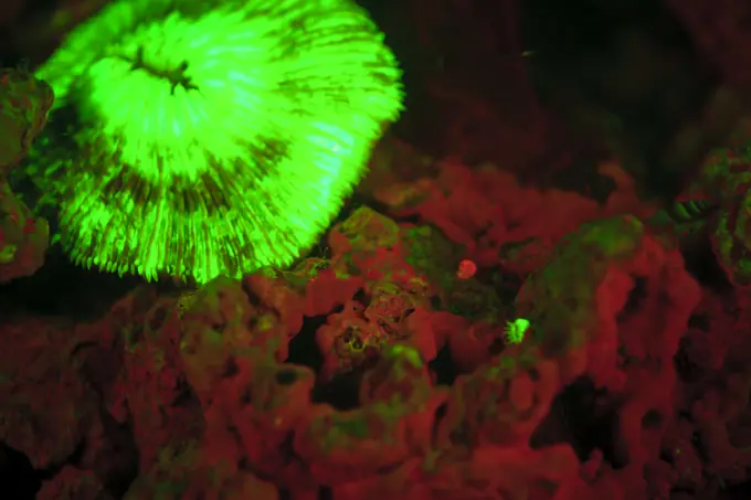
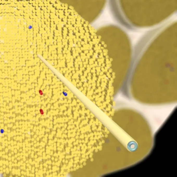
Microbial and Viral Studies

This cluster features a variety of microscopic and close-up images depicting viruses, fluid patterns, and chemical substances with vibrant colors.

Turquoise gel
Turquoise gel
298 assets in this story
4378-1168
Microscopic styled visualization of tapeworms (Cestoda).
1525-22327556
Close-up of blood cells
1525-22302508
Close-up of bubbles in water
824-57659514
Under 1200X magnification, this 1973 photomicrograph depicted a sample of mouse brain tissue stained using the hematoxylin-eosin (HE) staining technique and revealed the presence of Acanthamoeba polyphaga protozoa. CDC/Dr. George R. Healy 1973
4378-1170
Human blastula viewed close-up in the style of a scanning electronic microscope.
1525-56517275
Close up of virus, medical health
1525-26274978
Pannus is an abnormal layer of fibrovascular tissue or granulation tissue. 3d illustration. Joint space pannus. of Rheumatoid Arthritis.
4128-20039394
ICAM-1 on the surface of a human cell, illustration
1848-20176227
Detail of the Rio Tinto (Red river) with its deep reddish hue due to oxidised iron minerals in its water, aerial view, drone shot, Huelva province, An...
1525-56517364
Close up of virus, medical health
1815R-13386158
Blood vessel, 3D Rendering
4128-30416632
Intestinal microvilli, illustration
1848-64402054
Bubble Ring Ascends towards the Sun, Underwater
1899-61460385
Colorized transmission electron micrograph of monkeypox virus particles (orange) cultivated and purified from cell culture
4128R-15653
Acinetobacter sp. bacteria, computer artwork.
4128R-13681892
Viruses, illustration.
6188-55603621
Gold particles are repulsed from the floor Gold particles are repulsed from the floor. 3D rendered Copyright: xZoonar.com/RomanxBudnikovx 10344122
5507-33975184
Corona virus flu ncov 2019 cell world health crysis
1525R-7995
close up of receiver of a telephone
4384-280
Histoplasma capsulatum Antigens
6188-58106038
loop rotation of Coronavirus COVID-2019 Coronavirus COVID-2019. rotation loop. 3D virus animation Copyright: xZoonar.com/StanislavxRishnyakx 15063465
4128-15665004
Human sperm, illustration
6145-45047456
Especially bright patches, bluish in enhanced color, are due to seasonal frost that is accumulating as this hemisphere approaches winter. This image of a sand dune field in a Southern highlands crater was acquired by NASA's Mars Reconnaissance Orbiter.
4128R-13387775
Illustration of red blood cells flowing inside an artery or vein. Red blood cells are biconcave, disc-shaped cells that transport oxygen from the lung...
1525-22717547
bright jellyfish floating in the dark sea water
4128-28968484
Haemorrhage, illustration
7205-70654689
Papua New Guinea, West New Britain, Kimbe Bay, unidentified spider crab on coral
4128R-7521
Nasal lining, TEM
4378-4627
Legionella Pneumophila
4378-1152
Scanning electromicrograph styled dust mite on carpet fibers.
4128-28968437
Bone marrow, illustration
1525-26275095
Langerhans cells are dendritic cells of the skin 3D illustration. Antigen-presenting immune cells, Largerhans cell
1849-66221493
Illustration of Emiliania Huxleyi phytoplankton
1848-61394098
Burgundy truffle (Tuber uncinatum), autumn truffle spores
6188-54819658
starfish ultra macro beam hand with suction cups. starfish ultra macro beam hand with suction cups. marine animals of the world ocean with a large zoom Copyright: xZoonar.com/MaximilianxBuzunx 13595517
4128-28681164
Pollen grains, illustration
4128R-12577577
DNA (deoxyribonucleic acid) strand molecular model, computer illustration.
4128R-14181665
Actinomycete bacteria, illustration
1815-18058225
Aerial view of brown acidic landscape of Rio¶ÿTinto¶ÿMines area
1525-26182254
In the layer adjacent to the dermis, the keratinocytes are divided and thrown into the upper layers. 3D illustration. In the layer adjacent to the dermis, the keratinocytes are divided and thrown into the upper layers.
824-66066005
Colorized transmission electron micrograph of mpox virus particles (red and yellow) found within infected VERO E6 cells (blue). The virus particles are in various stages of maturity, which accounts for differences in shape. This image also features virus factories (brown), which are inclusions within infected cells where virus replication, maturation, and assembly occurs. Credit: NIAID.
1848-66201350
Air and oil bubbles inside water base form patterns
4128-V58568185
1525-26430411
Gut bacteria, intestine microbita 3d illustration
1815R-14323759
3D Rendered Illustration, visualisation of Tuberculosis Bacteria in an organism
6188-63307686
Smoked garlic Smoked garlic detail showing cross section of black cloves Copyright: xZoonar.com/PaulxFleetx 5441911
4128R-14181767
Microtubules, illustration
4128R-13446646
Computer illustration of Treponema pallidum, the bacterium which causes syphilis.
4128-V58568610
4128R-15516276
Illustration of a cancer cell being attacked by white blood
4378-1819
A magnification of filiform papillae on a human tongue.
7202-70643909
Natural occuring Fluorescence in Corals at night using special filters and powerful underwater lights. Hol Chan Marine Preserve, Belize Barrier Reef-2nd Largest in the World
4128R-14057185
CRISPR-CAS9 gene editing complex, illustration
4378-2748
Parasitic head louse clinging to a hair follicle.
6188-67149217
Nahaufnahme von blauen flüssigen kosmetischen Gel- oder Serum-Makeup-Mustern mit glänzender, glänzender Textur auf einem glatten blauen Hintergrund Close-up shot of blue liquid cosmetic gel or serum makeup swatches with a glossy, shiny texture on a smooth blue background Copyright: xZoonar.com/IrinaxKvyatkovskayax 22816345
4128-28970714
Contents of a pimple, illustration
6145-29245447
Embryo showing polarisation of membranes
1899-61460847
This scanning electron microscope (SEM) image revealed some of the ultrastructural features observed on the surface of a protozoan Acanthamoeba polyphaga. Note the amorphous nature of the organisms exterior from which the pseudopodia can extend. These pseudopodia allow the amoeba to move and grasp objects in its environment. CDC/Catherine Armbruster Margaret Williams 2009
1848-49303888
Strings of toadspawn from common toad, European toad (Bufo bufo) and frogspawn
1525-26182260
In the layer adjacent to the dermis, the keratinocytes are divided and thrown into the upper layers. 3D illustration. In the layer adjacent to the dermis, the keratinocytes are divided and thrown into the upper layers.
824-63184376
Influenza virus. Image produced using high-dynamic-range imaging (HDRI) from an image taken with transmission electron microscopy. Viral diameter ranges from around 80 to 120 nm.
4128R-9772
Skimmia pollen, SEM
1525-27519784
Infectious Disease and Health Medicine of Bacteria. Infectious Disease
6139-V30021850
6188-58176025
Extreme macro of the fruit kiwi seeds in blue color as backdrop Extreme macro of the fruit kiwi seeds in blue color as backdrop Copyright: xZoonar.com/LenaxIliopouloux 21367830
4128-20239816
Milk ducts, illustration
4128-V58577101
4378-3724
Structure of human DNA or deoxyribonucleic acid (PDB 1BNA).
6177-V53561592
4378-4632
Borrelia burgdorferi
4384-422
Microscopic Detail of a Carpenter Bee's Eye
4378-20014030
Intestinal Villi
4128R-14638098
Microbes in space, illustration
1525-19897645
Urea fertilizer
1848-58226573
Fluorescence light of a Hard Coral Madreporaria
4128R-13575733
Dendritic cell, illustration. Dendritic cells are antigen-presenting cells of the immune system. Their main function is to process antigen material an...
4128-18182076
Immune response to viral infection, illustration
4128R-14057528
Macrophage, illustration
1525-26182247
In the layer adjacent to the dermis, the keratinocytes are divided and thrown into the upper layers. 3D illustration. In the layer adjacent to the dermis, the keratinocytes are divided and thrown into the upper layers.
4070-48286278
Candy stripe shrimp (Lebbeus grandimanus) sheltering beneath tentacles of its host, Crimson anemone (Cribrinopsis fernaldi). Browning Pass, British Columbia, Canada. North East Pacific Ocean. July.
7203-70646280
Underwater sea life near airport and Laha Village, Ambon Island, Banda Sea, Indonesia
1899-61460855
This scanning electron microscope (SEM) image revealed some of the ultrastructural features observed on the surface of two Acanthamoeba polyphaga protozoa. The amoeba on the right is in its trophozoite stage, capable of moving using pseudopodia which it extends from its exterior. The amoeba on the left entered a cystic stage, after which its cell wall doubled in thickness, protecting it from adverse environmental conditions, and a state of dormancy set in on the organism. CDC/Catherine Armbruster Margaret Williams 2009
4239-18641401
Microscopic view of taste buds on tongue. Taste buds contain the receptors for taste. They are located around the small structures on the upper surface of the tongue, soft palate, upper esophagus
1525-22197444
cancer cell
4128-V58569100
1848-53877847
Mould, known as isolate, cultured in the laboratory for research purposes, Germany, Europe
4239R-8263
A black swarm of H5N1 avian flu viruses are attacked by antibodies (the three legged elements), which mark the virus for destruction.
4128R-11286517
Flu virus particles, computer artwork. In the virus' envelope are two types of protein spike, haemagglutinin (H, peg-like) and neuraminidase (N, squar...
1525-26235681
Pancreatic islet cells are the main source of insulin and glucagon, which are produced by cells and cells, respectively. 3D illustration. Functional Anatomy of the Endocrine Pancreas.
824-63227173
Ebola virus isolated in November 2014 from patient blood samples obtained in Mali. The virus was isolated on Vero cells.
7202-70643602
Fluorescence emitted in corals, captured using special barrier filter.  Tawali Resort, Milne Bay, Papua New Guinea
49-316A
Pill Coating Tumbler
1899-13186
Anatomy of a nerve: with close up of nerve fiber
4128R-15516435
Illustration of gluten damaging intestinal villi
4372-261
Fluorescing Coral Polyps
1899-53513337
Exploring and searching for a Covid-19 drug.
1525-26235663
Pancreatic islet cells are the main source of insulin and glucagon, which are produced by cells and cells, respectively. 3D illustration. Functional Anatomy of the Endocrine Pancreas.
7203-70647282
Natural occurring Green Fluorescence in Stony Corals (Lobophillia hemprichii), captured with special ultraviolet light-blocking filters, Palau, Micronesia, Rock Islands, World Heritage Site, Western Pacific
4201-77889
Coho Salmon (Oncorhynchus kisutch) eggs at ten weeks after spawning, Washington
4128-V58569601
PREVIOUS
of 3
NEXT
2401 S. Ervay, Suite 206
Dallas, Texas 75215
United States
Get Started
Free ResearchMy BoardsMy Cart
For Creators
How To License Your ContentContributor PortalFrame of Mind
Resources
API accessPricing
Contact
+1 866 236 0087help@viewpictures.co.uk Contact form
©2026 View Pictures. All Rights Reserved. -A
This site is protected by reCAPTCHA and Google Privacy Policy and Terms of Service apply.