My Boards
View Pictures
  • My Boards

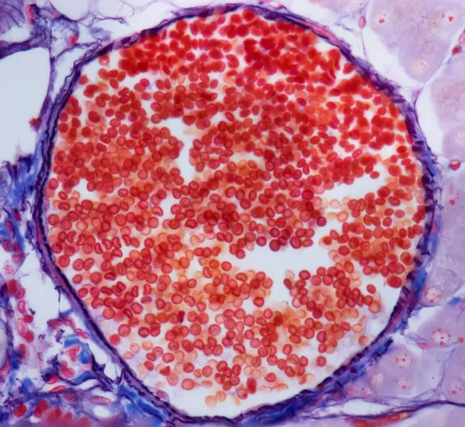
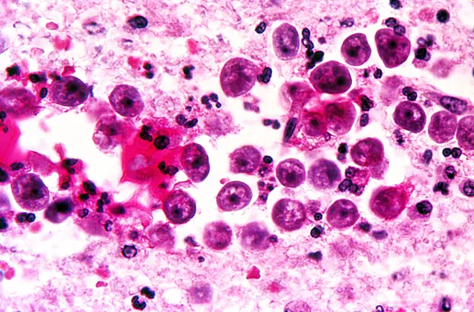
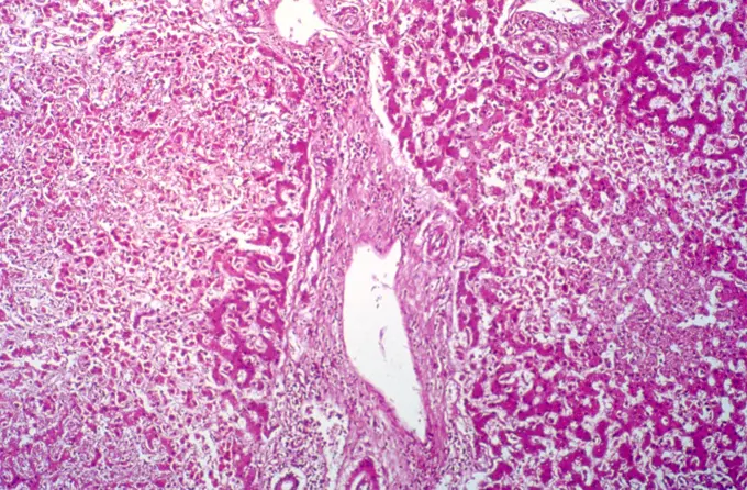
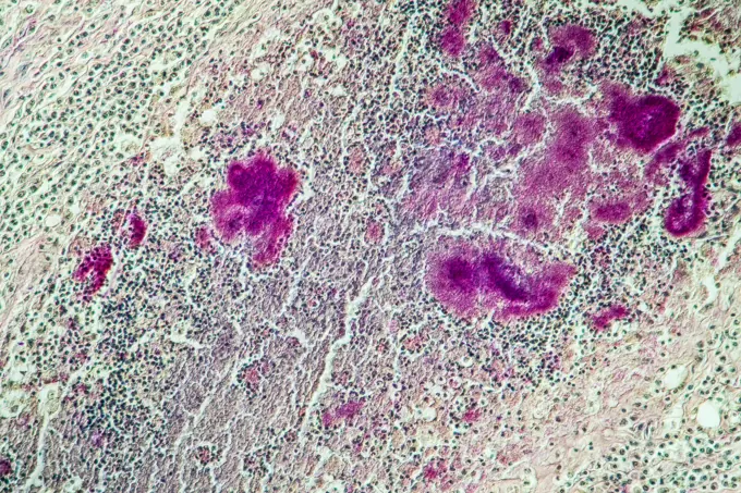
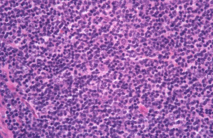
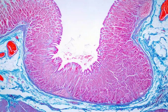
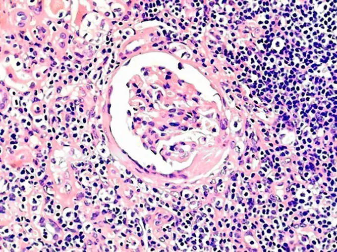
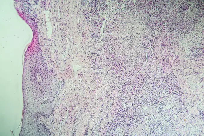
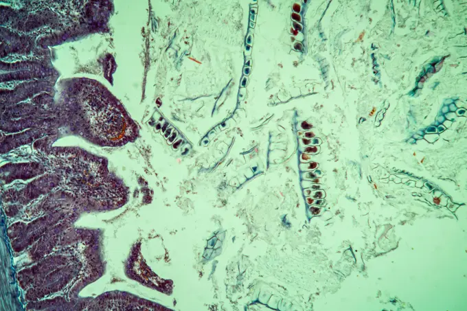
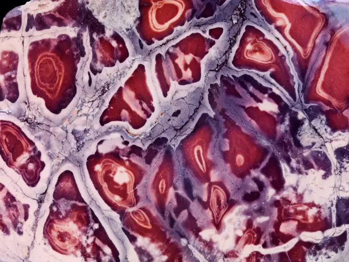
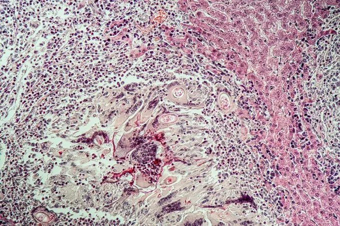
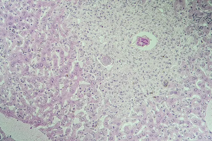
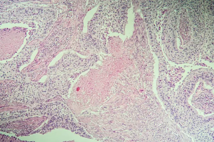
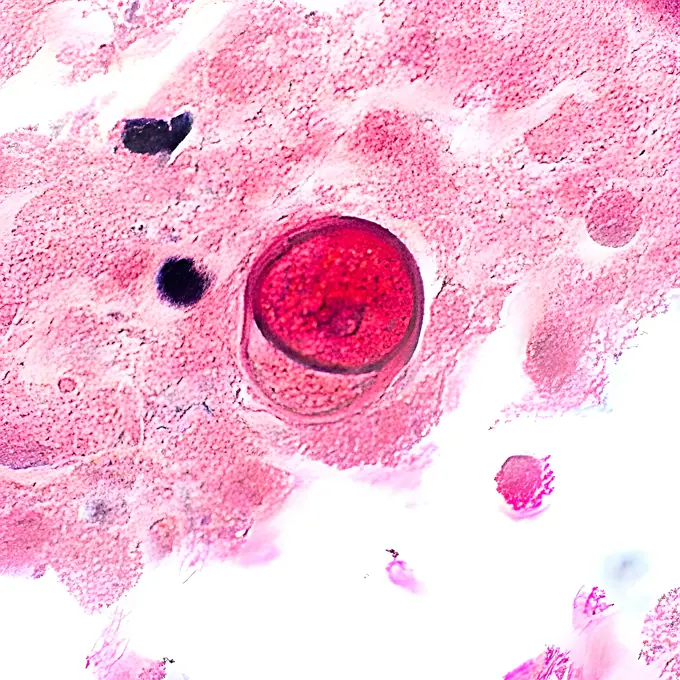
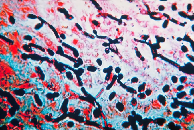
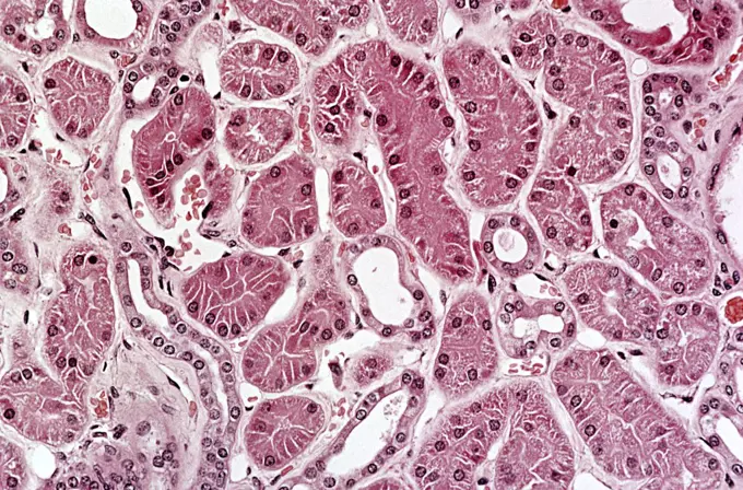
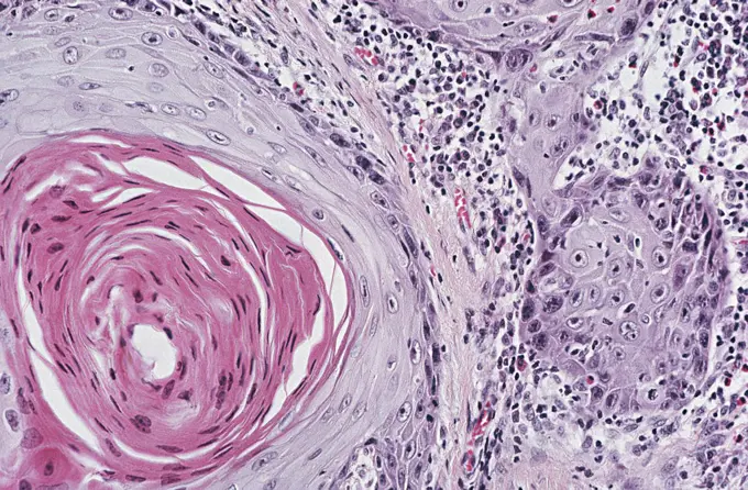
Microscopic Medical Conditions

Detailed microscopic views of various diseases, including leprosy and cancer, highlighting medical research.

Prostate
Prostate
340 assets in this story
6188-55644653
Blind intestine with parasite appendix with parasites oxiuris, maggot worm in the intestine, 100x copyright: xzoonar.com/dr.xnorbertxlangex 13747258
6188-55644058
Lilie Lilie Stamm quer 100x
6188-55644170
Liver from the pig pork pork cut 200x copyright: xzoonar.com/dr.xnorberbertxlangex 13698998
4128R-23748
Kidney disease. Light micrograph of a section through an inflamed kidney showing a glomerulus coiled capillaries, purple. It is surrounded by vascular...
6188-55644334
Hods of the rat rat hotel cross -section 100x copyright: xzoonar.com/DR.XNORBERBERTXLANGEX 13712918
6188-55643882
Cherry cherry blossom with pollen lengthways 100x copyright: xzoonar.com/dr.xnorberbertxlangex 13660684
6188-55644088
Mais Mais Stamm quer 100x Copyright: xZoonar.com/Dr.xNorbertxLangex 13688315
4128R-22532
Secondary heart cancer. Light micrograph of a section through cardiac muscle showing a metastatic seminoma. This is a germ cell tumour that originated...
6188-55644219
Tongue toad toads cross -section through the tongue 200x copyright: xzoonar.com/dr.xnorberbertxlangex 13702095
4128-18680648
Liver cirrhosis, light micrograph
4269-7452
Brain tissue with amebic infection with Naegleria fowleri, a free-living amebae responsible for primary amebic meningoenceohalitis. Light micrograph.
1788-111173554
Section of a human lymph node seen under a microscope.
824-63227261
National Institutes of Health researchers and international colleagues have discovered a genetic cause and potential treatment strategy for a rare immune disorder called CHAPLE. This light microscope image shows the intestinal tissue of a child with CHAPLE's disease. The large white areas in the lower right corner are enlarged lymphatic vessels, which may contribute to bowel distress.
1899-61460553
Balamuthia mandrillaris is a free-living amoeba (a single-celled living organism) naturally present in the environment. Balamuthia can cause a rare and serious brain infection called amoebic granulomatous encephalitis
4128R-3317
Cardiovascular syphilis, light micrograph. The media of the aorta contains numerous thin walled blood vessels white spaces containing red blood cells ...
4128-18929799
Ovarian follicle, LM
6145-29267465
Schistosomiasis: large intestine
6188-55644561
Wilmstumor Wilmstumor erkranktes Gewebe unter dem Mikroskop 100x Copyright: xZoonar.com/Dr.xNorbertxLangex 13735500
6188-63377881
Colon Cancer cells Microscope view of Colon Cancer cells in tissue culture showing walls, nucleus and organelles. Copyright: xZoonar.com/PaulxHakimatax-xwww.hakimata.comx 2322006
6188-55644240
Darm Frosch Frosch cross -section through the Darm 200x
6145-29268961
Amoebiasis: amoebic abscess in liver
6188-55643861
Mushroom disease in the cattle pine fungal disease in the cattle pine fabric 200x copyright: xzoonar.com/DR.XNORBERBERTXLANGEX 13660621
1788-111174474
Cross section of mammal lung tissue, injected to highlight the capillaries under a microscope.
6145-29269799
Sickle cell disease: liver
4269-25200
Sarcoidosis. Granuloma and giant cell Ganglion benign disease Light microscopy
1788-20761628
Example of a monolayer culture of colon cells viewed under a microscope.
6145-29276482
Lung: lymphoid interstitial pneumonitis
1788-111173867
Cross section demonstrating the structure of a human sciatic nerve.
4128-18498502
Colon biopsy, light micrograph
6145-29269320
Leprosy: skin section
4298-1070
Note the collecting tubules on the left side of the image as contrasted with the right side of the photo where the inflammatory cells (blue) have destroyed most of the tublules.  Magnification 400x
4269-20408824
Detail of one human immunodeficiency virus (HIV) virus particle, or virion, Transmission Electron Micrograph (TEM).
1525-20607957
Pine needle cross-section under the microscope, background, (Pinus)
4128-18929708
Jejunum, LM
1439R-1081446
Venous thrombosis
4297-1776
Negatively-stained transmission electron micrograph of numerous herpes simplex virions, members of the Herpesviridae virus family
6188-55644912
Lippen Lippen Karzinom unter dem Mikroskop 100x Copyright: xZoonar.com/Dr.xNorbertxLangex 13772989
6145-29293819
Liver: disseminated histoplasmosis
4128R-22521
Nerve cells. Light micrograph of Purkinje neurons nerve cells, blue in the cerebellum. These cells form the junction between the granular and molecula...
6188-55644267
Rehbock with worms roebuck lungworm under the microscope 200x copyright: xzoonar.com/dr.xnorbertxlangex 13706790
1773R-87685
Helicobacter pylori in antral crypt
4297-1781
Transmission electron micrograph of Lagos bat virus (LBV) virions and an intracytoplasmic inclusion body in a tissue sample
1788-111177863
Section of small blood vessels in human salivary gland, at x170 magnification.
1788-111173660
Cross section of human uterine tubes seen under a microscope, at x100 magnification.
1439R-1081424
Bone marrow
6188-55643632
Amöbenruhr amöbenruhr diseased intestine 100x copyright: xzoonar.com/DR.XNORBERBERTXLANGEX 13628270
6188-55643582
Rainfarn Rainfarn Blütenstand quer 100x Copyright: xZoonar.com/Dr.xNorbertxLangex 13623044
4128-17857789
Hidradenoma papilliferum, light micrograph (LM) of a section of affected vulva. Hidradenoma papilliferumis a type of benign cystic papillary growth (l...
1899-85597
Adenocarcinoma
6188-55643724
Oral mucosa with bacteria Epithelia cells with bacteria from the oral cavity 200x
1788-111173804
Section of granulomatous leison from human actinomycosis seen under a microscope.
4269-6705
Photomicrograph of human metastatic melanoma cells magnified to 320x.
4332-2519
Porcelain Jasper slab from the State of Sonora, Mexico.
6188-55644257
Rehbock with parasites roebock parasite egg in the tissue 100x copyright: xzoonar.com/dr.xnorberbertxlangex 13706765
6188-55644036
Liver mouse, bilharziosis liver mouse, bilharziosis diseased tissue 100x copyright: xzoonar.com/dr.xnorberbertxlangex 13683648
6188-55644596
Plattenzellkarzinom Plattenzellkarzinom erkranktes Gewebe unter dem Mikroskop 100x Copyright: xZoonar.com/Dr.xNorbertxLangex 13735569
1899-61460551
Acanthamoeba is a microscopic, free-living amoeba, or amoeba (single-celled living organism), that can cause rare but serious eye, skin, and central nervous system infections. The amoeba is found worldwide in the environment in water and soil. The amoeba can spread to the eyes through the use of contact lenses, cuts or wounds in the skin, or by being inhaled into the lungs. Most people will be exposed to Acanthamoeba in their lifetime, but very few will become ill from this exposure
4128R-6830
Bald scalp, light micrograph.
1788-111173840
Section of malignant connective-tissue tumour from a human seen under a microscope.
1439R-995045
Follicles of colloid in thyroid
1788-111173875
Histology image of the liver in a case of cirrhosis of the liver from alcoholism.
4298-1054
A section of human appendix showing Crypts of Lieberkuhn in cross section.  PAS stain.  Magnification 100x
6188-55643989
Fibromyoma, good -natured tumor of the uterus fibromyoma, good actual tumor of the uterus fabric 100x copyright: xzoonar.com/DR.XNORBERTXLANGEX 13674847
4128R-5129
Stomach lining, light micrograph. The stomach lumen is lined by mucous cells. At bottom centre is an accumulation of lymphocytes forming a lymphatic n...
4128R-22562
Islet of langerhans, light micrograph. This structure centre, found in the pancreas, secretes the hormones insulin and glucagon, which control blood s...
1788-111174435
Microphotograph of a section of gland from a newt (genus Triturus), Salamandridae, with histological staining, at x200 magnification.
4384-282
Nipah Virus Infection
824-57659284
Under 500X magnification, this 1973 photomicrograph of a trichrome-stained sample of human brain tissue revealed the presence of Naegleria gruberi, free-living amoebae, in a case of primary amoebic meningoencephalitis (PAM). CDC/Dr. George R. Healy 1973
4269-26717
Hemochromatosis.
1899-61460862
This photomicrograph of a sample of mouse brain tissue revealed the presence of some of the histopathology caused by an amoebic Acanthamoeba polyphaga infection. Note that there were a number of cysts of A. polyphaga in the field of vision. CDC/Dr George Healy 1973
6188-55644025
Möhre Möhre Wurzel quer 100x Copyright: xZoonar.com/Dr.xNorbertxLangex 13683626
824-63123786
Eastern Equine Encephalitis (EEE) virus. Electron micrograph of the Eastern Equine Encephalitis (EEE) virus in a mosquito salivary gland.
1773-97768
Light micrograph of Candida in esophagus mucosa
1788-111175511
Microphotograph of a cross section of a large bronchiole with counterstaining, at x80x1.2 magnification.
6188-55643746
Oral mucosa with bacteria Epithelia cells with bacteria from the oral cavity 200x
4269-6835
Transmission Electron Micrograph (TEM) of a cell containing variola (smallpox) viruses.
824-63195676
Using immunohistochemical (IHC) technique, this photomicrograph revealed some of the cytoarchitectural histopathologic changes associated with a Nipah virus infection, in this case affecting a human patient, resulting in the formation of a syncytial cell, ie giant, multinucleated endothelial cell, in the center of the image. Infection with Nipah virus has been associated with an encephalitis (inflammation of the brain) characterized by fever and drowsiness and more serious central nervous system disease, such as coma, seizures, and inability to maintain breathing. Nipah virus, a member of the family Paramyxoviridae, is related but not identical to Hendra virus. Nipah virus was initially isolated in 1999 upon examining samples from an outbreak of encephalitis and respiratory illness among adult men in Malaysia and Singapore. Its name originated from Sungai Nipah, a village on the Malaysian Peninsula where pig farmers became ill with encephalitis.
6188-55643873
Cherry cherry blossom with pollen lengthways 100x copyright: xzoonar.com/dr.xnorbertxlangex 13660646
6188-55643736
Maus Gehirn Maus Gehirn Gewebe unter dem Mikroskop 100x Copyright: xZoonar.com/Dr.xNorbertxLangex 13644924
4297-1756
Light microscope histological section showing ebola virus
824-66066019
A Coccidioides fungal spherule (yellow) found in a tissue sample. This organism causes coccidioidomycosis, also known as Valley Fever. Credit: micrograph by CDC colorization and visual effects by NIAID See related September 20, 2024 NIAID Now article, &quotFungal Disease Awareness Week Fighting Fungi at NIAID,&quot at.
1439R-1081411
Myometrium
6145-29292839
Mycobacterium avium-intracellulare infection and HIV
1439R-1081434
Kidney
1558-15241719
Microphoto of lungs of Domestic Cat (Felis silvestris catus)
824-63190246
Pneumococcus (streptococcus pneumoniae). Pneumococcus is the bacterium that causes pneumonia, bronchial pneumonia, purulent pleurisy, bacterial meningitis, ear infections, sinusitis and conjunctivitis. Optical microscopy x 1000.
6188-55643657
Gallenblasentumor Gallenblasentumor Gewebe unter dem Mikroskop 100x Copyright: xZoonar.com/Dr.xNorbertxLangex 13633443
4298-20535932
A section of human appendix showing Crypts of Lieberkuhn in cross section.  PAS stain.  Magnification 100x
6188-55644192
Liver from the pig pork pork cut 100x copyright: xzoonar.com/dr.xnorberbertxlangeex 13699035
1788-111176114
Section of skin with visible layer of sebum coating connective epithelium tissue, seen under a microscope.
4128-18929698
Glomeruli, LM
4128-17810101
Nerve cells, light micrograph
6188-63377884
Colon Cancer cells Microscope view of Colon Cancer cells in tissue culture showing walls, nucleus and organelles. Copyright: xZoonar.com/PaulxHakimatax-xwww.hakimata.comx 2322022
1439R-1081447
Squamous cell carcinoma
6188-55644644
Amyloid deposits in the tongue amyloid deposits in the tongue sick tissue under the microscope 200x copyright: xzoonar.com/DR.XNORBERBERTXLANGEX 13747203
1439R-1081394
Ileum
6188-55644349
Hoden der Ratte Rattenhoden querschnitt 200x Copyright: xZoonar.com/Dr.xNorbertxLangex 13712946
4269-6787
Transmission electron micrograph (TEM) of os a section of a glomerulus. The round shapes at center and right are blood vessels (two red blood cells are visible at right). The external surface of the vessels are covered with diverticuli that are ramifications of the podocytes which filter the blood and produce urine. Magnification x4000.
6188-55644647
ZebraBärbling zebraBärbling optic nerve of the fish 200x copyright: xzoonar.com/dr.xnorberberxlangeex 13747218
4128R-13447204
Venule. Light micrograph (LM) of red blood cells (erythrocytes, red) in a venule. These vessels arise from capillaries and are characterized by a wide...
PREVIOUS
of 4
NEXT
2401 S. Ervay, Suite 206
Dallas, Texas 75215
United States
Get Started
Free ResearchMy BoardsMy Cart
For Creators
How To License Your ContentContributor PortalFrame of Mind
Resources
API accessPricing
Contact
+1 866 236 0087help@viewpictures.co.uk Contact form
©2026 View Pictures. All Rights Reserved. -A
This site is protected by reCAPTCHA and Google Privacy Policy and Terms of Service apply.