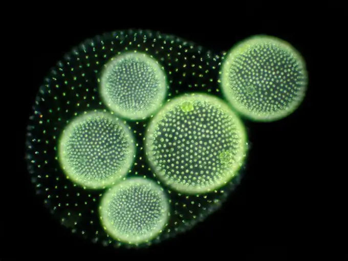
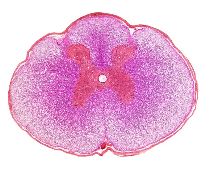
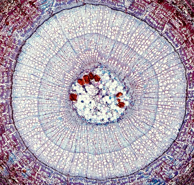
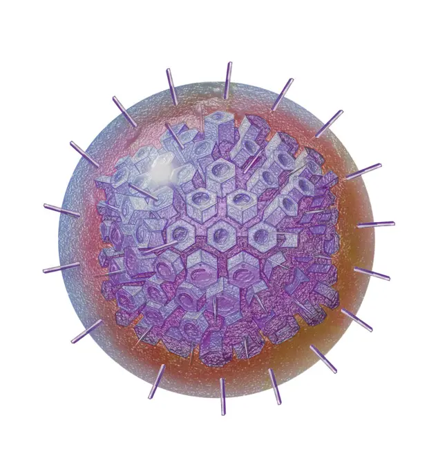
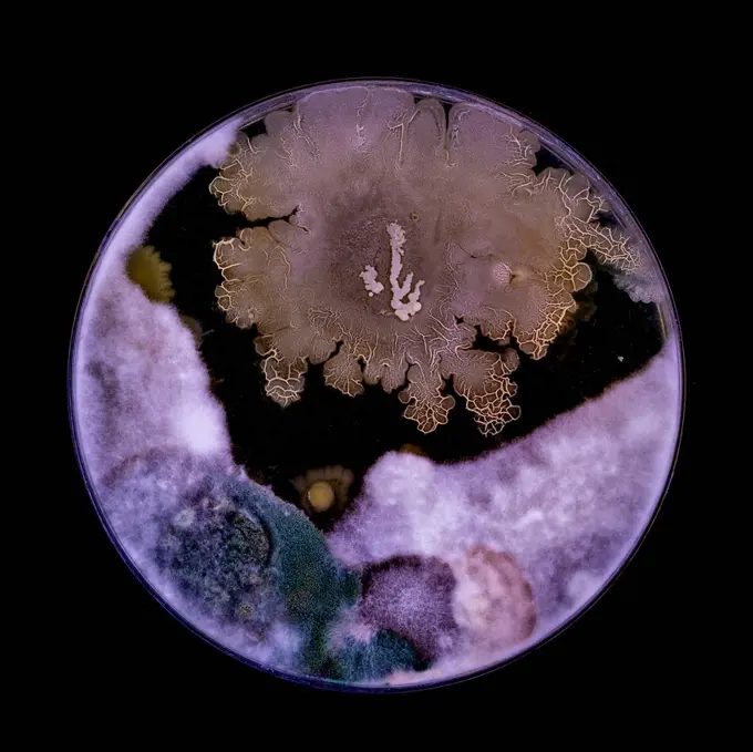
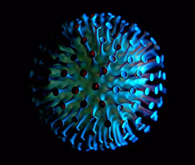
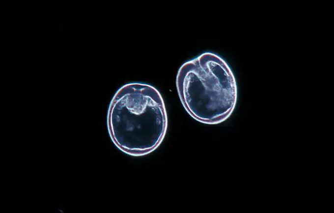
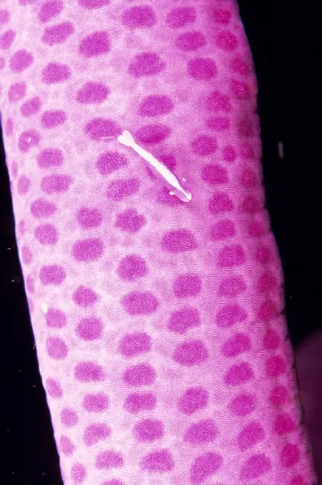
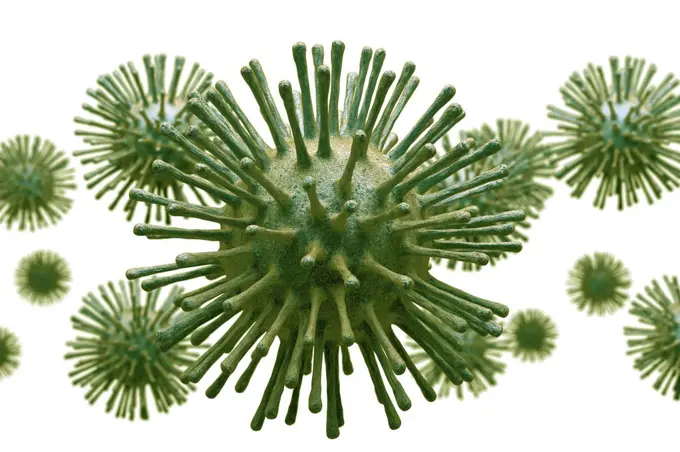
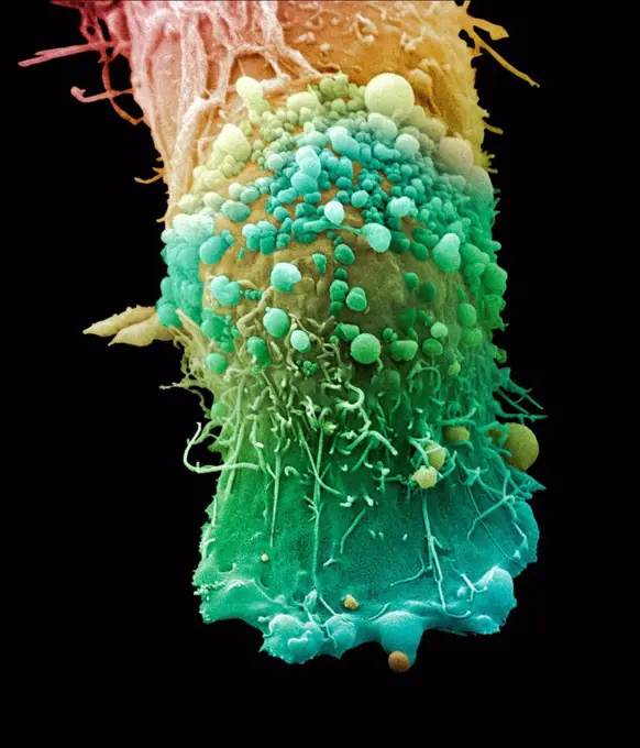
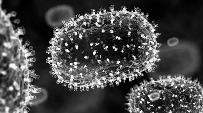
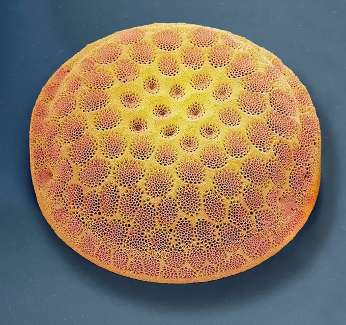
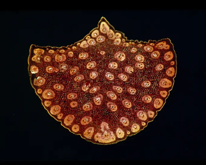
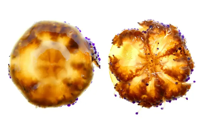
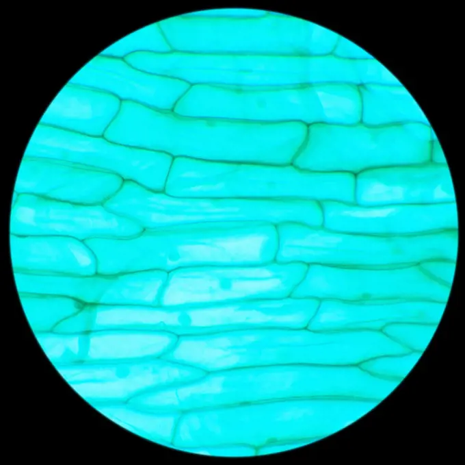
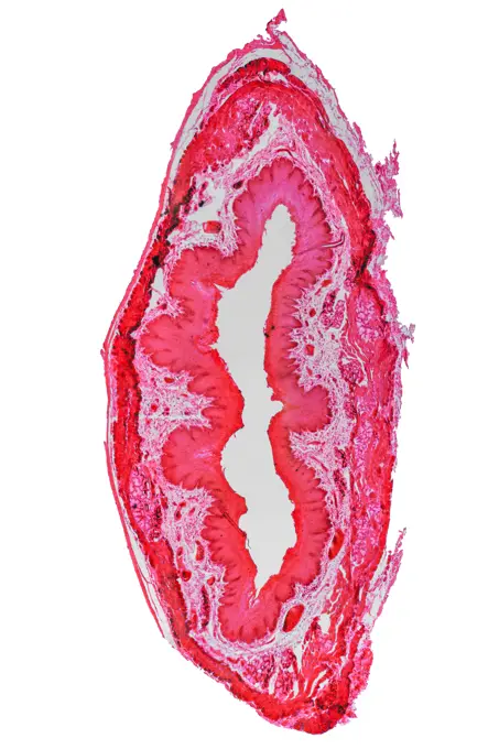
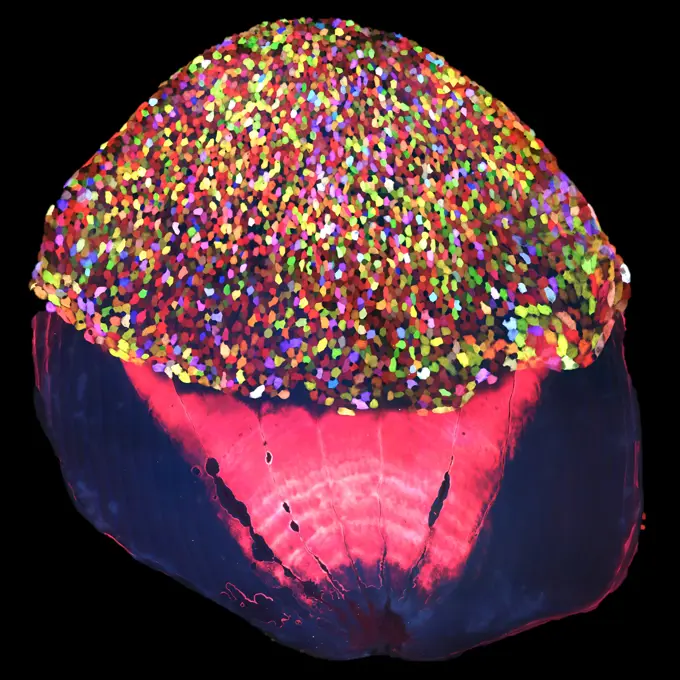
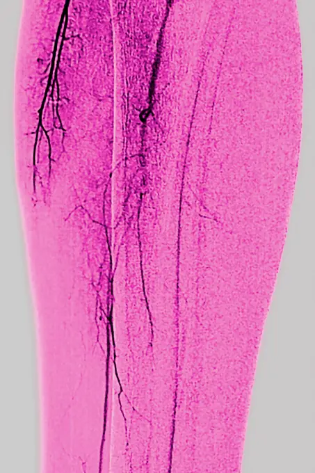
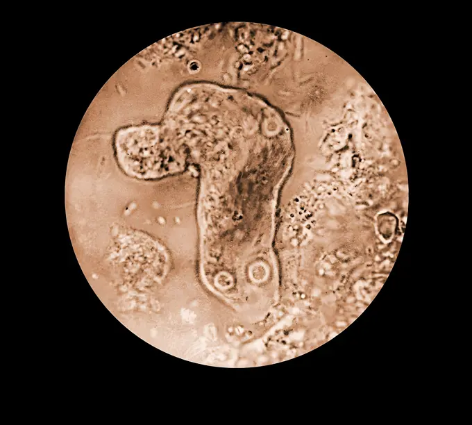
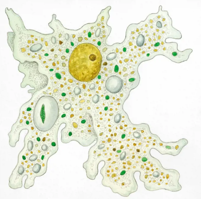
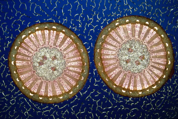
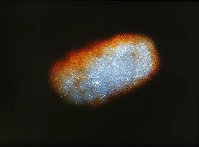
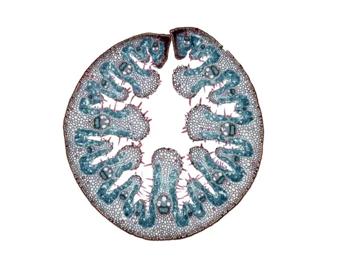
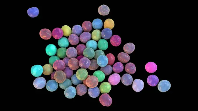
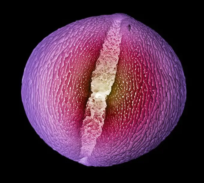
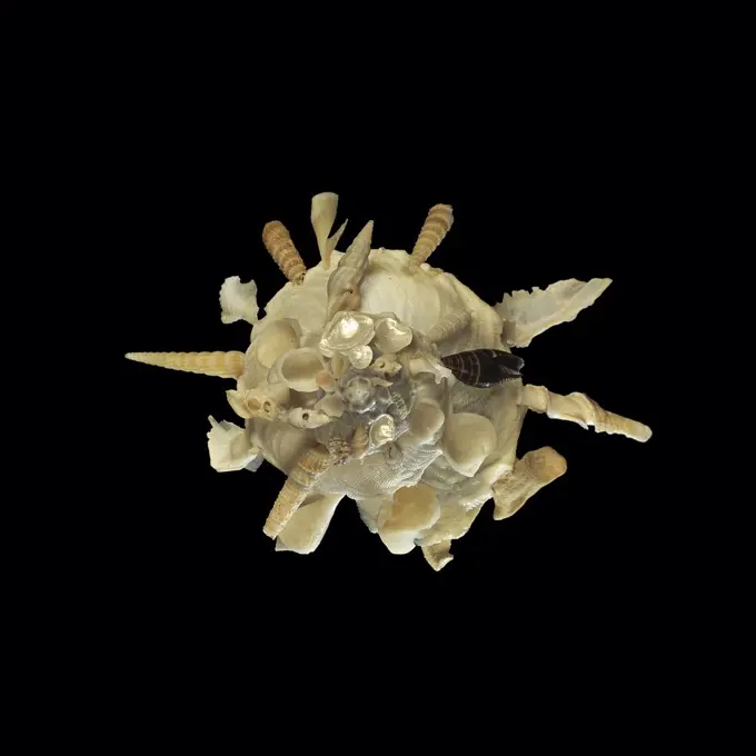
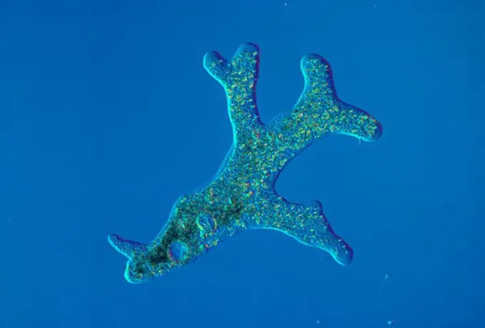
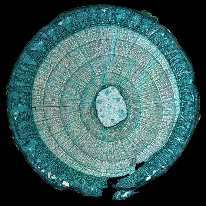
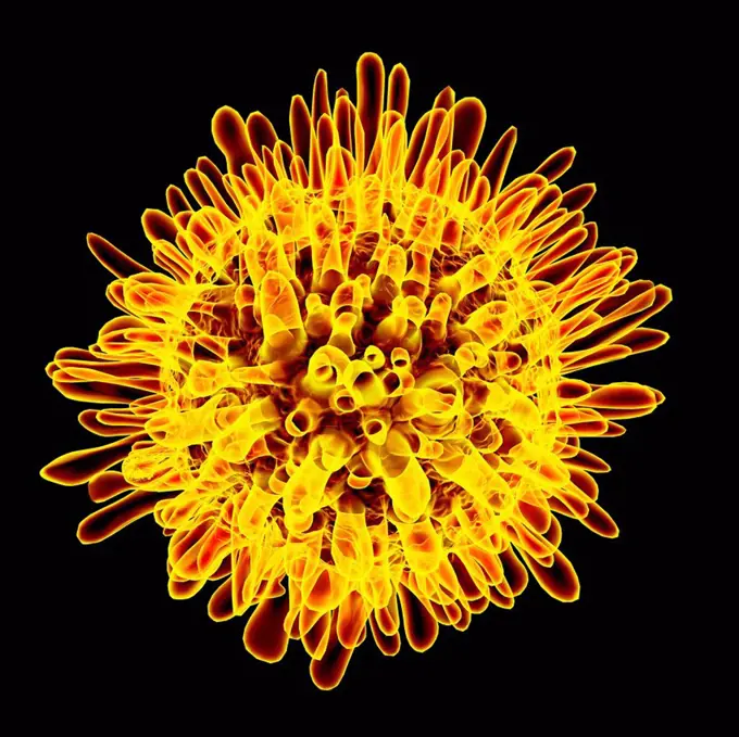
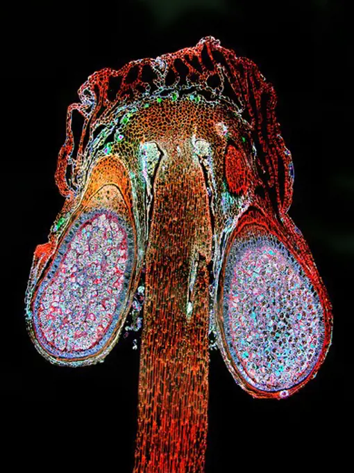
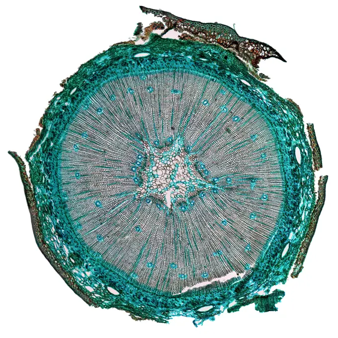
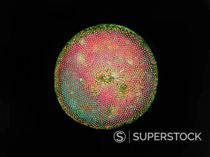
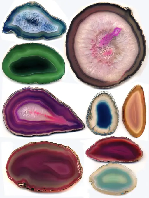
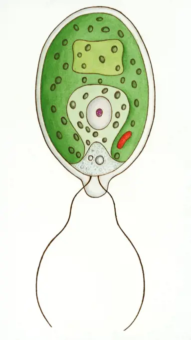
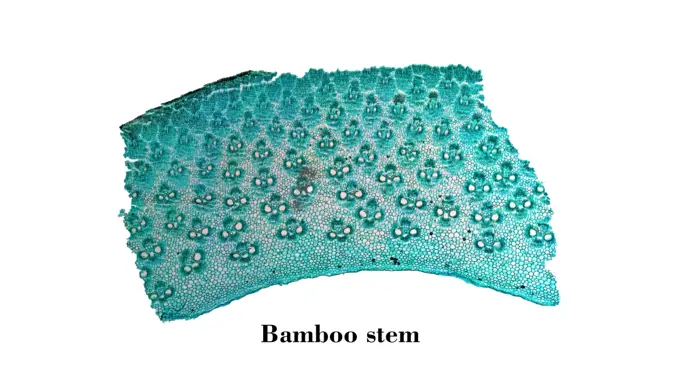
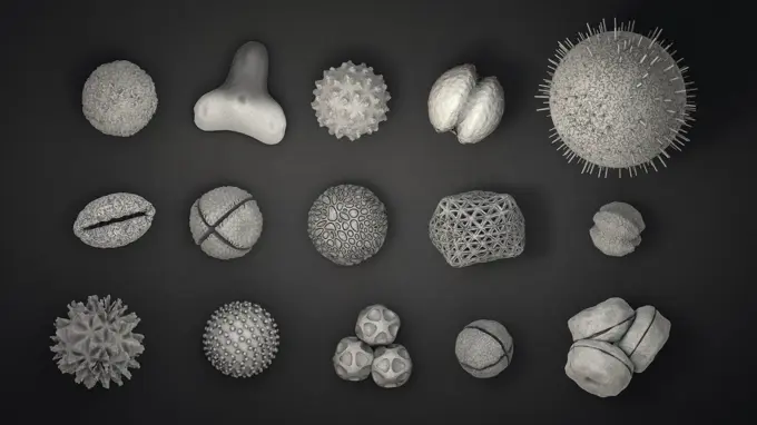
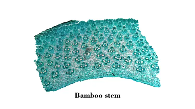
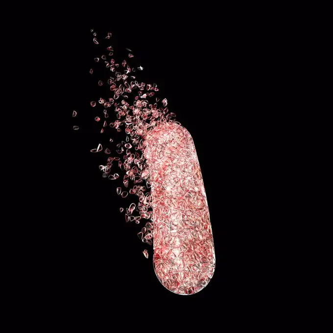
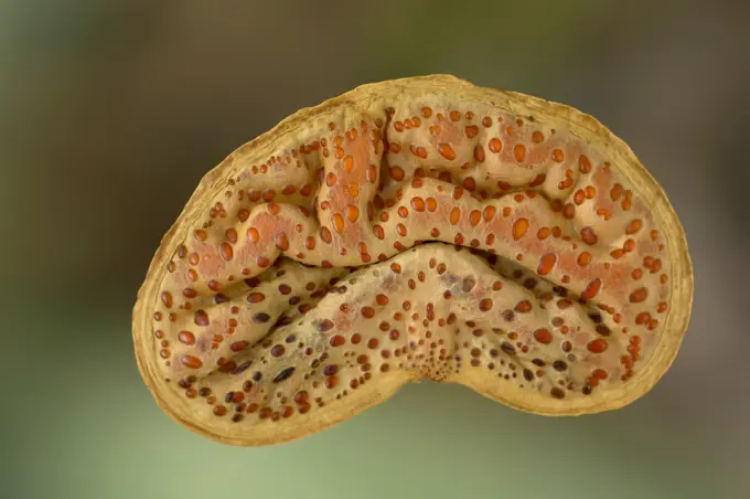
My Boards
View Pictures
  • My Boards

Microscopic Plant Cross-Sections

Detailed microscopic images of plant cross-sections, highlighting cellular structures in various species, showcasing their intricate designs and colors.

Volvox
Volvox
202 assets in this story
4128-18929723
Spinal cord, LM
4128-20042962
Escherichia coli bacteria in a net, conceptual illustration
4128R-15536332
Illustration of an abstract plexus brain
4128-20244126
Cell membrane lipid bilayer, illustration
4128R-13760
Tobacco necrosis virus. Computer model of the capsid of the tobacco necrosis virus.
4413-109791
Microscopic view of Sporangia of Riccia
6188-55551664
Germs of tuberculosis, vintage engraving. Germs of tuberculosis, vintage engraved illustration. Copyright: xZoonar.com/PatrickxGuenettex 10656390
4141-111428946
Light micrograph cross section of a Lime (Tilia species) second year growth stem section, CS, abstract, UK.
4239-20481133
Single virus particle.
1439-57952416
Mysterious microsphere with intricate patterns against a dark background
4141-32783
dinoflagellata showing flagella ceratium hirudinella anoptal contrast x80 at 1/3000th second
4128-28515238
Bacteria and fungi cultured on Petri dish
4128-16007337
Virus, illustration
1848-50837180
Black walnut (Juglans nigra), shoot t.s., colouring Wacker 3A
1848-61419908
Amber with inclusions, Amber Museum in Nida, Curonian Spit, Lithuania, Nida, Lithuania, Europe
4128R-14841
Honeybee mite Varroa destructor, artwork, artwork.
4128R-12571812
Diatom. Coloured scanning electron micrograph (SEM) of a single diatom (cylotella sp.). Diatoms may be extremely abundant in both freshwater and marin...
4128R-13941181
Bottlebrush pollen, illustration.
6188-55645584
Zieralge unter dem Mikroskop 100x Zieralge unter dem Mikroskop 100x Copyright: xZoonar.com/Dr.xNorbertxLangex 13916040
4141-32748
freshwater phytoplankton phase contrast 0.003 secs. northern ireland, uk
4332-2463
Slab of Ocean Jasper found on the near the village of Marovato on northwest coast of Madagascar.
1848-51647860
The European european edible sea urchins (Echinus esculentus) or common sea urchin, is a species of marine invertebrate in the family Echinidae. Gastrulal, two stages of invagination 40hours
4128-V58573295
6188-55598496
Mulberry micrograph image Light photomicrograph of Mulberry seen through microscope Copyright: xZoonar.com/ClaudioxDiviziax 11565716
1848-65076378
Mediterranean Jellyfish Cotylorhiza tuberculata Cassiopea isolated on white up and down views
1760-4801
Indonesia, closeup of shrimp (Periclimenes sp) on a pink seastar, detail A91E
4128R-13666980
Viruses against white background, illustration.
4128R-13047382
Skin cancer cell, coloured scanning electron micrograph (SEM). Cultured melanoma cell showing the numerous blebs and microvilli characteristic of canc...
4220-21334547
Light Micrograph: Tapeworm.     Date:
4413-88885
Cross section of a seed Fennel flower
6188-55645585
Kugelalge unter dem Mikroskop 100x Kugelalge unter dem Mikroskop 100x Copyright: xZoonar.com/Dr.xNorbertxLangex 13916045
4128-28575149
Mpox virus particles, illustration
4128R-12573014
Diatom. Coloured scanning electron micrograph (SEM) of a single diatom. Diatoms may be extremely abundant in both freshwater and marine ecosystems; it...
1890-106257
Light Micrograph LM of a transverse section of a stem of a Palm, magnification x12
1525-24978522
Mediterranean Jellyfish Cotylorhiza tuberculata Cassiopea isolated on white up and down views
1525-19842949
Rhode flagellate protozoa under a microscope (Allium Scale Euglena W.M.), 100x
4128R-15221387
Microbes growing on agar plate
1525-23804094
Vector set of abstract dirty green cocci bacteria isolated on dark background. Set of cocci bacteria
6188-55644995
Conifer needle cross -conifier needle cross -section in the dark field 100x copyright: xzoonar.com/dr.xnorbertxlangeex 13778382
4378-20014112
Basophil Cell
6145-30258254
Laterna Magica picture, astronomical image moon with a detailed surface, Newton, realization
1525-27680068
Golden exploded fireworks display,  isolated on black background
6188-55585655
Epithelium micrograph High resolution light photomicrograph of stratified flat epithelium section seen through a microscope Copyright: xZoonar.com/ClaudioxDiviziax 8018096
824-63227397
A freshly dissected scale from a Skinbow Zebrafish. The epithelial cells covering the exposed part of the scale are characterized in several colors.
6188-55644805
Stechpalme Stechpalme Blatt Querschnitt 100x Copyright: xZoonar.com/Dr.xNorbertxLangex 13762727
6188-55588017
Stoma micrograph Light photomicrograph of Stoma seen through microscope Copyright: xZoonar.com/ClaudioxDiviziax 8800676
6188-55644857
We's We's We's Weighing
1525-20952170
Wing dragonfly close up isolated on white
824-63224526
Arteritis in a lower limb (progressive occlusion of the arteries supplying the limbs and organs). Angiography of a lower limb.
824-57659472
This photomicrograph revealed the presence of an Entamoeba histolytica trophozoite, which was photographed here while alive. This organism exhibited an explosive extrusion of a pseudopodia, pointing it to the left. CDC/Armed Forces Pathology Institute (AFIP) 1964
1788-111173987
Pseudopods of Amoeba proteus, Protozoan. Drawing.
1890-105656
Light Micrograph LM of a transverse section of a stem of a Pepper, magnification x12
1788-21803456
White blood cells, human body, drawing.
1848-61332221
Historic illustration, Melethallia (Melethallia), tablet 34, Ernst Haeckel, Kunstformen der Natur, "Artforms of Nature"
1746-21120474
A single polyp of Fungia sp.
6145-44498019
Close-up of Prometheus, Io (color)
4220-21829860
Though seemingly uninteresting  to look at, this UFO or dim blob of light was seen to change both its shape and colour as it hovered over Hessdalen, Norway.  Mysterious lights appear regularly, and have never been explained.      Date: 1982
1890-105655
Light Micrograph LM of the transverse section of a leaf of Marram Grass Ammophila sp., magnification x 15
4128-28575380
MRSA bacteria, illustration
1788-58437
Lithostrotion sp, genus of the Tetracoralla (Rugosa), an extinct order of coral. Drawing.
4220-20149171
Glow Worm - female glowing at night (Lampyris noctiluca). Lower Saxony, Germany. Bioluminescent.
4297-1266
Scanning electron microscopic image of a pollen grain of a Petunia
4128R-11287667
Diverticulitis. Coloured X-ray of the colon of a patient with diverticulitis. A diverticulum is a small pouch-like structure that protrudes out of wea...
1788-111174482
Unicellular organism: Nostoc, Ceratium, Micrasterias, Closterium, Cymbella, Triceratium, drawing.
4070-9488
Shell of The shell collector Xenophora pallidula} with shells of other species attached to increase its own shell strength and rigidity
1746-19671738
The volcano called Prometheus, found on Jupiter's moon Io. Galileo.
4141-32761
amoeba amoeba proteus living magn x40, 1/3000th sec. nomarski interference contrast
6188-55585079
Tilia stem micrograph High resolution light photomicrograph of tilia stem cross section seen through a microscope Copyright: xZoonar.com/ClaudioxDiviziax 7887043
4201-70597
Hydroid (Pycnoclavella detorta) and Ascidians, Solomon Islands
4128R-11474595
Hepatitis c, computer artwork.
4128-V58573549
4128-V58573394
6145-44526025
Europa's Frozen Surface
6145-44665747
Saturn's moon Hyperion appears to tumble toward Cassini in this movie that shows variations in color across the moon's surface
4298-1006
Liverwort (Conocephalum) sporophyte, magnification 40x
6188-55582932
Pine Wood micrograph High resolution light photomicrograph of pine tree wood cross section seen through a microscope Copyright: xZoonar.com/ClaudioxDiviziax 6549527
1760-28567511
The Blue lined chiton (Tonicella undocaerulea) is found on the West Coast from California to Alaska; British Columbia, Canada
4409-20926095
NERVOUS CELL OF HORSE INTERVERTEBRAL GANGLES - SCIENTIFIC DRAWING. Author: SANTIAGO RAMON Y CAJAL. Location: PRIVATE COLLECTION.
4298-20535938
Diatom found in pond water near Santa Barbara, California, USA.  Image enhanced to show structure.  Magnification 400x
1848-49565848
Rhodochrosite Stalactite Cross Section, Argentina, South America
4128-16224685
Lotus seed head, X-ray
4413-106646
Gradual transformation of the fruit of Sacred Lotus
4128R-12885
Diatom, coloured scanning electron micrograph SEM. The diatoms are a group of photosynthetic, single_celled algae containing about 100,000 species. Ma...
4421-25012
Jellyfish - Common (Aurelia aurita) two, stranded on seashore
1848-50836636
Halved passion fruit (Passiflora edulis)
4128R-2952
Foraminiferans, light micrograp
6188-55644649
Kiesel algae Different types of pebbles, 200x copyright: xzoonar.com/dr.xnorbertxlangeex 13747222
6145-29811814
Stamp seal ca. late 3rd-early 2nd millennium B.C. Bactria-Margiana Archaeological Complex. Stamp seal. Bactria-Margiana Archaeological Complex. ca. late 3rd-early 2nd millennium B.C.. Copper alloy. Bronze Age. Bactria-Margiana
4128R-13620098
Coloured scanning electron micrograph (SEM) of Staphylococcus aureus, Gram-positive, MRSA, coccus prokaryote (dividing); causes food poisoning, toxic ...
6145-51411889
Raoulia, circa 1910, by Fred Brockett.
6243-73130476
Geodes - geological secondary sedimentary structures which occur in sedimentary and volcanic rocks. Geodes are spherical masses of minerals that were deposited syngenetically within rock formations.  Inside the chalcedony shell minerals are such as calcite, pyrite, kaolinite, sphalerite, millerite, barite, dolomite, amethyst, smithsonite and quartz.
824-63225858
Transmission electron micrograph of SARS-CoV-2 virus particles (UK B.1.1.7 variant), isolated from a patient sample and cultivated in cell culture. Image captured at the NIAID Integrated Research Facility (IRF) in Fort Detrick, Maryland.
1788-111173971
Diagram of Chlamydomonas angulosa, Flagellated Protozoan. Drawing.
6145-52608215
A close-up image of the endangered Mission Blue butterfly egg. Subjects: Endangered species; Insects. Location: California.
1525-19789674
Bamboo stem micrograph. Light photomicrograph of Bamboo Stem cross section seen through microscope
6188-65540193
3d-Illustration von vielen verschiedenen Pollenkörpern in schwarz-weiß 3d illustration of many different pollen bodies in black and white Copyright: xZoonar.com/ChristophxBurgstedtx 22576301
6188-55583357
Bamboo stem micrograph Light photomicrograph of Bamboo Stem cross section seen through microscope Copyright: xZoonar.com/ClaudioxDiviziax 6676398
4128R-14323926
Destruction of a bacterium, illustration
4441-4560
Mopane Tree (Colophospermum mopane) seed kernel that looks similar to the human brain. Northern Tuli Game Reserve. Botswana
4269-24602
Karyotype. Light microscope
PREVIOUS
of 3
NEXT
2401 S. Ervay, Suite 206
Dallas, Texas 75215
United States
Get Started
Free ResearchMy BoardsMy Cart
For Creators
How To License Your ContentContributor PortalFrame of Mind
Resources
API accessPricing
Contact
+1 866 236 0087help@viewpictures.co.uk Contact form
©2026 View Pictures. All Rights Reserved. -A
This site is protected by reCAPTCHA and Google Privacy Policy and Terms of Service apply.