My Boards
View Pictures
  • My Boards
← Medical & Healthcare

MRI Scans and Medical Imaging

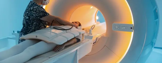
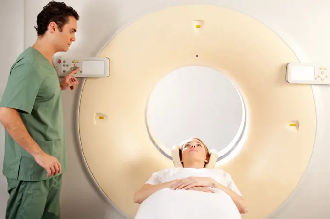
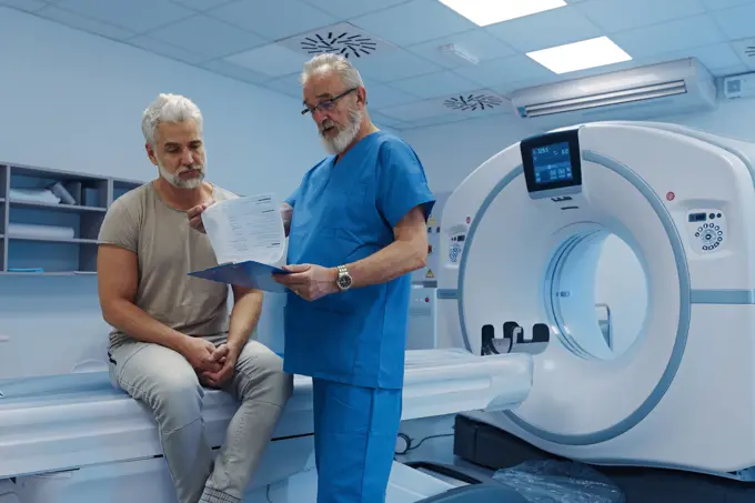
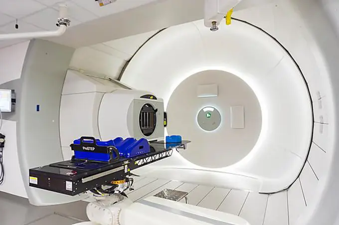
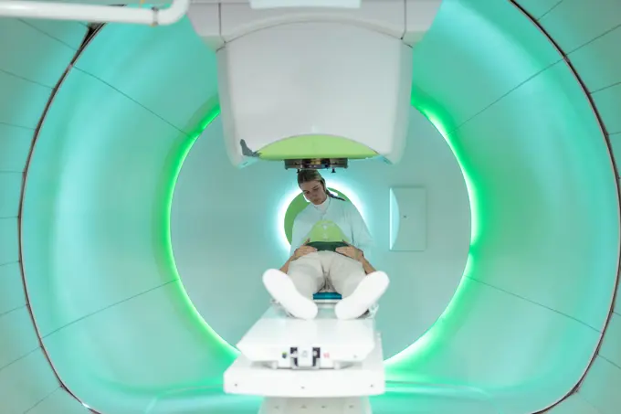
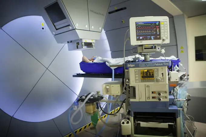
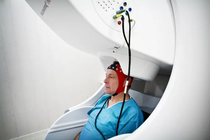
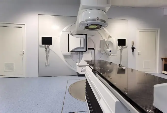
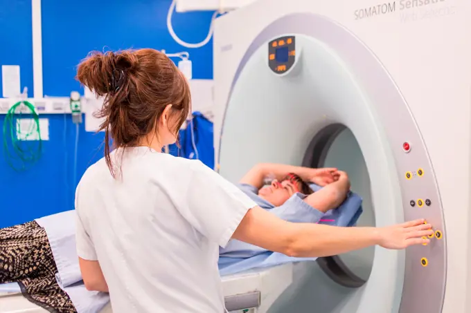
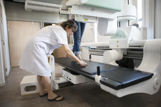
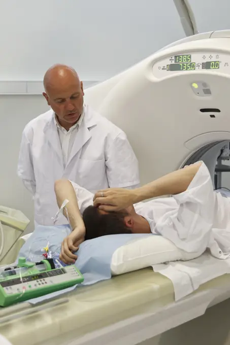
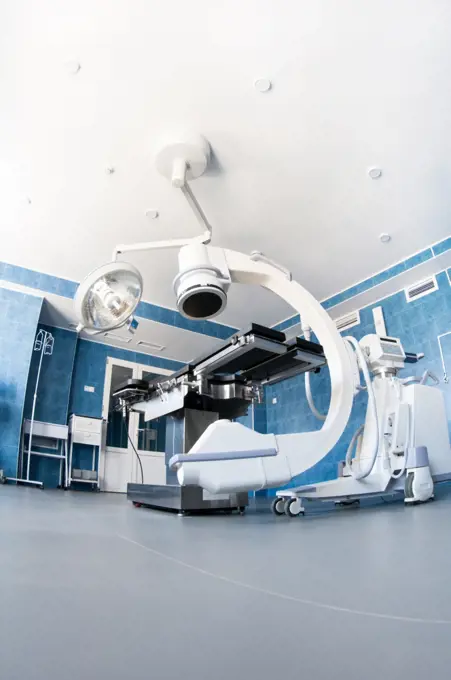
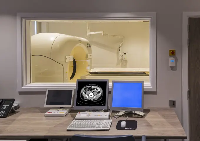
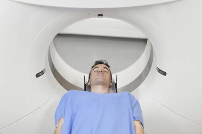
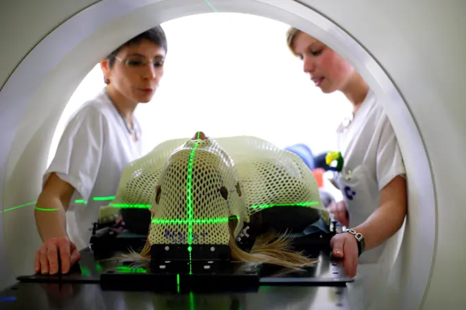
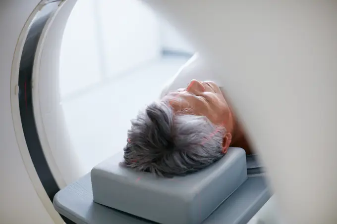
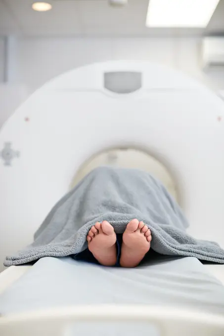
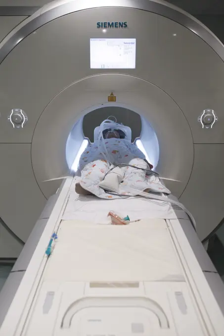
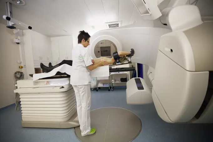
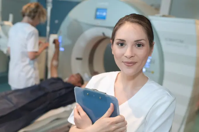
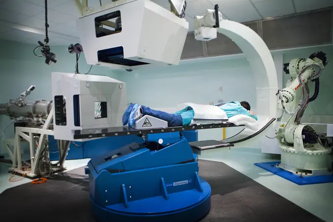
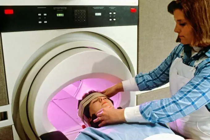
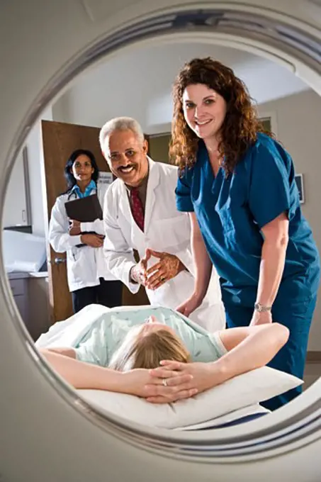
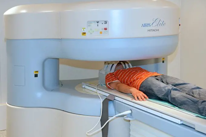
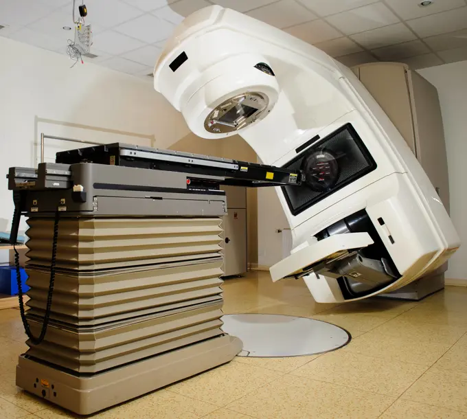
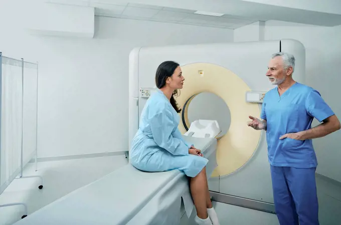
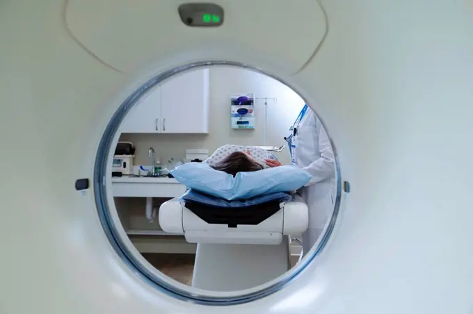
Visuals depicting patients undergoing MRI scans, with medical staff assisting in a clinical setting focused on diagnostic imaging.

Caucasian doctor comforting patient near scanner
211 assets in this story
1525-57384357
doctor giving instructions to technician for carrying out mri scan
1660R-54940
Male x-ray technician wearing protective glasses
1848-50946606
Onco Ray
4269-3171
Young woman undergoing a gamma scan examination of her bones at the department of nuclear medicine, Angoulème Hospital, France.
1660R-54933
Woman undergoing x-ray in lab
824-29055911
Digital medical imaging center, mammography room.
6188-60030523
Doctor or nurse and patient with tomography CT or MRI Scan in hospital. Interior of radiography department. Technologically advanced equipment in white room. Magnetic resonance diagnostics machine Doctor or nurse and patient with tomography CT or MRI Scan in hospital. Interior of radiography department. Technologically advanced equipment in white room. Magnetic resonance diagnostics machine. ,model released, Symbolfoto Copyright: xZoonar.com/MariyaxLvovax 17882722 ,model released, Symbolfoto ,property released
1899-66171
Close up of EpiPen
1848-49845953
Ruhr area. Compertomograph (CT) of the years 1987-8
1525-25072326
A CT Scanner Technician preparing a patient for scanning
824-63197526
Reportage in Pitié Salpêtrière hospital in Paris, France.
1589R-14623261
Doctor comforting patient at scanner
824-63210940
Report on PET imaging. Positron Emission Tomography, or PET Scan, is used for the diagnosis and monitoring of cancer patients. This method makes it possible to detect a tumor thanks to a radioactive product which accumulates in an excessive way in the cells presenting a pathological hypermetabolism.
6188-60030396
Medical CT or MRI Scan in the modern hospital laboratory. Interior of radiography department. Technologically advanced equipment in white room. Magnetic resonance diagnostics machine Medical CT or MRI Scan in the modern hospital laboratory. Interior of radiography department. Technologically advanced equipment in white room. Magnetic resonance diagnostics machine. Copyright: xZoonar.com/MariyaxLvovax 17861734
6188-68131613
Doctor explaining CT scan results to patient in hospital setting Doctor explaining CT scan results to male patient in hospital setting model released
1848-50946609
Onco Ray
4286-43439
A patient being placed into an MRI (magnetic resonance imaging) machine for a scan test.
1815-69224368
Patient undergoing proton therapy in a medical facility with advanced technology
1525-27067733
Interior of operating room in modern clinic,operating room with modern equipment.
1848-53791504
Patient lying in a linear particle accelerator for radiation treatment, Germany, Europe
824-63210928
Report on PET imaging. Positron Emission Tomography, or PET Scan, is used for the diagnosis and monitoring of cancer patients. This method makes it possible to detect a tumor thanks to a radioactive product which accumulates in an excessive way in the cells presenting a pathological hypermetabolism.
824-63205932
Reportage at the Rinecker Proton Therapy Center in Munich, Germany. The centre has the latest equipment for proton therapy treatment. Proton therapy involves irradiating cancer cells by concentrating a proton beam on the heart of the tumour, while preserving the healthy surrounding tissue. Settings on software which allow the tumour to be targeted precisely. To treat tumours on organs that move during breathing, the patient is placed under artificial apnea.
824-63179574
Report from the Neuro Imaging Research Center located at the Pitié Salpêtrière in Paris. Magnetoencephalography (MEG) platform. MEG makes it possible to detect the variations in the magnetic field that accompanies the various cerebral activities. It is used to study normal or abnormal functioning of the brain. The recording of magnetic fields induced by the electrical activity of neurons requires the use of ultra-sensitive sensors called SQUIDs (Superconducting Quantum Interference Device). The 306 sensors are spread over 102 sites, which makes it possible to measure near and far magnetic fields, allowing a better view of the deep structures of the brain. Here the subject wears an EEG helmet (Electroencephalography) which makes it possible to combine MEG and EEG.
1525-23830920
Medical advanced linear accelerator in oncological cancer therapy in a modern hospital.
1815-15679466
Patient in hospital during CT examination and radiologist
4269-39804
Female patient undergoing a CT scan, Angoulême hospital, France .
824-63208918
Reportage in a radiology centre in Haute-Savoie, France. A technician carries out a foot x-ray on a patient who had an operation on a hallux valgus.
824-63173363
Ct scanner examination
4269-6297
Department of Medical Imaging, Centre Cardiologique du Nord, St Denis, France. The Lightspeed VCT 64-Slice CT Scanner produces 64 slices per rotation, and reduces radiation exposure by 70%.
4269-4489
Patient undergoing Positrons Emission Tomography scan (PET scan) of the brain.
1525-21754326
operating room with X-ray medical scan.
4294R-1469
Doctor with patient at CT scanner
1801-62846679
Lyca Health Clinic at Westferry Circus, London Docklands. MRI and CT scanner suite. LycaHealth Centre Westferry Circus, London, United Kingdom. Architect: James Park Associates, 2015.
4269-21551552
Treatment of breast cancer by radiotherapy, Angouleme hospital, France.
4493-V111115380
1773R-84538
Patient laying in CT scanner in hospital
6145-54193558
HONIARA, Solomon Islands (Sept. 1, 2022) - Hospital Corpsman 1st Class Jed Rodriguez, from Los Angeles, left, gives a presentation of a computerized tomography (CT) to medical professionals from the National Referral Hospital in Honiara aboard the Military Sealift Command hospital ship USNS Mercy (T-AH 19) during Pacific Partnership 2022. Now in its 17th year, Pacific Partnership is the largest annual multinational humanitarian assistance and disaster relief preparedness mission conducted in the Indo-Pacific. Pacific Partnership is a unifying mission that fosters enduring friendships and cooperation among many nations. The years mission in Solomon Islands will include participants from the United States, Japan and Australia.
4269-20408074
CT scan-assisted biopsy, Saint-Louis hospital, Paris, France.
824-63205941
Reportage at the Rinecker Proton Therapy Center in Munich, Germany. The centre has the latest equipment for proton therapy treatment. Proton therapy involves irradiating cancer cells by concentrating a proton beam on the heart of the tumour, while preserving the healthy surrounding tissue. Settings on software which allow the tumour to be targeted precisely. To treat tumours on organs that move during breathing, the patient is placed under artificial apnea.
1525-24502167
Patrient being prepared for mri scan
4269-5316
In January 2007, the Institut Curie, Paris, France, was the first hospital in France to treat cancer with tomoTherapy. This image-guided intensity-modulated radiation therapy improves the precision of the treatment and reduces radiation exposure to healthy surrounding tissue. A thermoformed mask maintains the patient still during exposure.
824-63181706
Digital medical imaging centre in Paris, France. Abdominal CT scan.
1525-24502173
medical equipment doctor and patient in mri room at hospital
1624R-4171
Portrait of adult patient sitting in front of cat scan
1801-64283621
London Clinic-A Medical X-Ray Ct Scanner , London, , Architect Unknown
1848-53791501
Linear particle accelerator in a radiotherapy practice, Germany, Europe
1525-24502174
Medical worker holding up scanner results
1589R-14623544
Doctor and nurse helping patient at scanner
4197-64120280
Doctor, mri and woman with patient in hospital ready to start scanner machine. Ct scan, examination and medical professional with senior person before cancer radiology test for healthcare in clinic.
824-63181479
Digital medical imaging centre in Paris, France. X-ray room.
4269-25875
Lung scintigraphy. Man undergoing a gamma scan of his lungs. The bell-shaped element above his head diffuses a gaz containing radioactive tracers, which accumulate in lungs tissues and are detected by a gamma camera.
1841R-111748
1525-25971082
Medics around man in scanner
824-63205936
Reportage at the Rinecker Proton Therapy Center in Munich, Germany. The centre has the latest equipment for proton therapy treatment. Proton therapy involves irradiating cancer cells by concentrating a proton beam on the heart of the tumour, while preserving the healthy surrounding tissue. Settings on software which allow the tumour to be targeted precisely. To treat tumours on organs that move during breathing, the patient is placed under artificial apnea.
4269-8940
Closed geometry Magnetic Resonance Imaging (MRI) unit, 3 tesla with a brain MRI scan on the control screen.
4269-3172
Young woman undergoing a gamma scan examination of her bones at the department of nuclear medicine, Angoulème Hospital, France.
1525-21156736
Woman lying in front of CAT scan machine
4197-73070584
Healthcare, mri machine or patient in clinic for medical procedure, radiology or brain scan for diagnosis. Senior woman, examine skull or tech for tumor screening, cancer detection or monitor growth
824-29055371
MRI in a hospital radiology department.
1815-69224402
Doctor assisting patient in a proton therapy room for cancer treatment
4197-73070583
Assessment, healthcare and MRI with feet of patient in machine for appointment or checkup. CT scan, diagnosis and medical equipment with person in hospital for brain anatomy, exam or function
1841R-92301
Patient getting MRI scan
824-63173337
Brain, mri
824-63196947
Reportage in a radiation oncology service in Switzerland. The service is equipped with the latest linear accelerator, the Truebeam. A technician sets a patient up for prostate cancer treatment.
824-63206546
Reportage in a radiology service in a hospital in Haute-Savoie, France. Scanner.
1525-57365268
female radiologist with colleagues standing by mri machine
6145-54192683
Bridget Moran, 81st Medical Group radiation therapist, operates radiation equipment for a patient, Terry Eddy, 81st MDG medical dosimetrist, at Keesler Air Force Base, Mississippi, Aug. 30, 2022. The primary mission of the 81st MDG is to maintain medical readiness for worldwide contingencies by providing health care for more than 22,000 enrollees, including almost 4,500 active-duty members.
1525-26417776
MRI scanning flat color vector illustration. Healthcare monitoring. Disease diagnostic. Ultrasound technology. Patient visiting doctor 2D cartoon characters with interior on background. MRI scanning at hospital flat color vector illustration
6145-54224346
Matt Sherwood, Wright State University Center of Neuroimaging and Neuro-Evaluation of Cognitive Technologies director, briefs Col. Christopher Meeker, 88th Air Base Wing and Wright-Patterson Air Force Base commander, on the schools research MRI scanner Dec. 5, 2022. Air Force Research Laboratory scientists will now have access to the machine, thanks to an agreement Meeker signed with WSU earlier that evening.
824-63197806
Reportage in the National Oncology Particle Therapy Centre in Milan, Italy (The CNAO - Centro Nazionale di Adroterapia Oncologica). Particle therapy is a state-of-the-art technique used to treat cancer. It consists of using hadrons, protons (proton therapy) and cabon ions (carbon-ion treatment) to irradiate tumours. It has two therapeutic purposes, a high ballistic accuracy which allows healthy tissue surrounding the tumour to be spared, and a greater efficiency at treating certain cancers. The CNAO is the only centre to own carbon-ion treatment technology in Europe. An x-ray is carried out to check that the patient is in exactly the right position.
6177-V54158632
824-63210923
Report on PET imaging. Positron Emission Tomography, or PET Scan, is used for the diagnosis and monitoring of cancer patients. This method makes it possible to detect a tumor thanks to a radioactive product which accumulates in an excessive way in the cells presenting a pathological hypermetabolism.
4391-165
A woman's head is being secured by a female technician, preparing the woman for magnetic resonance imaging (MRI).
4128-20038226
Man having CT scan
824-63181481
Digital medical imaging centre in Paris, France. X-ray room.
824-63181654
Digital medical imaging centre in Paris, France. CT scan of the abdomen.
1785-18809
Healthcare workers preparing patient for CAT scan
4286-67746
magnetic resonance tomography
824-63206554
Reportage in a radiology service in a hospital in Haute-Savoie, France. Scanner.
1525-23918275
Vector flat cartoon doctor character in protective suit disinfects MRI table in clinic lab-coronavirus covid prevention,protection vs pandemic viral infection,medical concept. Flat cartoon doctor character,coronavirus covid-19 prevention vector illustration concept
824-63161360
Oncological hadrontherapy for the treatment of cancer with hadrons, protons or carbon ions.
824-63210231
Reportage on a mountain rescue doctor working in both the Carroz and de Flaine ski resorts in Haute-Savoie, France. He treats skiers and snowboarders who have been injured on the slopes. A nurse x-rays a young patient who has a fractured wrist and has just had it plastered.
1589R-12919186
Hispanic doctor and nurse using digital tablet in MRI room
824-63128992
Medical imaging center in Paris, France.
1660R-54956
Woman having an MRI
6188-64821516
Linear Accelerator at hospital Linear Accelerator at hospital. Linear accelerators are the newest radiation technology available today. Copyright: xZoonar.com/ALEXxTIHONOVx 3952203
824-63196928
Reportage in a radiation oncology service in Switzerland. The service is equipped with the latest linear accelerator, the Truebeam. This machine carries out intensity modulation radiation therapy with one or mutiple arcs (RapidArc), adapted to all areas and types of cancer. It also allows stereotactic radiation therapy. A scanner known as the Cone Beam CT has been integrated into the TrueBeam in order to optimise treatment. It allows images to be captured during treatment in order to follow the movement of the tumor during irradiation, and to adapt the treatment in real time. Thanks to the recordings of breathing movement, the TrueBeam allows the most mobile organs and tumors to be treated while limiting uncertainties on the volume to be covered during treatment.
824-63197462
Reportage in the Rinecker Proton Therapy Centre in Munich, Germany. The centre is equipped with the latest protontherapy treatment technology. Protontherapy consists of irradiating cancer cells by concentrating a beam of protons on the core of the tumour, while preserving healthy neighbouring tissue.
4128-20038245
Woman having CT scan
4128-24794773
Patient talking to radiologist
1848-51025463
Doctor monitors patient on monitor at CT, computed tomography, Karlsbad, Czech Republic, Europe
1815-69224383
Doctor and patient in a proton therapy room for cancer treatment
6145-58888369
Corpsman 3rd Class Johndenzel Macatuno, from Dededo, Guam, conducts a CT scan on a local patient aboard the hospital ship USNS Mercy (T-AH 19) anchored off of Chuuk, Federated States of Micronesia, as part of Pacific Partnership 2024-1, Jan. 16, 2024. Pacific Partnership, now in its 19th iteration, is the largest multinational humanitarian assistance and disaster relief preparedness mission conducted in the Indo-Pacific and works to enhance regional interoperability and disaster response capabilities, increase security stability in the region, and foster new and enduring friendships.
4286-16875
A medical lab technician checks the I.V. of a patient who is laying on a table preparing to have a CAT scan.
824-63181643
Digital medical imaging centre in Paris, France. Dental x-ray.
4269-4485
Patient undergoing Positrons Emission Tomography scan (PET scan) of the brain.
824-29055343
Proton therapy irradiates cancer cells with a beam of protons in the center of the tumor.
1848-54953244
Electrophysiological cardiac catheterization, EPU, for cardiac arrhythmia, in a hospital in Germany, Europe
824-29055916
Digital medical imaging center, cranial scanner.
1589R-14622688
Futuristic nurses repairing cyborg
PREVIOUS
of 3
NEXT
2401 S. Ervay, Suite 206
Dallas, Texas 75215
United States
Get Started
Free ResearchMy BoardsMy Cart
For Creators
How To License Your ContentContributor PortalFrame of Mind
Resources
API accessPricing
Contact
+1 866 236 0087help@viewpictures.co.uk Contact form
©2026 View Pictures. All Rights Reserved. -B
This site is protected by reCAPTCHA and Google Privacy Policy and Terms of Service apply.