My Boards
View Pictures
  • My Boards

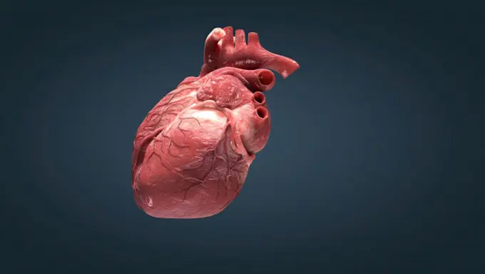
3D Heart Anatomical Illustrations

Detailed 3D representations of the human heart, showcasing its chambers and blood vessels with realistic textures and colors.

Human Heart
Human Heart
234 assets in this story
4239R-20483368
Conceptual image of human heart.
4128R-11313596
Human heart and lungs, computer artwork.
1525-26244446
As the heart beats, it pumps blood through a system of blood vessels, called the circulatory system. The vessels are elastic, muscular tubes that carry blood to every part of the body. 3D illustration. Heartwork system. Pumps Blood Through The Human Body
1525-23339132
DNA and heart. 3d illustration
1525-26243949
Heart and circulatory system are our bodys lifeline, delivering blood to the bodys .The heart then sends the blood to the lungs to pick up more oxygen.3D illustration. This features the heart and circulatory system and how they work.
1525-21341722
Human heart beats
1525-23455521
Human heart. Human heart with cog and gear mechanisms
1899-53510756
Anatomy of the adult heart in section in anterior view and pericardium. The heart provides blood circulation. It pumps blood from the veins to return it to the organs through the arteries. The heart is enveloped by a membrane, the pericardium shown in gray. The heart is a hollow muscle containing two parts separated by a septum, the septum. There is the left heart and the right heart. Each part is subdivided into two chambers, an atrium and a ventricle. Each cavity is separated by a valve shown in gray. The mitral valve separates the atrium from the left ventricle. The tricuspid valve separates the atrium from the right ventricle. Large vessels open into the chambers of the heart. The upper and lower vena cavae open into the right atrium (return of deoxygenated blood). While from the right ventricle opens the pulmonary trunk through the pulmonary valve. From the pulmonary trunk opens the right and left pulmonary arteries to the lungs. The pulmonary veins open into the left atrium (oxyg
1899-12939
Conducting system of the heart III
4128R-28212
Atherosclerosis. Computer artwork of a narrowed artery, due to a cholesterol plaque.
4269-2887
3D Computed Tomography (CT) reconstruction scan of a normal heart. The left coronary artery at center separated the right ventricle (at left) and the left ventricle (at right). Also seen the left and right atrium. Image obtained with a 64-Slice CT Scanner. Producing 64 slices per rotation, this advanced equipment is the first in France used only for heart diagnostic. The very high definition 3D images produced in less than 10 secondes are a usefull non invasive technique to observe a mobile orga
4128R-11542228
Human heart, computer illustration.
1296R-197
Close-up of a human heart
1525-23236457
Human heart. Human heart image with mechanisms. Health and medicine
4128R-26130
Healthy heart, computer artwork.
4128-30419449
Heart, illustration
4128R-15515534
Illustration of a fatty heart
1525-21337770
Human heart
1525-26244086
Human heart, realistic anatomy 3D illustration. Human heart, realistic anatomy
824-72488532
Anatomy of the heart.
1525-56722631
Atrial Septal Defect, Blood Flow 3D rendering. Atrial Septal Defect, Blood Flow
1525-23167790
Human heart beats. Virtual image of human heart with cardiogram
1525-23440558
Human heart beats. Virtual image of human heart with cardiogram
4378-4334
Coronary Vessels
1525R-14977057
Drawing sketch style illustration of a human heart anatomy set isolated white background.. Human Heart Anatomy Drawing
4128R-15515535
Illustration of human heart
1525-23274930
Human heart beats. Virtual image of human heart with cardiogram
4378-825
Human heart viewed from the front.
4128-19248733
Human fetus at week 40, illustration.
1525-26243929
As the heart beats, it pumps blood through a system of blood vessels, called the circulatory system. The vessels are elastic, muscular tubes that carry blood to every part of the body. 3D illustration. Heartwork system. Pumps Blood Through The Human Body
4128-19248096
Human heart, illustration.
4128R-24344
Human heart, computer artwork.
1422R-1027
Realistic plastic human heart model
1525-56323157
Human heart vector image
1525-23176971
Human heart beats. Virtual image of human heart with cardiogram
1525-23558417
Human heart. Human heart image with mechanisms. Health and medicine
1428R-249
Close-up of a human heart
1525-21331243
Human heart beats
1525-26244456
Systole, period of contraction of the ventricles of the heart that occurs between the first and second heart sounds of the cardiac cycle. 3d illustration. Systole causes the ejection of blood into the aorta and pulmonary trunk.
6188-62298978
Human heart cross section. Human heart cross section, with detailed internal structure. 2 D digital illustration, on white background, with clipping path. Copyright: xZoonar.com/Pixelchaosx 9888023
4128R-24483
Human heart, computer artwork.
4128R-14057458
Normal Heartbeat, illustration
4239-18642326
Human heart cross-section with detailed internal structure, on white background.
1525-23423256
Human heart. Human heart with cog and gear mechanisms against black background
4128R-11313406
Human spleen, cut-away computer artwork.
1848-77402231
3D Illustration Concept of Human Fetus Baby in Womb Anatomy
6188-55647642
Cardiac anatomy with veins and coronary vessels heart anatomy with veins and coronary vessels copyright: xzoonar.com/dr.xnorberbertxlangex 14363462
1525-27648773
Sickle cell heart circulation blockage and anemia as a disease with normal and abnormal hemoglobin in a human artery anatomy with heart cardiovascular medical illustration concept with 3D illustration elements.
824-63165993
Heart and right coronary artery, 3D CT scan.
4378-3931
Front view of the heart with coronary blood vessels visible set against an atmospheric white backdrop.
1525-22197388
human heart
4239-20481126
Posterior view of human heart on photo of blood pressure cuff.
4378-1664
Wireframe diagram of a heart.
1428-1331
Cross sections of human heart and major vessels on gray background
1574R-018799
Close-up of a human rib cage
4128R-28731
Balloon angioplasty. Computer artwork of a stent being placed in a narrowed blood vessel.
1525-23190581
Human heart beats. Virtual image of human heart with cardiogram
1525-56183570
The circulatory system or cardiovascular system is the organ system that ensures the circulation of substances throughout the body. 3d illustration. circulatory system or cardiovascular system
1849-66219949
Pulse trace, target and anatomical illustration of the heart over male human body
1525-21335635
Human heart
1525-21357748
Human heart
1525-56190173
Each time your heart beats, electrical signals travel through your heart. These signals cause different parts of your heart to expand and contract. 3D illustration. Cardiac Conduction The heart conduction system is the network of nodes, cells and signals that controls your heartbeat.
1899-53513170
Heart, illustration.
824-63217340
Illustration showing the coronary sinus which produces the great cardiac vein, the small cardiac vein and the left posterior interventricular artery. The coronary arteries are visible in the background.
6243-71428021
Medical - Victorian Anatomical Illustrations - on a white background for cut out.
824-63218266
Frontal cut of the heart showing the 4 cardiac cavities. right and left atria, separated from the right and left ventricles by the tricuspid and mitral valves. Entering the large vessels: the aorta, the pulmonary trunk and the superior and inferior vena cava.
4128-30417770
Heart, illustration
4128R-13387479
Human heart and medication, illustration.
1525-26243947
Heart valve The valves keep blood moving through the heart in the right direction 3D illustration. Heart valve The valves keep blood moving through the heart in the right direction
1525-75959099
Heart vector design. Realistic anatomy pictures. Human body internal organs
1525-21362353
Human heart beats
4128R-28592
Human heart, computer artwork.
1848-54708926
Human heart, illustration
4306R-10400
An anatomical model of a heart.
1525-19835638
Mechanical Heart on a black background
6188-58105257
loop 3d rendered medically accurate animation of the human lung loop 3d rendered medically accurate animation of the human lung Copyright: xZoonar.com/StanislavxRishnyakx 14600768
824-63188671
Illustration of the stages of formation and setting off of a myocardial infarction, or heart attack. An artherosclerotic plaque forms in the lining of coronary arteries which forms a blood clot causing an arterial occlusion. When the artery is completely blocked, the myocard no longer receives oxygen and starts to die, causing a heart attack with pain that is most often located on the left side near the left arm.
1525-56183884
The heart and the blood vessels 3D illustration. The heart and the blood vessels
4128R-28793
Human heart, computer artwork.
4128R-14637688
Human heart pulmonary valve, illustration
4128-V58562497
1525-26243903
The right atrium receives blood from the veins and pumps it to the right ventricle. 3D illustration. The right atrium receives blood from the veins and pumps it to the right ventricle.
4128-19249334
Atherosclerosis, illustration.
1746-30002701
Human heart.
1525-21346107
Human heart
1899-53508457
3D CT scan of normal coronary arteries.
4128R-14060232
Child's heart, illustration
1525-26243932
As the heart beats, it pumps blood through a system of blood vessels, called the circulatory system. The vessels are elastic, muscular tubes that carry blood to every part of the body. 3D illustration. Heartwork system. Pumps Blood Through The Human Body
4269-25414
Calcifying pericarditis. 3D computed tomographic (CT) scan reconstruction of a heart affected by calcifying pericarditis (armoured heart) seen from above. Pericarditis is the inflammation of the pericardium, a thin layer of tissue that covers the outer surfaces of the heart. Here, the pericardium is covered by a fibrous calcarious rigid bag (white coat) and can not complete its natural mouvements.
5507-45883347
Heart with normal and partly blocked blood vessel
4378-3977
Three-quarter view of the heart and its coronary blood vessels.
4128R-28594
Human heart, computer artwork.
5507-37452228
heart anatomy and ecg background
1525-25984113
Heart care concept. Close up of man hand holding human heart
4128-19248727
Human fetus at week 39, illustration.
4220-21935774
Illustration of a human heart - top of a layered anatomical fold-out in a medical book.     Date: c.1900
1428-1325
Human heart and major vessels with metallic chrome effect on black background
4128R-11314504
Human heart and liver, computer artwork.
1525-56211132
The heart is the most important muscle in your body. There are two sides, the right and left, and four chambers. 3D illustration. The heart is the most important muscle in your body. There are two sides, the right and left, and four chambers.
4378-1697
Wireframe diagram of a heart.
PREVIOUS
of 3
NEXT
2401 S. Ervay, Suite 206
Dallas, Texas 75215
United States
Get Started
Free ResearchMy BoardsMy Cart
For Creators
How To License Your ContentContributor PortalFrame of Mind
Resources
API accessPricing
Contact
+1 866 236 0087help@viewpictures.co.uk Contact form
©2026 View Pictures. All Rights Reserved. -A
This site is protected by reCAPTCHA and Google Privacy Policy and Terms of Service apply.