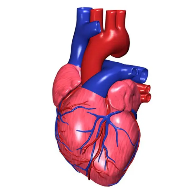
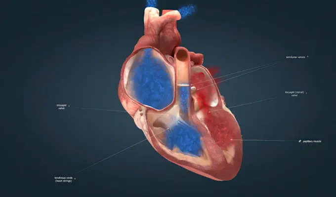
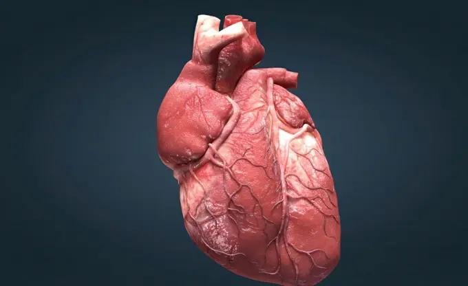
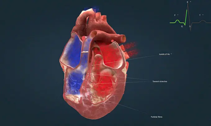
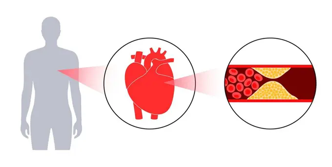
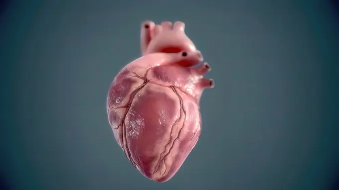
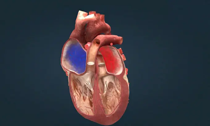
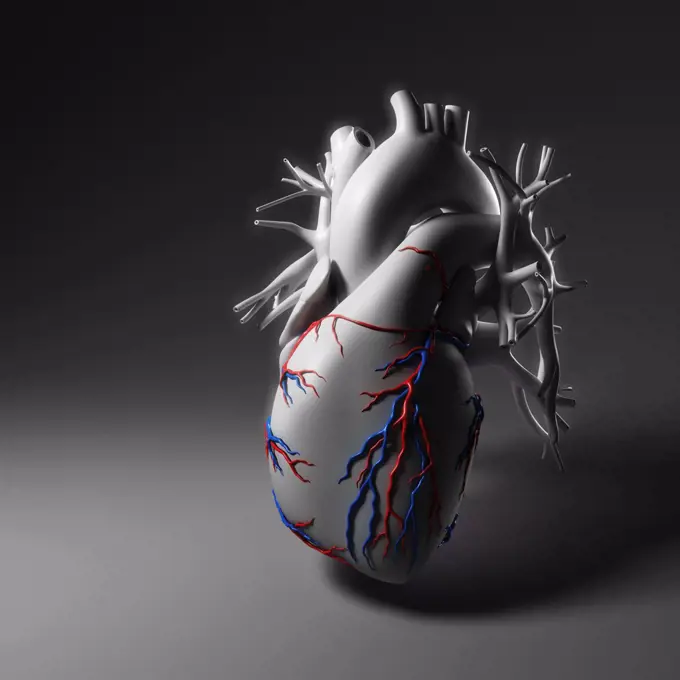
My Boards
View Pictures
  • My Boards

3D Heart Anatomical Illustrations

Detailed 3D representations of the human heart, showcasing its chambers and blood vessels with realistic textures and colors.

Human Heart
Human Heart
234 assets in this story
1525-26244027
3D Animated right-sided heart failure - beating heart 3D illustration. 3D Animated right-sided heart failure - beating heart
1525-26244114
As the heart beats, it pumps blood through a system of blood vessels, called the circulatory system. The vessels are elastic, muscular tubes that carry blood to every part of the body. 3D illustration. Heartwork system. Pumps Blood Through The Human Body
1525-56211287
The four valves are the aortic valve, mitral valve, pulmonary valve and tricuspid valve. A heart murmur is often the first sign of a heart valve problem. 3D illustration. As your heart pumps blood, four valves open and close to make sure blood flows in the correct direction.
1525-28312634
Cartoon style illustration of a human heart anatomy, an organ that pumps blood throughout the body via the circulatory system on isolated white background.. Human Heart Cartoon
1525-56190136
The heart is the most important muscle in your body. There are two sides, the right and left, and four chambers. 3D illustration. The heart is the most important muscle in your body. There are two sides, the right and left, and four chambers.
1525-56198029
Each time your heart beats, electrical signals travel through your heart. These signals cause different parts of your heart to expand and contract. 3D illustration. Cardiac Conduction The heart conduction system is the network of nodes, cells and signals that controls your heartbeat.
1899-53511461
Myocardial infarction. Visualization by electrocardiogram.
4128-19249323
Atherosclerosis, illustration.
1525-26244045
Human heart, realistic anatomy 3d model of human heart on the monitor, visual heart beating. Human anatomy, cardiovascular system. 3D illustration. Human heart, realistic anatomy 3d model of human heart on the monitor,
1849-66219770
Human heart and pulse trace with walking man
4128R-29119
Human heart, computer artwork.
1525-56190188
Each time your heart beats, electrical signals travel through your heart. These signals cause different parts of your heart to expand and contract. 3D illustration. Cardiac Conduction The heart conduction system is the network of nodes, cells and signals that controls your heartbeat.
4128R-15290292
Bradycardia, illustration
1570-18379224
Human heart organ on blue background
1525-20582724
Slow heart rate concept as a snail shell on human anatomy from an unhealthy body as a medical health care symbol of problems with the inner cardiovascular organ.
1525-70016995
Diagram of human heart with data annotations, vaccine bottle, and verification checkmark. Ideal for medical research, healthcare, cardiology, vaccination awareness, science education, medical app. Human heart with annotations showing medical data and vaccine bottle alongside verification checkmark
4128R-11314254
Human liver and gall bladder, computer artwork.
1296R-192
Close-up of an electrocardiogram
4128-111580858
Human heart, computer illustration.
1525-19835731
Mechanical Heart on a white background
4128R-15061609
Heart, illustration
1525-56183826
The four valves are the aortic valve, mitral valve, pulmonary valve and tricuspid valve. A heart murmur is often the first sign of a heart valve problem. 3D illustration. As your heart pumps blood, four valves open and close to make sure blood flows in the correct direction.
4378-4254
Coronary Vessels
4128R-15290296
Ventricular fibrillation, illustration
4128R-11313189
Human respiratory system, computer artwork.
4186-17678
1980S October 1988 Seismograph With Seismogram Recording Hurricane Gilbert
5513-18677151
The map of the heart.Blood is pumped through the heart through the aorta and connects arteries to the trunk of body after supplying food and oxygen it returns through the Veins passes to the lungs for oxygen. July 16, 1938.
1525-28131827
Electrocardiogram graph, heartbeat shown on monitor, 3D rendering
4128-17927802
Electrocardiagram, illustration
824-63188669
Illustration of the stages of formation and setting off of a myocardial infarction, or heart attack. An artherosclerotic plaque forms in the lining of coronary arteries which forms a blood clot causing an arterial occlusion. When the artery is completely blocked, the myocard no longer receives oxygen and starts to die, causing a heart attack with pain that is most often located on the left side near the left arm.
1525-27228801
Atherosclerosis of heart vessels flat infographics with labeled parts vector illustration
1525-26200267
Atherosclerosis. Fibrous plaque formation. Medical human, health graphic, thrombus in vessel. Vector illustration infographic. Atherosclerosis. Fibrous plaque formation. Vector infographic
4409-28578941
Infographic about the human heart; an extraordinary muscle that makes blood circulate through the body. Adobe InDesign (.indd).
4128R-15290275
Heart attack, illustration
PREVIOUS
of 3
NEXT
2401 S. Ervay, Suite 206
Dallas, Texas 75215
United States
Get Started
Free ResearchMy BoardsMy Cart
For Creators
How To License Your ContentContributor PortalFrame of Mind
Resources
API accessPricing
Contact
+1 866 236 0087help@viewpictures.co.uk Contact form
©2026 View Pictures. All Rights Reserved. -B
This site is protected by reCAPTCHA and Google Privacy Policy and Terms of Service apply.