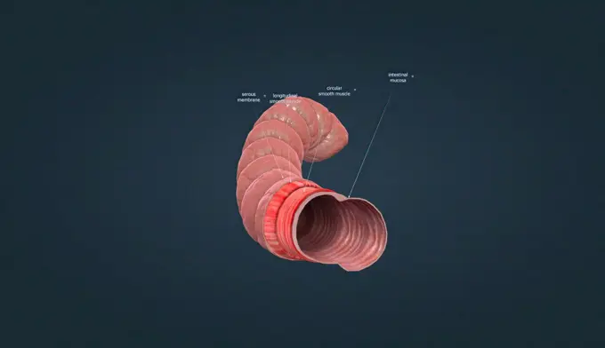
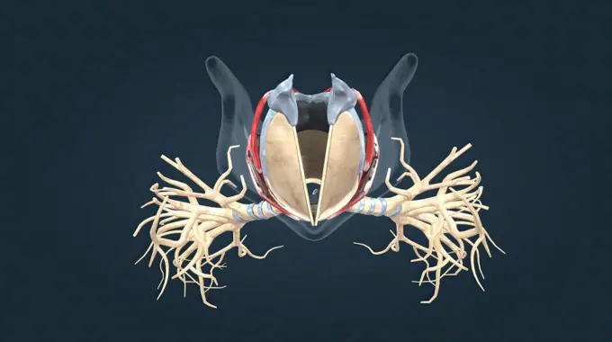
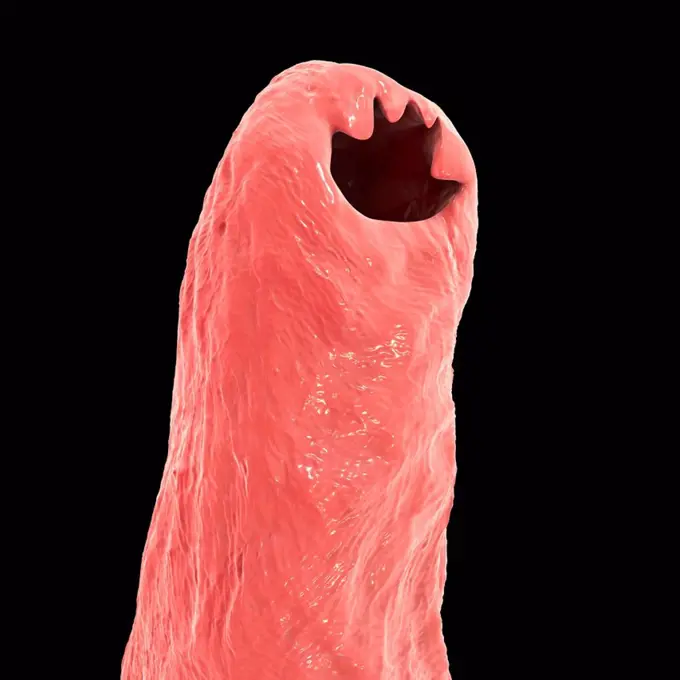
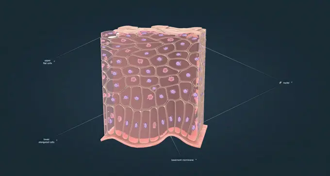
My Boards
View Pictures
  • My Boards

3D Medical Illustrations

Detailed 3D renderings of human anatomy, depicting stages of embryonic development, cancer, cardiovascular issues, and respiratory systems.

Human heart, illustration
152 assets in this story
1525-56139657
The small intestine consists of four main layers of tissue, which are (from the outside to the centre). 3d illustration. The human intestines function to absorb digestive products and have special structures to perform this function.
4128R-13409985
Human brain anatomy, illustration
1525-56149791
intestinal villi (singular: villus) are small, finger-like projections that extend into the lumen of the small intestine 3d illustration. intestinal villi (singular: villus) are small, finger-like projections that extend into the lumen of the small intestine
4378-1757
A human model showing pregnancy between 6-8 days.
1525-56211216
The adrenal glands produce corticosteroid hormon and epinephrine, which maintain blood pressure and regulate metabolism 3D illustration. The adrenal glands produce corticosteroid hormon and epinephrine, which maintain blood pressure and regulate metabolism
824-63189353
Illustration of a healthy uterus (at the top), and a view of the base of the cervix (bottom).
824-63189056
Illustration of the anatomy of the hypophysis or pituitary gland. -the posterior lobe of hypophysis (beige), the neurohypophysis make up the neurological part. -the anterior lobe of hypophysis (blue), the adenohypophysis, make up the glandular part. The two lobes are covered by the infundibulum. Above that is the hypothalamus (beige/brown).
1570R-141301
Four petri dishes with pink liquid
1525-56139660
The small intestine consists of four main layers of tissue, which are (from the outside to the centre). 3d illustration. The human intestines function to absorb digestive products and have special structures to perform this function.
1525-23789028
blood clot formation 3d illustration. blood clot formation 3d medical
1525-26182241
Structure and Working of Human Eye 3D illustration. 3D illustration Medical of Anatomy of the Eye
1525-56190189
The larynx is an organ in the upper part of the neck that protects the trachea against breathing, sound production, and food aspiration. 3D illustration. The larynx is an organ located in the anterior neck.
1525-23016352
Artificial human heart model isolated on black background
4128R-11292096
Human muscular structure, computer artwork.
1428R-357B
Close up of the human mouth and human tongue with a hot pepper on it
4128R-13763862
Hookworm head. Computer illustration of the head of the parasitic hookworm Ancylostoma duodenale. This is one of the commonest causes of hookworm infe...
1525-26235685
The importance of macrophage in the immune system 3D illustration. The importance of macrophage in the immune system
1525-56190180
A non-keratinized stratified squamous epithelium consists of cells arranged in layers upon a basal membrane. 3D illustration. A non-keratinized stratified squamous epithelium consists of cells arranged in layers upon a basal membrane.
824-63189395
Rear view illustration of the prostate with the seminal vesicles at the back.
4128R-11322999
Human fetal development (week 4), computer artwork.
1525-26220678
The green background digestive system 3D illustration. The green background digestive system
824-63219036
Human urinary system, anterior view, anatomy. The human urinary system seen anterior with the right kidney seen in section to show the fibrous capsule which envelops the cortex of the kidney. The pyramids abut in the pelvis which is extended by the ureter which opens at the bottom of the bladder at the level of the ostium of the ureter. Between the osmium, the bladder trigone. Directly under the bladder, the prostate passes through the urethra with the mouth of the seminal vesicles at the level of the veru montanum.
1525-56183981
long bone microscopic view anatomy 3d illustration. long bone microscopic view anatomy
4413-67356
Udder of a Holstein cow and mammal vein
4378-5280
Rat Brain Anatomy
4128-20044554
Anatomical model of the male urinary and reproductive system
824-28705997
Anatomical mannequin for teaching human organs
1525-26275125
The thigh bone is the large upper leg bone that connects the lower leg bones to the pelvic bone 3D illustration. Leg skeletal anatomy.Leg joint
824-28705995
Anatomical mannequin for teaching human organs
4239-18641591
Osteoclast breaking down bone tissue.
4128-V58569442
1525-23789224
The vagina is a muscular, hollow tube that extends from the vaginal opening to the uterus.3d illustration. A female’s internal reproductive organs are the vagina, uterus, fallopian tubes, and ovaries.
4269-24722
Pneumothorax. Pneumothorax (bottom right) Axial coloured Computed Tomography (CT) scan of a pneumothorax on the left lung.A pneumothorax is a gazeous effusion in the virtual space included in the visceral pleura and the parietal pleura (black), compressing the left lung reduced (red).
4128R-13763864
Computer illustration of the head of the parasitic hookworm Ancylostoma duodenale. This is one of the commonest causes of hookworm infestation in huma...
824-28705982
Anatomical mannequin for teaching human organs
1899-53508695
Anatomical mannequin for teaching human organs
4128-28769267
Buruli ulcer and Mycobacterium ulcerans, illustration
1525-56234239
The Overview of Nephron Function 3D rendering. The Overview of Nephron Function
6145-44302255
Scarab. Unknown
4128R-11313401
Human spleen, cut-away computer artwork.
4128-30416876
Reproductive system, illustration
4128R-13574344
Illustration of human brain structures.
1525-23789223
The vagina is a muscular, hollow tube that extends from the vaginal opening to the uterus.3d illustration. A female’s internal reproductive organs are the vagina, uterus, fallopian tubes, and ovaries.
1525-56184003
The humerus also known as the upper arm bone is a long bone that runs from the shoulder and scapula to the elbow. 3d illustration. The humerus also known as the upper arm bone is a long bone that runs from the shoulder and scapula to the elbow.
1525-23016347
Hand shows inside of human heart model isolated on black background
824-63186211
Reportage in Descartes University, Paris, France where Vizua 3D is based. Vizua provides real-time 3D visualisations for doctors using imagery from patients. Doctors can then print these visualisations in 3D. Heart printed with a 3D printer.
4239R-8548
Front view of human mouth with teeth and gums.
4128R-25339
Two-cell embryo, computer artwork.
1525-26235650
An inner layer, the synovial membrane or inner layer of the joint, which is an epithelium like structure 3D illustration. Joint capsule isolating and covering the entire joint
4239R-20483364
Conceptual image of human eye anatomy.
1525-56183838
The heart is the main organ of your cardiovascular system, a network of blood vessels that pumps blood throughout our body. 3D illustration. Heart is the main organ of your cardiovascular system, a network of blood vessels that pumps blood throughout our body.
6145-45251057
Some of the earliest concerns about fluid behavior in microgravity was the management of propellants in spacecraft tanks as they orbited the Earth. On the ground, gravity pulls a fluid to a bottom of a tank (ig, left). In orbit, fluid behavior depends on surface tension, viscosity, wetting effects with the container wall, and other factors. In some cases, a propellant can wet a tank and leave a large gas bubbles in the center (ug, right). Similar probelms can affect much smaller experiments using fluids in small spaces.
PREVIOUS
of 2
NEXT
2401 S. Ervay, Suite 206
Dallas, Texas 75215
United States
Get Started
Free ResearchMy BoardsMy Cart
For Creators
How To License Your ContentContributor PortalFrame of Mind
Resources
API accessPricing
Contact
+1 866 236 0087help@viewpictures.co.uk Contact form
©2026 View Pictures. All Rights Reserved. -A
This site is protected by reCAPTCHA and Google Privacy Policy and Terms of Service apply.