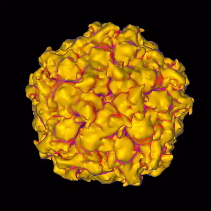
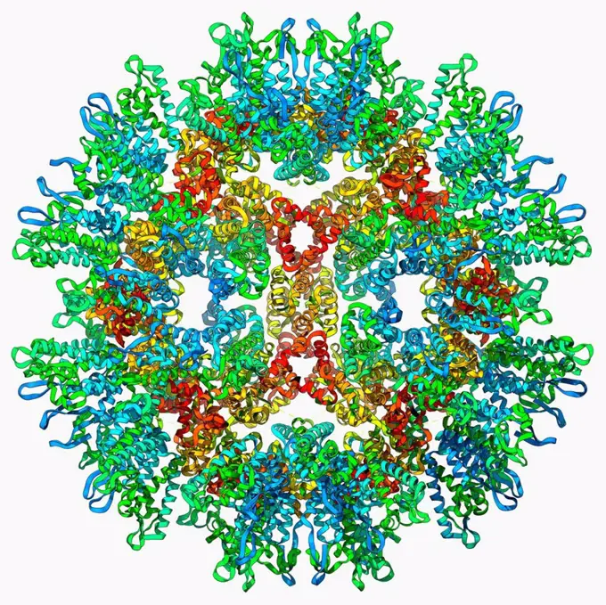
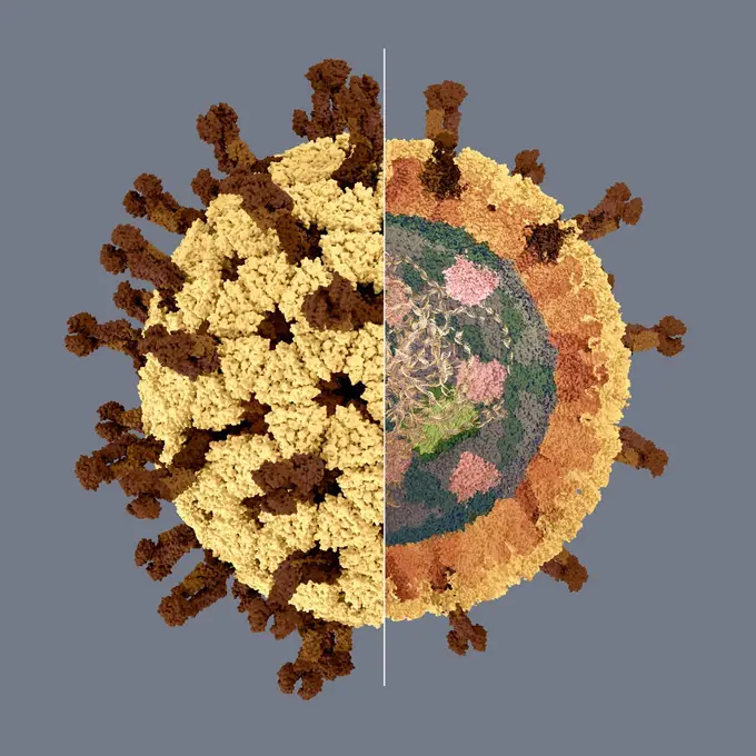
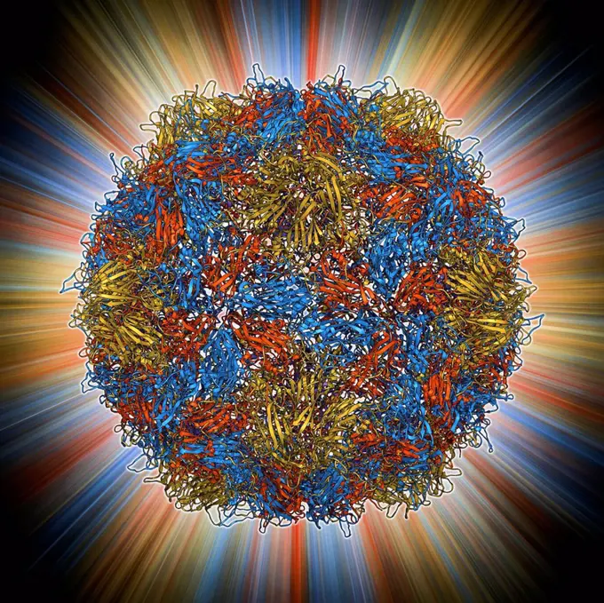
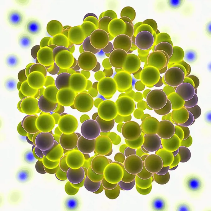
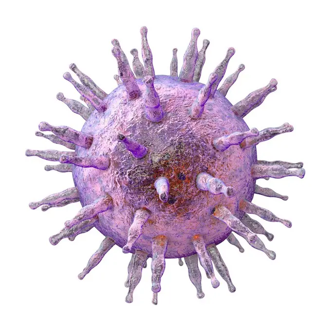
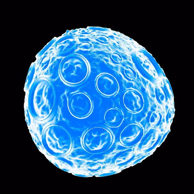
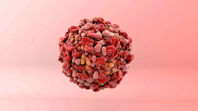
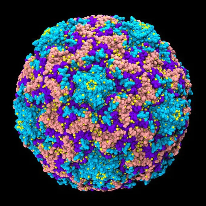
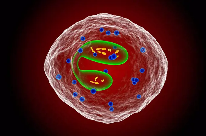
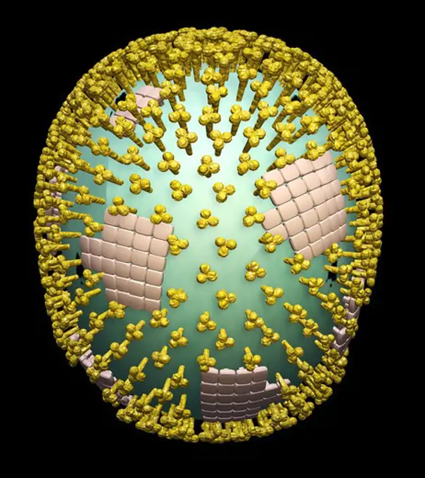
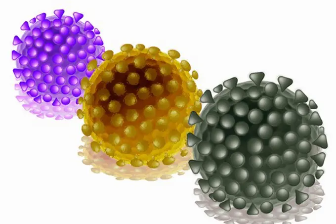
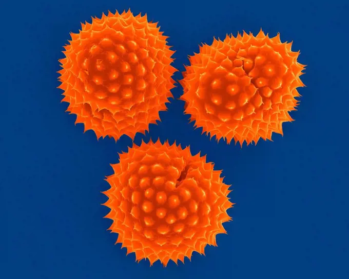
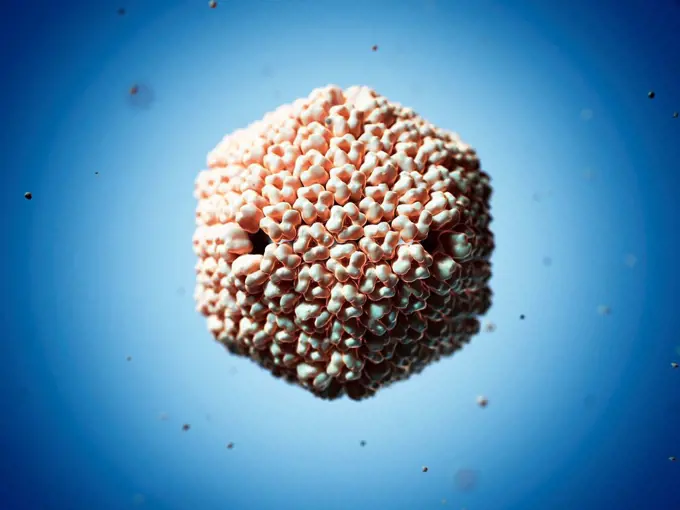
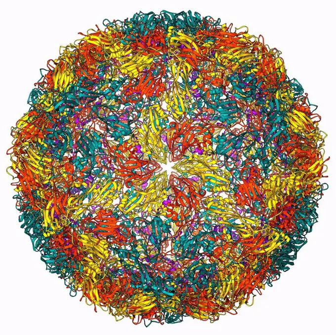
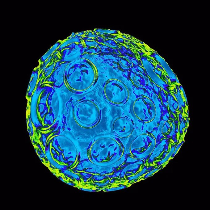
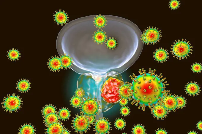
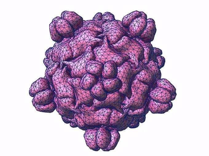
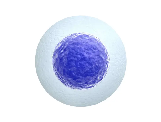
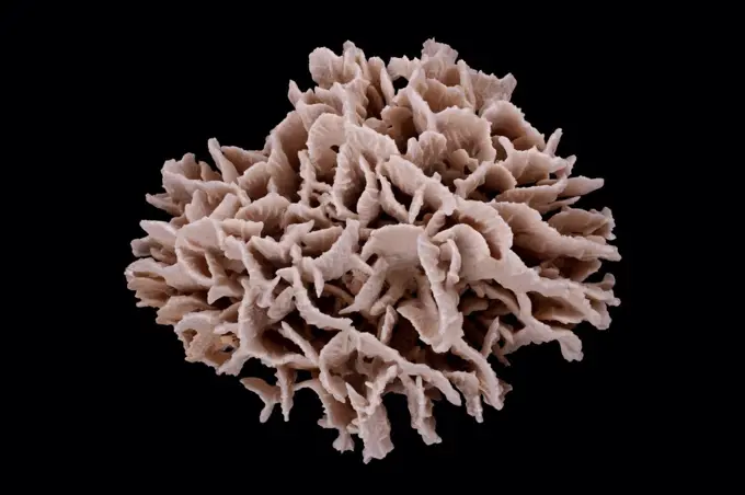
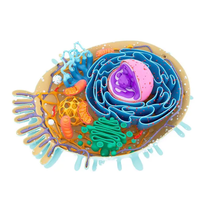
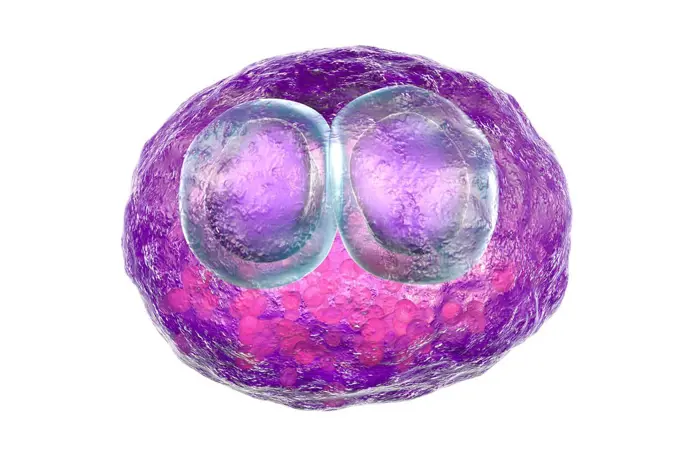
My Boards
View Pictures
  • My Boards

3D Virus Illustrations and Models

Computer-generated artworks representing different viruses. Each visual depicts intricate designs and vibrant colors, emphasizing their microscopic complexity.

Satellite panicum mosaic virus, computer artwork.
279 assets in this story
4128R-11313452
Satellite tobacco necrosis virus particle, computer artwork.
4128-V58573741
4128R-34114
Rous sarcoma virus capsid, molecular model. In viruses, the capsid is the protein shell that encloses the genetic material. A capsid consists of subun...
4128-28767013
Rotavirus structure, illustration
4128R-11313635
Equine rhinitis A virus, computer artwork.
4128R-11471594
Poliovirus type 3 capsid, molecular model. This enterovirus causes poliomyelitis (polio) in humans, which affects the nervous system, sometimes leadin...
4128R-11313685
L-A virus, computer artwork.
4128R-11474189
Rice dwarf virus (RDF), computer artwork.
4128-111493547
Nanoparticles, conceptual illustration.
4128R-13575546
Epstein-Barr virus (EBV), computer illustration. EBV, also known as human herpes virus 4, is 1 of 8 herpes viruses that infects humans. It is best kno...
4128R-11474047
Infectious salmon anaemia virus (ISA), computer artwork.
4128R-11473246
Nano gears, computer artwork.
4128-V58569460
824-63227213
The image on the left is the procapsid (outer protein coat) of herpes simplex virus type 1 (HSV1), the virus that causes cold sores. The image on the right is the mature capsid of the same virus. The major capsid proteins form hexons (lighter blue) and pentons (darker blue) connected by triplexes (green). As the virus matures, the holes in the procapsid ferment to stabilize the mature capsid. Understanding this process can help researchers determine how the virus infects cells and may lead to new treatments.
4128R-11284817
Basophil white blood cell, computer artwork. Basophils are the smallest and least common of the white blood cells. They are involved in allergic and i...
4128R-11474724
Human t-cell leukemia virus, computer artwork.
4128-111495171
Cancer cell, illustration.
4128-28515054
Bacteria cultured on Petri dish
4297-1649
3D computer generated model of the rhinovirus virion based upon the Protein Database entry 4RHV
4128R-11313734
Herpes virus, computer artwork.
4128R-13844622
Eosinophil white blood cell, illustration.
4128R-10687
Viral research, conceptual artwork
4128R-6825
Flu virus particle, artwork.
6188-64820021
Grape cluster covered with bubbles Grape cluster covered with bubbles Copyright: xZoonar.com/ALEXxTIHONOVxAlexanderxTihonovx 2908396
4297-1651
Illustration of swine flu virus showing the structure of the influenza virion including the hemagglutinin and neuraminidase proteins on the surface of the particle
4128R-11313565
Norovirus particle, computer artwork.
4269-24729
AIDS virus. Cutaway illustration of an AIDS virus Artwork
4128R-15365972
Immune response activation, illustration
1890-105657
Light Micrograph LM of a transverse section of a Helianthus stem, magnification x 18
1848-49194862
Illustrationsvirus
4128R-11473977
Hantavirus, computer artwork.
4128R-13413
Flu virus particles, computer artwork.
4128R-13575642
Colon cancer cell, computer illustration.
4128R-11474040
Ichnovirus, computer artwork.
1848-58229085
Fluorescence light of a Hard Coral Madreporaria
4128-16073915
Coronavirus infecting human cell, illustration
1773R-87982
Colored virus atoms
4128R-13575567
Herpes simplex virus, computer artwork.
824-63222941
Coronavirus (CoV). viruses of the Coronaviridae family and the Orthocoronavirinae subfamily. It is a pathogen of respiratory syndromes. Viewed from a transmission electron microscopy (TEM) image. Viral diameter 80nm to 100nm.
4128R-13620163
Ragweed (Ambrosia psilostachya) pollen, coloured scanning electron micrograph (SEM). This pollen is an allergen. Ragweed is a main cause of weed aller...
4128-V58573749
4128R-11313310
Human papilloma virus, computer artwork.
6188-67449438
Covid virus, model. Coronavirus, COVID-19, 3D rendering Covid virus, model. Coronavirus, COVID-19, 3D rendering isolated on white background Copyright: xZoonar.com/natatravelx 22984936
4128-16224206
Blackberries and blueberries, X-ray
1525-21938049
Classic soccer ball made of scattered 3D particles for Brazil
824-63194673
Colorized transmission electron micrograph showing particles of Middle East respiratory syndrome coronavirus that emerged in 2012.
4128R-13941173
Bladder cancer cells, illustration.
4409-17348607
Basophile.
1525-25012460
small green planet. Isolated 3d rendering
4128R-11313398
Sputnik virophage particle, computer artwork.
4128R-34017
Foot-and-mouth disease virus. Molecular model of the foot-and-mouth disease (FMD) virus (Aphtae epizooticae) protein coat (capsid). FMD affects cloven...
4128R-11313587
Foot and mouth disease virus particle, computer artwork.
4128R-11474054
Infectious salmon anaemia virus (ISA), computer artwork.
4128R-15516287
Illustration of a cancer cell
4128R-11474615
Herpes simplex, computer artwork.
5514-67119077
Botanical specimen unidentified and housed in a Kansas herbarium. The photograph was back lit then Inverted.
4128R-14768526
Viral etiology of prostate cancer, conceptual illustration
4128R-11543393
Pollen grain, computer illustration. The characteristic surface of pollen grains is used by botanists to recognise and classify plants. Each pollen gr...
4128-16102477
Virus, illustration
1848-49194839
Carrier virus and tumour or metastasis
824-63223111
3D print of a SARS-CoV-2 also known as 2019-nCoV, the virus that causes COVID-19 virus particle. The virus surface (blue) is covered with spike proteins (red) that enable the virus to enter and infect human cells. The spikes on the surface of coronaviruses give this virus family its name  corona, which is Latin for crown, and most any coronavirus will have a crown-like appearance. For more information, visit the NIH 3D Print Exchange at 3dprint.nih.gov. Credit: NIH
4128-28769454
DNA cube, molecular model
1795-51174264
Model of yellow Coronavirus against black background
1525-20622692
macro X-ray shot of fuzzy mold on white background
4128R-13745
Bacteriophage alpha 3. Computer model showing the structure of the protein capsid of bacteriophage alpha 3. Bacteriophages are viruses that infect bac...
4239R-20483442
Conceptual image of a group of platelets.
4128-19052507
Blood clot, illustration
4128R-11312853
Parvovirus, computer artwork.
4128R-13682630
Human egg, illustration.
4128R-11473968
Granulosis virus, computer artwork.
4201-98345
Coral (Pachyseris sp)
4128R-13941147
Cell structure, illustration.
824-63223068
3D printing of HIV. The surface of the virus (yellow) is covered with proteins (purple) that allow the virus to enter and infect human cells.
4128R-13682556
Unhealthy cell, illustration.
4128R-15366522
Cytomegalovirus infection, illustration
4128-30422598
Streptococcus pneumoniae bacteria, illustration
4128R-11312849
Brome virus, computer artwork.
4128R-14181801
Pollen grains from different plants, illustration
4128R-11291504
Artwork of an antibody structure.
PREVIOUS
of 3
NEXT
2401 S. Ervay, Suite 206
Dallas, Texas 75215
United States
Get Started
Free ResearchMy BoardsMy Cart
For Creators
How To License Your ContentContributor PortalFrame of Mind
Resources
API accessPricing
Contact
+1 866 236 0087help@viewpictures.co.uk Contact form
©2026 View Pictures. All Rights Reserved. -A
This site is protected by reCAPTCHA and Google Privacy Policy and Terms of Service apply.