My Boards
View Pictures
  • My Boards
← Science

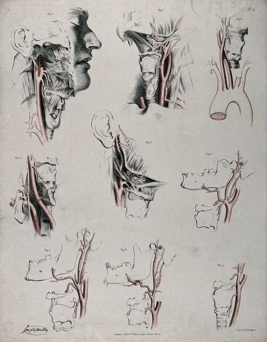
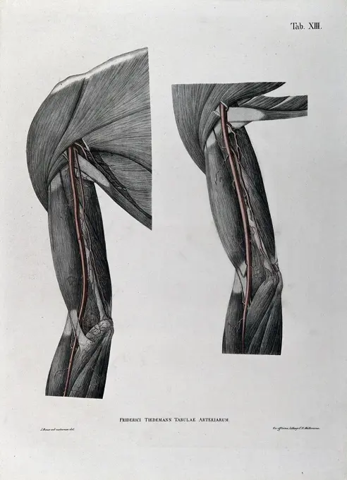
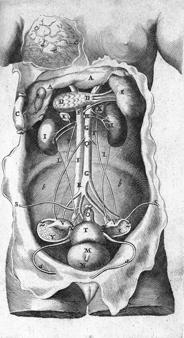
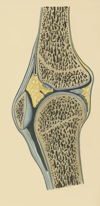
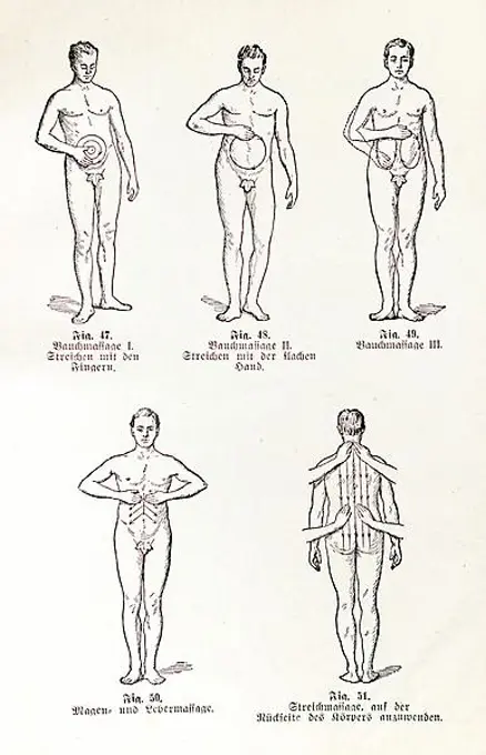
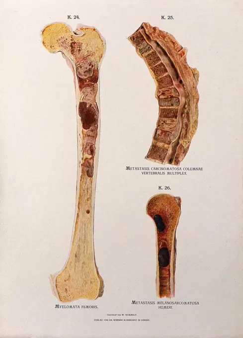
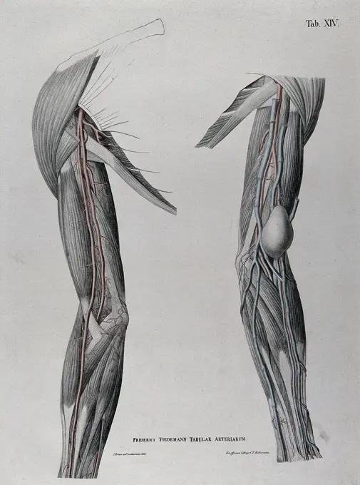
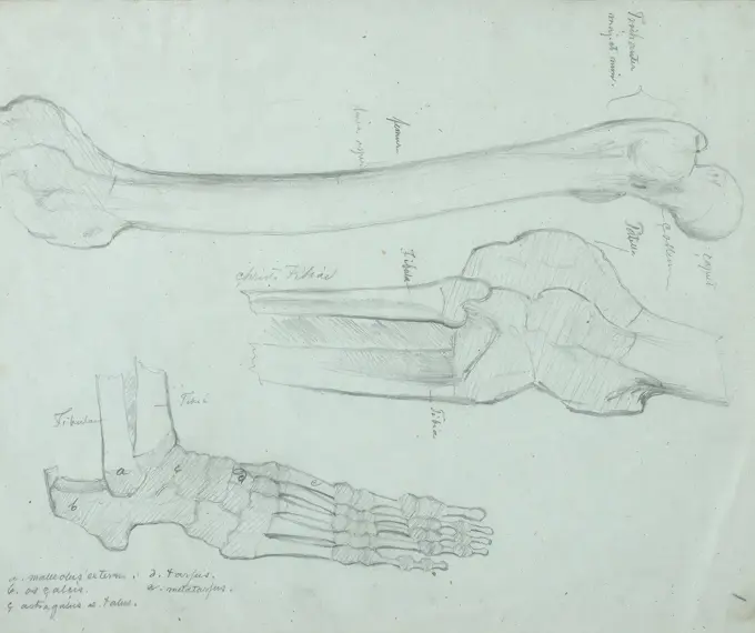
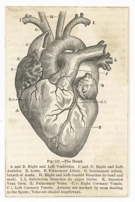
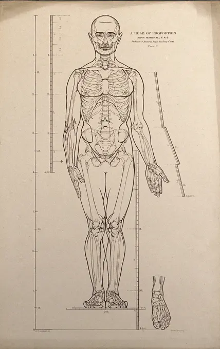
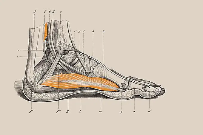
Anatomical Illustrations of Humans

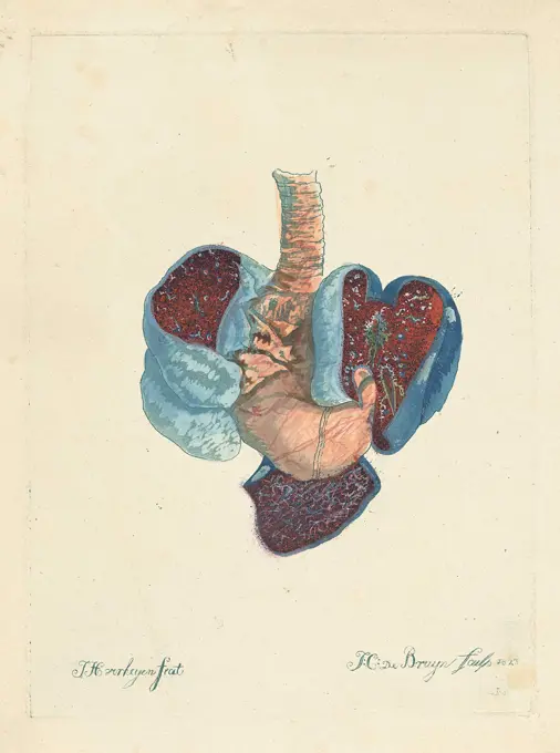
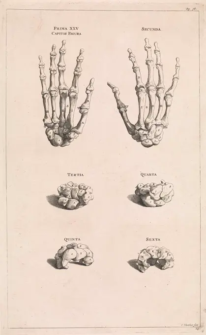
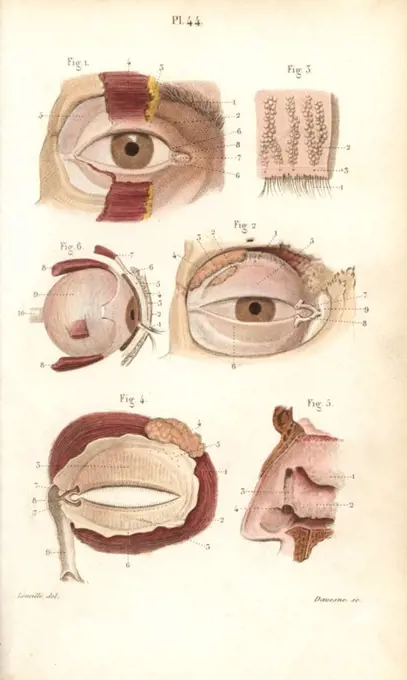
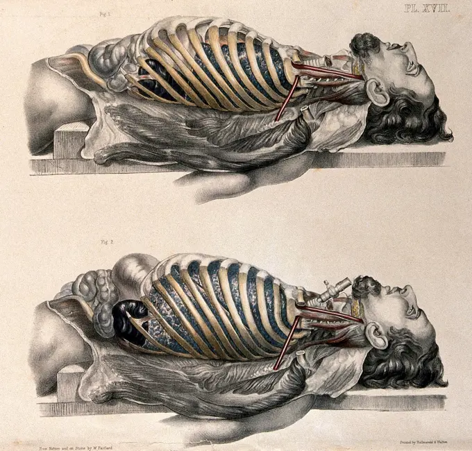
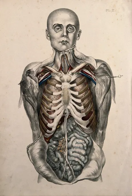
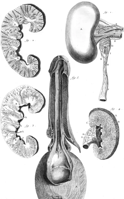
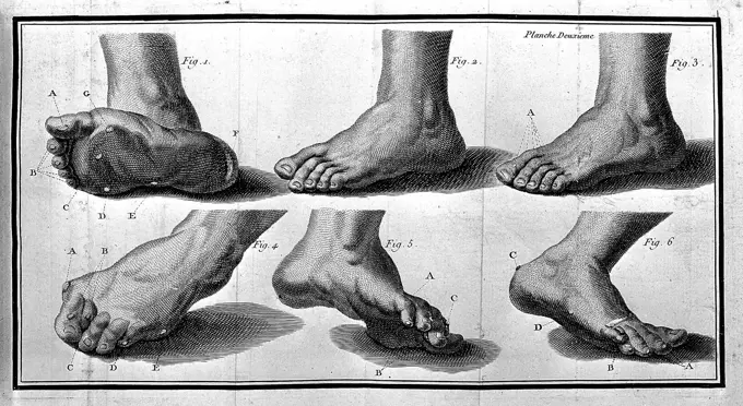
Detailed color lithographs and engravings depicting human anatomy, focusing on muscles, blood vessels, and internal organs.

Netherlands, Weissenbruch, Isaac, Part of the human body with an abnormality, print, prints, height 95 mm, width 60 mm, Dutch, 1826-08-27 - 1912-11-13, print maker, printmaker, 1836 - 1912, paper, papier, houtgravure
180 assets in this story
6145-29280027
The circulatory system: dissections of the neck and jaw of a man, with arteries and blood vessels indicated in red. Coloured lithograph by J. Maclise, 1841/1844.
6145-29265119
Dissections of the shoulder and arm; two figures, with the arteries and blood vessels indicated in red. Coloured lithograph by J. Roux, 1822.
6145-29255475
Gaspare Asellius, De lactibus sive lacteis venis
6145-29245790
T. Bartholin, parles muliebres generationi..
6145-29286830
Left, anterior view of the nerves of the leg; centre, surgical tools (); right, posterior view of the nerves of the leg. Coloured engraving, 1834-1837.
6145-29294493
Diagram of a Hernia bandage on male subject
6145-29279578
Head, neck, shoulder and chest of a dissected male écorché, with arteries and blood vessels indicated in red. Coloured lithograph by J. Roux, 1822.
1788-21803407
Knee joint, section, human body, drawing.
4409-17332981
TOPOGRAPHISCH- ANATOMISCHER ATLAS, 1872.
6145-45637620
Anatomy of the lungs and stomach. Print with anatomical data from the lungs and stomach.
1788-39566
Medicine - Tapeworm (Taenia echinococcus). Drawing.
6145-29256423
Muscles around eye from Darwin's Expression of the Emotions...
1525-28038672
Carotid, vintage engraved illustration. Dictionary of words and things - Larive and Fleury - 1895.
6176-67061931
Circulation of the blood, 1628. Artist: Unknown.
4443-21167973
restoration, medical equipment, surgical instrument, history of medicine
6145-30089208
Botten van de handen; Prima XXV Capitis Figura.Six illustrations of the Carpale and Meta Carpale Bones from the hands.
1848-55072654
Medicine, anatomy, illustration of a section through the human head with mouth, nose and throat in black and white, frontal sinus, nasal septum with olfactory nerves, canal to the nasal cavity, incisors, tongue muscle, vocal cords, sphenoid sinus, choanae, hard and soft palate, oral cavity, uvula, pharyngeal cavity, epiglottis, hyoid bone, vertebral body, cricoid cartilage, oesophagus, historical illustration in black and white from Meyers encyclopedia 4. Edition 1889 and 1890, Germany, Europe
4409-71708
Sections of the eye socket and nasal cavity. Handcolored steel engraving by Davesne of a drawing by Leveille from Dr. Joseph Nicolas Masse's "Petit Atlas complet d'Anatomie descriptive du Corps Humain," Paris, 1864, published by Mequignon-Marvis. Masse's "Pocket Anatomy of the Human Body" was first published in 1848 and went through many editions.
1525-28067568
Mid section through the head of a European, vintage engraved illustration. From the Universe and Humanity, 1910.
1848-72924435
Medical illustration, clinical picture, venereal disease, gonorrhea, female reproductive organs altered by gonorrhea, cervicitis, uterus, endometritis, fallopian tubes, salpingitis and ovaries, oophoritis, school picture, authentic reproduction of a school mural, around 1900, exact date unknown, historical
6145-29275339
Muscles of the thorax and abdomen, with the bones of the ribcage, clavicle and upper arm: écorchés figure. Colour mezzotint by J. F. Gautier d'Agoty after himself, 1745/1746.
6145-29255192
The body of a man lying down with the trunk dissected: two figures showing the lungs after breathing out (above) and after breathing in (below, simulated by inflating the lungs). Coloured lithograph by William Fairland, 1869.
6145-29287482
The body of a standing man with his head shaved and his trunk dissected to reveal the ribs and viscera. Coloured lithograph by William Fairland, 1869.
1899-61459136
Medicine, human anatomy, femoral hernia, femoral hernia, also called femoral hernia, refers to hernia gaps immediately below the so-called inguinal ligament. The femoral hernia occurs where the large blood vessels of the leg enter the abdominal cavity. Colored lithograph by J. Maclise, 1841.
6145-29252092
Artistic poses, plate VII: a man écorché aiming a spear. Lithograph by Robert J. Colenso.
4220-21905089
sections of the kidneys and  the bladder.         Date: Circa 1760
1848-50784927
Deforming (disfiguring) gout, Praktischer Hausschatz der Heilkunde, II. vol., 1930, Heinrich Killinger, Nordhausen
4443-21168030
excision, medical equipment, surgical instrument, history of medicine
1848-50784451
Massage, Praktischer Hausschatz der Heilkunde, II. volume, 1930, Heinrich Killinger, Nordhausen
4409-17511521
A Comparative Anatomical Exposition of the Structure of the Human Body with that of a Tiger and a Common Fowl: Human Figure, Posterior View, Undissected (Finished Study for Table VII). Date/Period: 1795 to 1806. Drawing. Graphite on moderately thick, slightly textured, cream wove paper. Height: 540 mm (21.25 in); Width: 406 mm (15.98 in). Author: George Stubbs.
6145-29293694
Sections through diseased femor, spinal and humerus bones: three figures. Chromolithograph by W. Gummelt, ca. 1897.
6145-45729705
Upper and lower jaws of an Hippopotamus amphibius (Hippopotamus).
1899-61459135
Medicine, human anatomy, dissection of the left groin of a standing man, veins, muscles, penis. Colored lithograph by J. Maclise, 1841.
6145-29278389
A prone écorché figure, seen from behind, resting on a table, with the bones and muscles of the trunk, pelvis and thighs indicated, and a sketch of an écorché leg. Pen and ink, with pink and grey watercolour washes, by C. Landseer, ca. 1815.
6145-59190613
Psoriasis gyrata du pli du coude. Arthur de Montmeja, photographer (French, born 1841) negative about 1867, print 1882 Close view of the inner elbow showing bands of psoriasis gyrata. (Recto, mount) upper center, printed in black ink: "DARTRES" Lower left, printed in black ink: "A. de Montmeja" Lower center, printed in black ink: "PSORIASIS GYRATA DU PLI DU COUDE" Lower right, printed in black ink: "Ad naturam fecit"
4220-21865919
Circulation of  the blood         Date: Late 19th Century
6145-29255230
Dissections of the shoulder and arm; two figures, one showing a tumour or growth (), with the arteries and blood vessels indicated in red. Coloured lithograph by J. Roux, 1822.
6145-29251480
Advertisment for a thigh splint: two figures, including an illustration showing the splint in place on a bandaged limb. Engraving with etching, 1830/1860
4443-73183106
Ekman, Robert Wilhelm, work, Skeleton exercise from the foot and lower leg, drawing, 33.5 cm x 27.5 cm
6145-30095252
Study of an arm ..
4409-48151058
Figures of male feet, toes, ankles, from paintings by an old master in an 18th century art tutorial book. Handcoloured copperplate engraving from Robert Sayers The Artists Vade Mecum, Being the Whole Art of Drawing, London, 1766.
4443-22012137
Fibroma Molluscum - After Operation; Charles B. Brigham (American, 1845 - 1903); Cambridge, Massachusetts, United States; 1876; Heliotype; 14.4 × 9.4 cm (5 11,16 × 3 11,16 in.)
6145-29293481
Page from article on distortions of the feet.
6145-52344360
Elle présente le diaphragme avec les muscles de la verge & ceux de l'anus, Pl. 15 Muscles of the diaphragm, also the anus, arteries, and urethra. Prints, Scientific illustrations. 1773. The Miriam and Ira D. Wallach Division of Art, Prints and Photographs: Print Collection. Human anatomy
1788-21803413
Scapula, body, drawing.
6145-29260230
Brain: horizontal section showing lateral ventricles. Watercolour after() W.H. Lizars, ca. 1826.
1525-28067766
The muscles of the arm of the man hand being lifted, vintage engraved illustration. From the Universe and Humanity, 1910.
4443-73182822
Kleineh, Oscar, work, Jalkaterä, academy practice, 1867, drawing, 28.5 cm x 39 cm
1848-78927604
Anatomy, image shows a detailed anatomical cross section of the human ear, divided into outer ear, middle ear and inner ear. It is used to illustrate the structures responsible for hearing and the sense of balance, reproduction of a historical school painting, mural or teaching board, authentic, exact date unknown
1848-19360039
Entwicklung des Menschen vor der Geburt in der Frau, Historisch, digitale Reproduktion einer Originalvorlage aus dem 19. Jahrhundert
4220-21856980
CHINESE THEORY OF PULSES The human hand, showing the  points where the pulse of the  blood flow can be accessed and  used for healing purposes      Date: circa 1680
6145-29263382
Post-partum haemorrhage. Lithograph after W. F. Victor Bonney.
6145-29294772
Bones of the skull, vertebrae and sacrum: six figures. Pencil drawing by J.C. Zeller ca. 1833 () after G. del Medico, 1811.
1746-30002483
John Snow's (1813-1858) inhaler for the administration of chloroform or ether. From The Lancet, London, 5 February 1848.
6176-67058403
Teeth of a horse as an indication of age, c1905 (c1910). Artist: Unknown.
6145-29284363
Cross-sections through the human face and jaw. Coloured lithograph by M. Hanhart after C. Heath after J.B. Léveillé.
1788-39569
Human muscle system, locomotor apparatus, illustration.
4220-21934076
Illustration depicting  the structure and internal mechanism of an artificial limb constructed by Bechard for the opera singer M. Roger     Date: 1860
6145-29252519
Acupuncture points. Woodcut by a Japanese () artist.
6145-29262688
The human head, divided according to the system of phrenology. Coloured lithograph by C. Ingrey, 1824.
1899-18722322
Arteries of the arm circa 1521.
1746-21105779
Illustration from a Japanese anatomical and medical text 'Complete notes on the dissection of cadavers' by Shinnin Kawaguchi, 1736-1811. Illustration by Shukuya Aoki published 1772.
6145-29251245
The body of Napoleon Bonaparte laid out after death, 1821. Lithograph after Captain Marryat.
6145-48492223
Church, school porta: final stones and cantilever stones (from: Atlas to the magazine for building, ed.
4220-21871432
the heart showing the  ventricles, auricles, aorta,  arteries, carotid branches  and other associated  vessels      Date: circa 1860
1848-78930252
Anatomy, human, the human organ of vision, eye, organum visus, reproduction of a historical school painting, mural or teaching board, authentic, exact date unknown
6145-29257922
Standing male figure, front view, with scales of proportion: illustration shows the skeleton and outline of the body and includes a detail of a foot. Lithograph by J.S. Cuthbert, 1789.
6145-29254715
Brain: dissection showing a section of the right hemisphere. Watercolour after() W.H. Lizars, ca. 1826.
1788-20761573
Medulla oblongata, anatomical plate.
6145-52804411
A study of arm muscles by Theodor von Hörmann, executed in pencil with red ink, highlighting muscular structure and anatomical details.
1848-54506294
Skeleton of a human foot, anatomical illustration
4220-21856981
CHINESE THEORY OF PULSES Using the wrist-pulse of  the human hand for healing  purposes       Date: circa 1680
6145-29292612
Right profile of head with depressed frontal lobes, divided up to show the location of all the lobes. Drawing, c. 1900.
4390-141
Drawing of Suspender Fastening
6145-29277711
Haemorrhages of pregnancy and parturition, and abnormalities of the placenta. Lithograph after W. F. Victor Bonney.
6145-29287267
Horse's teeth: showing development from six years old to extreme old age: ten views with five detailed images of youthful teeth. Coloured engraving attributed to T. Cowan after a drawing attributed to B. Herring, ca. 1860.
1848-54752522
Respiratory organs of humans, illustrations from the book 'Dr. F. Koenig's Ratgeber in gesunden und kranken Tagen', German for "Dr. F. Koenig 's book with advice for the healthy and sick days", 1928
6145-29269406
Plate showing the feet from La Forest, L'art de soigner les pieds.
6145-29249828
Mechanisms of occipito-anterior positions and rectified occipito-posterior positions of the foetus. Lithograph after W. F. Victor Bonney.
4391-318
A line drawing of a woman's pelvic area showing the fallopian tubes, ovaries, uterus, cervix, and vagina, and the method for performing a pelvic exam.
PREVIOUS
of 2
NEXT
2401 S. Ervay, Suite 206
Dallas, Texas 75215
United States
Get Started
Free ResearchMy BoardsMy Cart
For Creators
How To License Your ContentContributor PortalFrame of Mind
Resources
API accessPricing
Contact
+1 866 236 0087help@viewpictures.co.uk Contact form
©2026 View Pictures. All Rights Reserved. -A
This site is protected by reCAPTCHA and Google Privacy Policy and Terms of Service apply.