Anatomical Illustrations of the Human Body

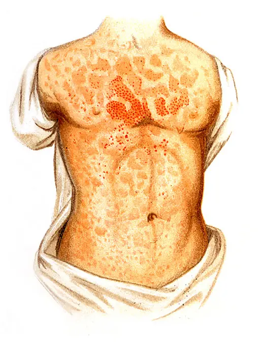
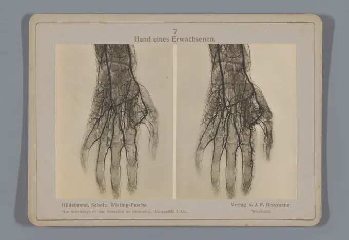
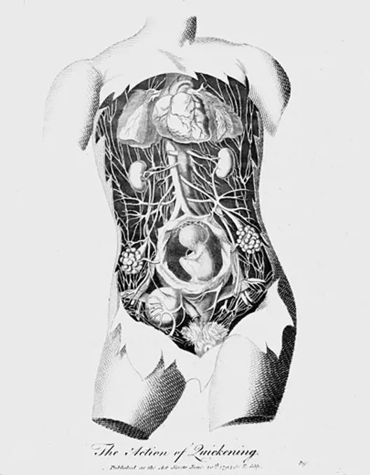
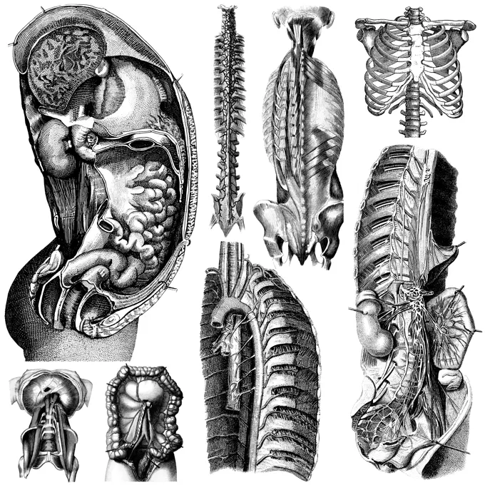
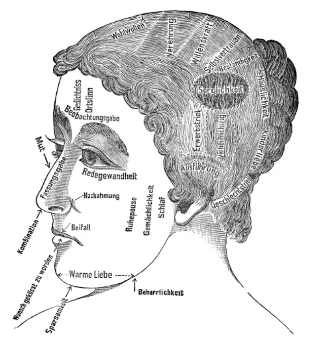
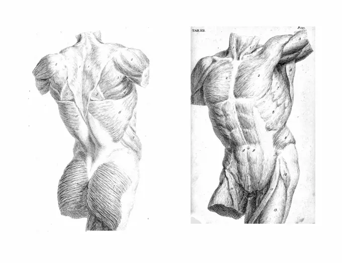
Detailed anatomical plates showcasing the human nervous and circulatory systems. Features hand-colored engravings and dissection studies, emphasizing musculature and vascular networks.

Plate IV. Surgical anatomy of the cervical and facial regions
Brain: base and meninges. Colour lithograph by Brocades Great Britain Ltd.Hand of an adult. .Foetus in uterus at time of quickening (16-18 weeks) when first fetal movements felt by mother. Engraving c1795. TintedAnatomical illustrations of the human respiratory system, depicting the lungs, the bronchial system, the trachea, the larynx and the thorax, reproduction of a historical school picture, mural or teaching board, authentic, exact date unknownMuscles of the abdomen and thigh. Coloured lithograph by N.H Jacob, 1831/1854.Medical - Victorian Anatomical Illustrations - on a white background for cut out.Illustration showing the general lines of the left hand of a female. The lines are created by the movement of the hand and joints. Dated between 1617-1619Medicine: Human anatomy, chest muscles. DrawingThe Chinese system of physiology - how your facial  characteristics reveal your  character       Date: 1912Medical illustration, The human digestive system, school picture, authentic reproduction of a school mural, around 1900, exact date unknown, historicalThe aortic arch and the arteries of the neck: eleven figures. Coloured lithograph by J. Roux, 1822.medicine, anatomy, physiognomy, character traits of a manageress, wood engraving, from: Friedrich Eduard Bilz, New Naturopathic Treatment, Leipzig, Germany, 1902,Woodcuts in a medical textbook, illustration of the nerves and arteries in the human headAnatomy of a mussel, probably a painter's mussel or river pearl mussel, in three different views: from the side with raised shell (1), in longitudinal section (2) and in cross-section, reproduction of a historical school picture, mural or educational panel, authentic, exact date unknownThe muscles of the arm of the man hand being lifted, vintage engraved illustration. From the Universe and Humanity, 1910.Measles, illustration, Meyers Konversations-Lexikon encyclopedia, 18972 Views of the human torso, muscles and internal organs  from The anatomy of the human body by  William Cheselden in 1763.Method of sterilising wounds using Dakin's sodium hypochlorite 0.05 per cent, devised by Dr Alexis Carrel of the Rockefeller Institute. Left and bottom, irrigator and sectional view of wound: right top, device applied to a leg wound. Dated 20th centuryEntwicklung des Menschen vor der Geburt in der Frau, Historisch, digitale Reproduktion einer Originalvorlage aus dem 19. JahrhundertAbnormalities of foetal position and abnormal descent of the cord in childbirth. Lithograph after W. F. Victor Bonney.Le Progres medical, Journal de medicine, 1888Brain sections. Drawing by Léonce Manouvrier, ca. 1900.Collage of spine, dumbbell, and food and milkHuman anatomy vector imageFour diagrams illustrating heads with severe wounds, demonstrating how to stitch the skin back together properly. Stipple engraving by J. Bell.Surgical human anatomy, title page of the cover of fascicle 1 of Joseph Maclise's work, 1851, human anatomy, medicine, historical, digitally restored reproduction from a 19th century original.Surgical human anatomy, title page of the cover of fascicle 1 of the work by Joseph Maclise (1851), human anatomy, medicine, historical, digitally restored reproduction from a 19th century originalThis set of photographs details Skylab's Human Vestibular Function experiment (M131). This experiment was a set of medical studies designed to determine the effect of long-duration space missions on astronauts' coordination abilities. This experiment tested the astronauts susceptibility to motion sickness in the Skylab environment, acquired data fundamental to an understanding of the functions of human gravity reception under prolonged absence of gravity, and tested for changes in the sensitivity of the semicircular canals. Data from this experiment was collected before, during, and after flight. The Marshall Space Flight Center had program management responsibility for the development of Skylab hardware and experiments.