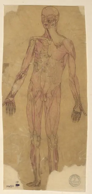
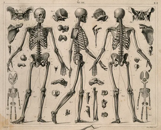
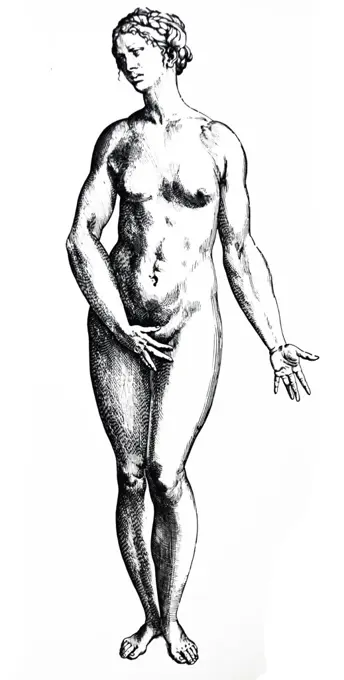
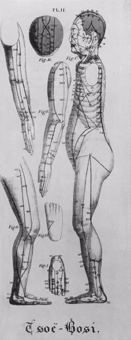
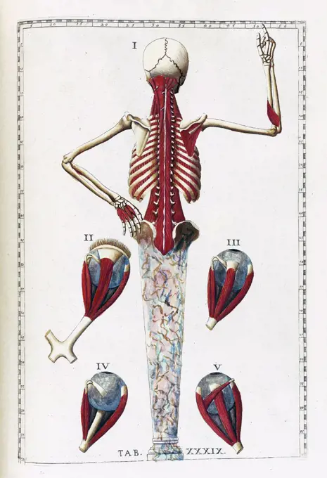
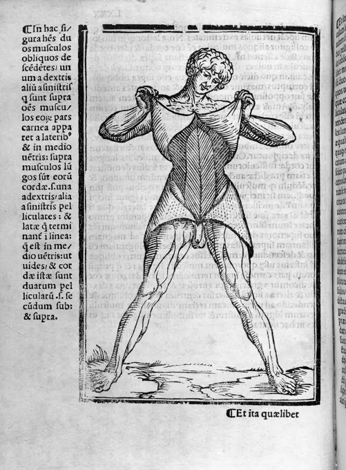
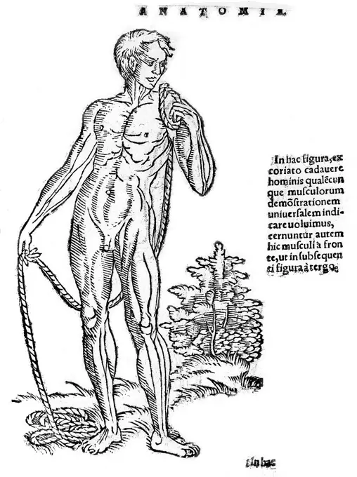
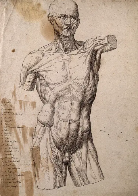
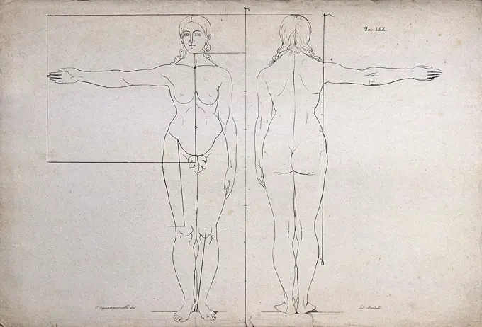
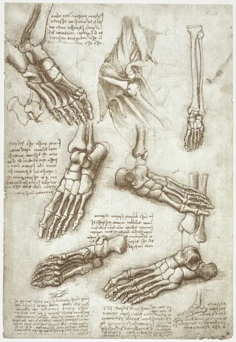
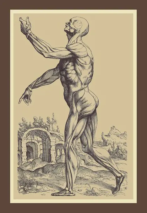
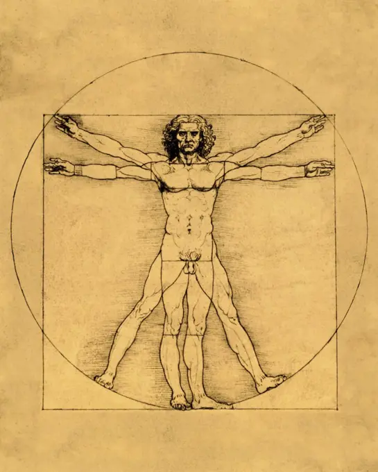
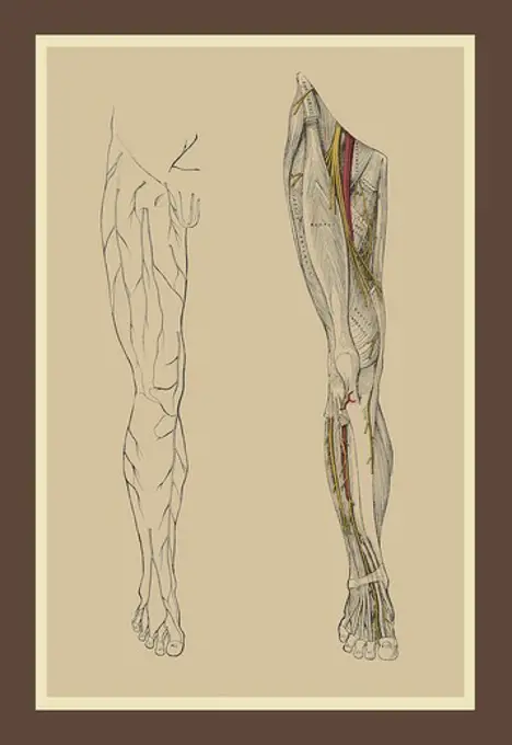
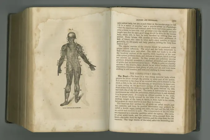
Anatomical Illustrations of the Human Body

A collection of detailed anatomical drawings showcasing the human muscular system from various angles and perspectives, in a classic artistic style.

View of the muscles in the  human body (from the front)
View of the muscles in the human body (from the front)
Anatomy van de Man, Cornelis Ploos van Amstel, 1781 print The print is loose in the back of an album. Amsterdam paper etching anatomical models (usually series of five figures)A human skeleton, seen from the front, with labels giving the latin names of the bones. Photograph after a woodcut after Hans Wechtlin, 1517.Muscles and ligaments of the hand: two figures of écorché hands. Lithograph by Martelli after C. Squanquerillo, 1839.Acupuncture points and meridians of human body (front view), engraving from modern edition of the Nei Tsing, earliest known manual of medicine, published 1000 AD by Chinese Emperor Hoang TiDesign Mannequin, from Encyclopédie 1762-1777 France . Benoît-Louis PrévostMale skeleton seen from the front, with left arm extended: two figures (one an outline drawing). Line engraving, ca. 1850.Anatomical drawing by Jehan Cousin ('le jeune,' or 'the younger') in Livre de pourtraiture. (Paris, 1608).Illustration from 'Tashrih-i badan-I insan Anatomy of the Human Body' a Persian medical text (ca. 1390); by Mansur ibn Muhammad ibn Ahmad ibn Yusuf ibn Ilyas. this illustrated treatise, often called Mansur's Anatomy (Tashrih-i Mansur-I), was dedicated to the sultan Ziya' al-Din Pir Muhammad Bahadur, grandson of Timur, known to Europeans as TamerlaneGiovanni Battista Bracelli, From Bizzarie di varie Figure, 1624 From Bizzarie di varie FigureAn écorché showing bones, with left arm extended to the side, seen from behind. Line engraving by Campbell, 1816/1821.The muscles of the human body seen from the back and the salivary glands of a dog. Engraving, 1686.Diagrams illustrating how to set a broken arm. Stipple engraving.anatomical study by Jacques Gautier d'Agoty 'Anatomie générale des viscères' Paris 1752.Skeleton of a horse, viewed from above with legs outstretched, showing the heart, muscles and blood vessels. Coloured engraving by J. Pass after Harguinier, 1805.Anatomical fugitive sheetsWoodcut, Monstrum bicorpor et monocephalon utriusq sexus, monster with two bodies and one head, Siamese twins, Aldrovandi, Historia Monstrorum, 1642, RenaissanceThe muscles of the arm of the man hand being lifted, vintage engraved illustration. From the Universe and Humanity, 1910.Anatomical Figure VI, Jan Luyken, 1680 - 1688 print Print at the top right marked: fol. 158. Amsterdam paper etching anatomical models (usually series of five figures). biologyThree figure studies by Alexander Rothaug on blue paper with pencil, chalk, and white highlights, emphasizing posture and anatomy.Left, muscles of the head and trunk (back view); right, muscles of the thigh and leg (back view). Coloured drawing, 18--.Unit of circulation, vintage engraved illustration.Rudolf Weyr's study of the human figure, showing anatomy, posture, and proportions, drawn with red and brown ink on transparent paper.Historia naturalis ranarum nostratium, Nürnberg, Gedruckt bey Johann Joseph Fleischmann, 1758, Anura, Engravings, Errata lists, Printing, Frogs, Germany, Handcoloring, Nuremberg, Pictorial works, August Johann Rösel von Rosenhof, August Johann Rösel von Rosenhof, Katharina Barbara Rösel von Rosenhof., A detailed anatomical illustration showcases a human figure displayed in a position that emphasizes the muscular and skeletal systems. The main figure is depicted with limbs extended outward, allowing for a comprehensive view of the musculature. Surrounding the central figure are smaller diagrams that delve into specific anatomical features, including detailed representations of the brain and other internal structures. Each labeled part is meticulously outlined, providing a valuable resource for studying human anatomy, particularly focusing on the intricacies of muscle groups and their connections. The illustration serves as an educational tool for medical students and professionals alikeAnatomical illustration by Amé Bourdon, from 'Nouvelles tables anatomiques'; Chambray: 1678.Relationship of the organs of the body, the Humours and signs of the Zodiac, 1508. Artist: UnknownHuman Body, Posterior View (Outline Drawing for Table VII), George Stubbs, 1724-1806, British, 1795 to 1806, Graphite and red chalk; verso: graphite and red chalk on cream, thin, smooth, wove paper, Sheet: 19 1/8 x 12 5/8 inches (48.6 x 32.1 cm), anatomical study, anatomy, back, figure study, manClaude Ambroise Seurat, a very thin man who was known as the 'Human Skeleton'. Drawing.Plate from Albrecht Durer's, VIER BUCHER VON MENSCHLICHER PROPORTION (Four Books on Human Proportion). Durer applied the science of human anatomical proportions to aesthetics. 1528 engraving.Skeletons and bones, including the skeleton of a foetus (). Line engraving by H. Winkles under the direction of J.G. Heck, 1830/1845.The Plates from the Epitome of the De Humani Corporis Fabrica by Andreas Vesalius, (1514-1564) Plate 82 - The figures reproduced in plates 82-85 were to be cut out and used in the construction, by superimposition of two manikins, one of which was to show the topography and relations of the viscera in the female and the other, in the male.Human figure shown in left profile with acupuncture points and meridians identified.  Also shown are views of the head, the arms, legs, and the top and bottom of a foot. Illustration from a 1825 French book on Chinese acupuncture by Jean Baptiste Sarlandiere.Illustration from 'Tabulae anatomicae'. (Rome: 1783) by Bartolomeo Eustachi (1500 or 1514 ñ 27 August 1574), (Eustachius), one of the founders of the science of human anatomy.James Ward, 1769-1859, British, Drawing of a Man's Skeleton, undated. Brown ink over graphite on medium, slightly textured, beige laid paper.Male figure holding the skin of his trunk open'Drawings of a Left Leg Showing Bones and Tendons', c1480 (1945). Artist: Leonardo da Vinci.Anatomical measurements, supine figure, Chinese woodcutTert. fig.: Integram totius magnæ arteriæ ab omnibus partibus liberæ delineationiem monstrat.. Geminus, Thomas, d. 1562. Prints, Illustrations, Scientific illustrations. 1545. Rare Book Division. Human anatomy, AnatomyRamon Llull (1235-1316). Spanish writer and philosopher. Practica Compendiosa Artis Raymundi Lulli, 1523. Book 8. Medicine subject. Description of the human skeleton.Vitruvian Man by Leonardo da Vinci, pen, brown Indian ink on paper, 1492, 1452-1519, Italy, Venice, Gallerie dell' Accademia, 34, 4x24, 5Musculature plate from 18th century anatomy treatise by Bernhard Albinus, with engravings by Jan Wandelaar (1690-1759). The elaborate background of architectural and botanical elements distinguished the 1747 book.The Venus de Medici from the front and back, and in both lateral views; with details of her head, arm and feet. Engraving by B.L. Prevost after G. Audran.Skeleton of an adult patient  afflicted with rickets  (rachitis)        Date: 1827Woodcut, Gemelli hermaphroditi tergore connati, bisexual Siamese twins, Aldrovandi, Historia Monstrorum, 1642, RenaissanceAnatomical fugitive sheetsSkeleton contemplating a skullAnatomical fugitive sheet,femaleDesign for an Oil Lamp, Giovacchino Belli, Italian, 1756 - 1822, Pen and black ink, brush and gray, green, blue wash, graphite on paper, Vertical rectangle. Design for an oil-burning lamp with alternative suggestions. A young Egyptian male figure stands upon a short piece of a fluted column. He carries upon his head a bowl, containing the oil. Upon it stands a vase, to which is fastened the eye shade, which is decorated with a sun. Two lamp pokers hang at a chain, which hangs through the rings of three lion masks at the body of the bowl. The alternative suggestions refer to a more or less rich ornamentation of the bowl and the vase. Base line below., Rome, Italy, late 18th century, lighting, Drawing, Drawing'Proportions of the human figure', c1492, (1936). Artist: Albrecht DürerFront of male figure, showing muscles.An emaciated naked man, displaying large swellings on his neck and groin, with smaller swellings visible on his chest, arms, abdomen and thigh. Process print, 18--.Illustration from Dürers Vier Bucher von Menslicher Proportion, Nuremberg, 1528 1528 Albrecht Dürer German. Illustration from Dürers Vier Bucher von Menslicher Proportion, Nuremberg, 1528 387805Liagno, Teodoro Filippo di, Hondius, Hendrick (I), Den Haag, Stock, Andries Jacobsz., Dierskeletten, Skeleton of an unknown mammal, The Hague, verso - stamped, The skeleton of an unknown quadruped in a landscape. At the bottom a two-line text in Latin. Print from a series with representations of skeletons by the printmakers Hendrick Hondius I (NewHollst.243-258), Andries Jacobsz. Stock (Hollst.4-10) and Simon Frisius., print, prints, lo: AS, mo in frame: Cum furtim hic subeas...poena datur. ro 18, height 97 mm, width 157 mm, Noord-Nederlands, 1582 - 1648, print maker, printmaker, 1625 - 1626, second quarter 17th century, paper, etching, etchings, engraving, engraving (printing process), 1573-06-09 - 1650-10-25, intermediary draughtsman, after a drawing by, Work conception, ItalianAnatomical fugitive sheetsAn écorché of a male torso and head, seen from the front. Pen and ink drawing.Proportion study of a boy of nine years, side view. Draughtsman: anonymous. Dating: 1700 - 1800. Measurements: h 208 mm × w 328 mm. Museum: Rijksmuseum, Amsterdam.Anatomical study of the torso of a man, seen from Opzij, Wenceslaus Hollar, After Leonardo da Vinci, 1645 print   paper etching torso. musclesAnatomical illustration from 'Vier Bücher von menschlicher Proportion' ' by Albrecht Dürer (1471-1528), Nuremberg, 1528. His collection of drawings were used to apply the science of human anatomical proportions to aestheticsSkeletons of child and foetus, 16th CenturyEngravings of techniques of blood transfusion from man to animal.A Doryphorus-type of the canon Polycleitus , Drawings, Sculpture. Nicholas Catsimpoolas CollectionDigital artistic Sketch of the human Anatomy, based on own 3D Rendering, Property Release not required.Woodcut, Homo syluestris, pygmy, Aldrovandi, Historia Monstrorum, 1642, RenaissanceProportions of the female body: front and back view of a female nude, with proportions marked. Lithograph by Martelli after C. Squanquerillo, ca. 1840.Anatomy Studies: The Bones of the Foot & Anatomy of the Shoulders Leonardo da Vinci (1452-1519 Italian) Pen & ink Royal Library, Windsor Castle, EnglandMuscles of the human body. Chromolithograph by Gustave H. Michel.Englishman John Lambert (22) exhibited his porcupine skin disorder, Ichthyosis hystrix, in Leipzig. John was a descendent of Edward Lambert, whose condition was first described by Machin in 1732. Handcolored copperplate engraving from G. T. Wilhelm's "Encyclopedia of Natural History: Mankind," Augsburg, 1804. Gottlieb Tobias Wilhelm (1758-1811) was a Bavarian clergyman and naturalist known as the German Buffon.Anatomical study of leaning man, Paulus Pontius, after Peter Paul Rubens, 1616 - 1657 print  Antwerp paper engraving parts of the human body (skeleton excepted)Illustration from 'Tashrih-i badan-I insan Anatomy of the Human Body' a Persian medical text (ca. 1390); by Mansur ibn Muhammad ibn Ahmad ibn Yusuf ibn Ilyas. this illustrated treatise, often called Mansur's Anatomy (Tashrih-i Mansur-I), was dedicated to the sultan Ziya' al-Din Pir Muhammad Bahadur, grandson of Timur, known to Europeans as TamerlaneAnatomical fugitive sheetsAnatomical study of blood vessels and blood circulation, Pieter van Gunst, After Gerard de Lairesse, 1685 print Anatomical study of blood vessels and blood circulation. At the top right numbered T. 23. Amsterdam paper engraving blood circulation. anatomical model of the arteriesPersian treatise composed by Manr ibn Muammad ibn Amad ibn Ysuf ibn Ilys, who came from a Persian family of scholars and physicians working in the city of Shiraz. His illustrated treatise, often called Manr's Anatomy (Tashr-i Manr-I), was dedicated to the sultan Ziyí al-Dn Pir Muammad Bahadur, in all likelihood referring to Pir Muammad ibn ëUmar ibn Timur, the ruler of the Persian province of Fars from 1394 to 1409 The skeleton, viewed from behind with the head hyperextended so tMuscle studies, late 15th or early 16th century (1954). Artist: Leonardo da VinciLeonardo Probes the Nature of Mankind. Muscles and tendons of the lower leg and foot, seen from the side: two figures showing both stylised outline and detailed drawing. Pencil drawing by or associated with A. Durelli, ca. 1837.Thomas Jones, a boy with icthyosis, a skin disease that resembles fish scales. Coloured lithograph by J. Perry, 1841.Myotomia reformata, by William CowperSecond Plate of the Muscles, Medical - AnatomyProportions of the Human Figure (Vitruvian Man) ca. 1485-1490 Leonardo da Vinci (1452-1519 Italian)  Pen and ink  Galleria dell'Accademia, Venice, ItalyAnatomical fugitive sheetsAnatomical fugitive sheet (female).Close up of Old anatomy drawings by Leonardo Da VinciSkeleton or bones of a horse. Handcoloured copperplate engraving by J. Pass after an illustration by Daniel Dodd from William Augustus Osbaldistons The British Sportsman, or Nobleman, Gentleman and Farmers Dictionary of Recreation and Amusement, J. Stead, London, 1792.Blood-vessels of the human body: nine figures. Line engraving by I. Taylor, 1790/1810().Tattooed armsAcupuncture/pressure points on the body seen in three-quarter profile to left. Woodcut by a Chinese artist.Musculature and nerves, artwork.Lumbar Plexus, Medical - AnatomyAnatomical fugitive sheetsOld book, vintage and anatomy of human body in literature, manuscript or ancient scripture against a studio background. History novel, journal or figure for the study of muscle, medicine or organsSkeleton (front view).Anatomical fugitive sheetsAnatomical drawing of a female body by Adriaan van den Spiegel (1578-1625). Spiegel was a Flemish anatomist who was born in Brussels and practiced medicine in Padua. He is known as one of the greatest physicians associated with the city.Leonardo DaVinci s Vitruvian Man, Uomo Vitruviano, illustrated Leonardo DaVinci s Vitruvian Man, Uomo Vitruviano, illustrated in sepia colours Copyright: xZoonar.com/RobertxBriggsx 16694912anatomical study by Bernardino Genga 'Anatomia per uso et intelligenza del disegno ricercata non solo su gl'ossi, e muscoli del corpo humano'. (Rome, 1691). Bernardino Genga (1620ñ1690) was a scholar of Classical medical texts, editing several works of HippocratesP. Mascagni, "Anatomia per uso...", 1816Anatomy, the division of the human body in man and woman, 1880  /  Anatomie, die Einteilung des menschlichen Körpers bei Mann und Frau, 1880, Historisch, historical, digital improved reproduction of an original from the 19th century / digitale Reproduktion einer Originalvorlage aus dem 19. Jahrhundert,. Anatomical fugitive sheet, female.Anatomical illustration from 'Vier Bücher von menschlicher Proportion' ' by Albrecht Dürer (1471-1528), Nuremberg, 1528. His collection of drawings were used to apply the science of human anatomical proportions to aestheticsMyologic structure of the 'Borghese Gladiator' statue: the figure is presented as an écorché. Ink and watercolour drawing by J.C. Zeller after J.G. Salvage, ca. 1833.Engraving of an ape-like cave man accompanying an article by Boitard in the French 'Magasin Universel' (April 1838). This is the earliest reconstruction of a pre-historic human. It is striking that it predates the Darwinian debates on human origins and antiquity by over 20 years.