My Boards
View Pictures
  • My Boards

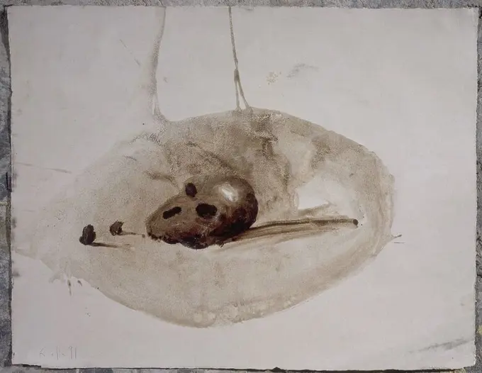
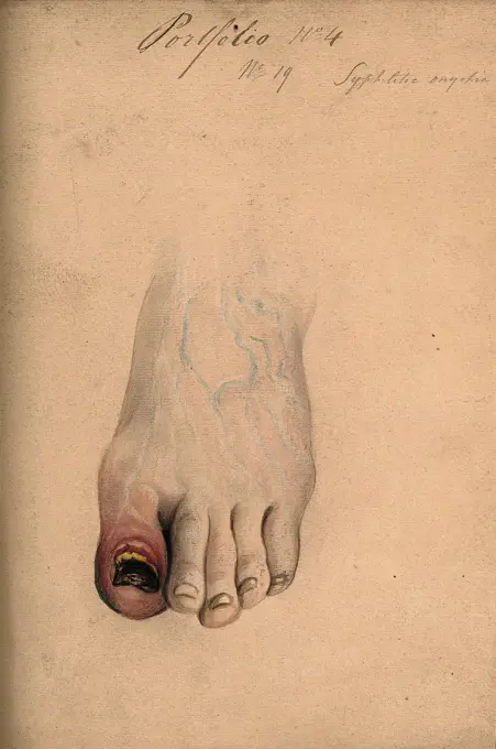
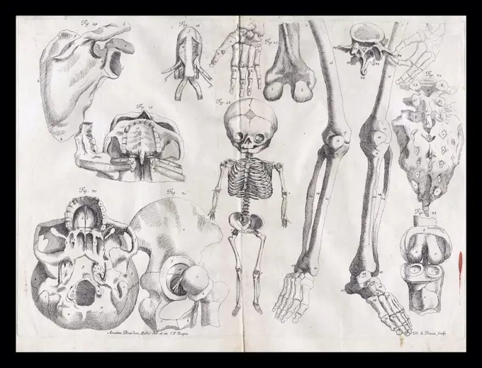
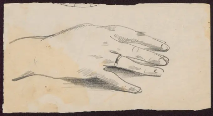
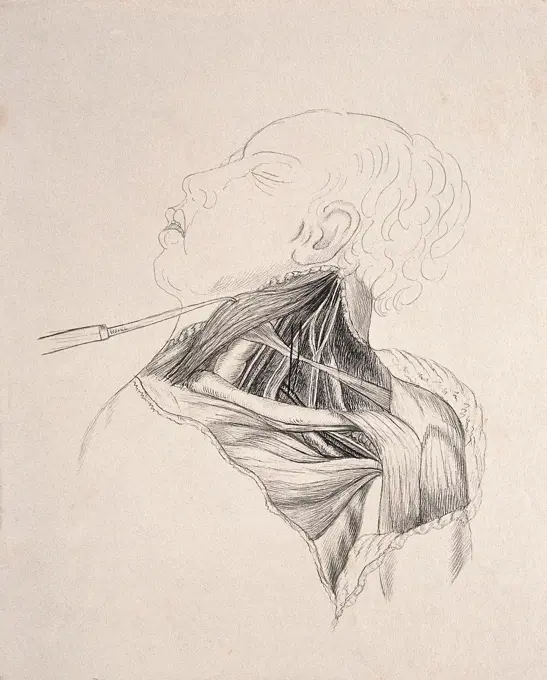
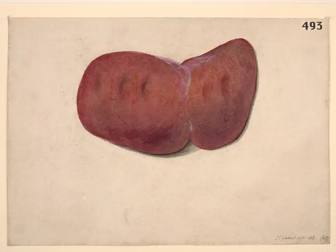
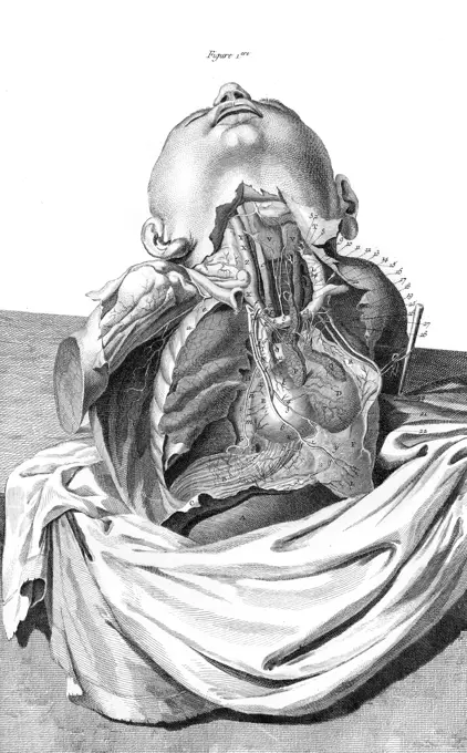
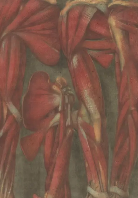
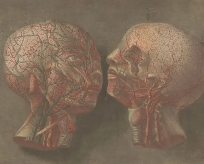
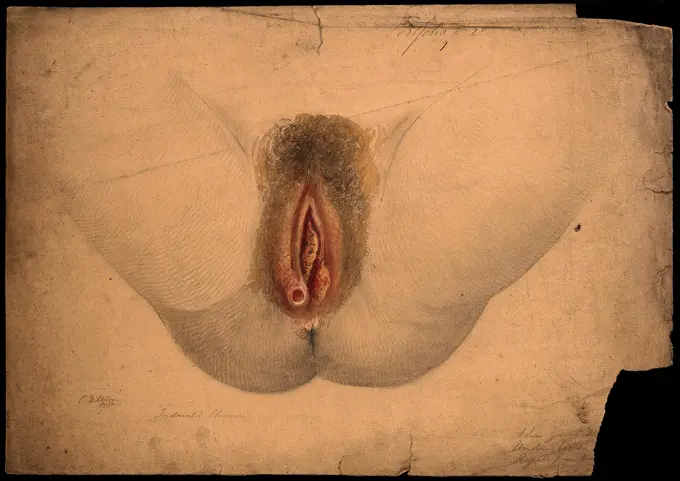
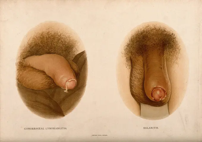
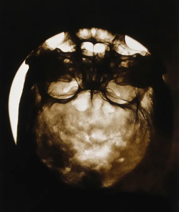
Anatomical Sketches and Illustrations

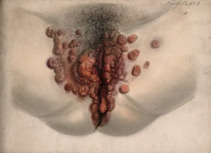
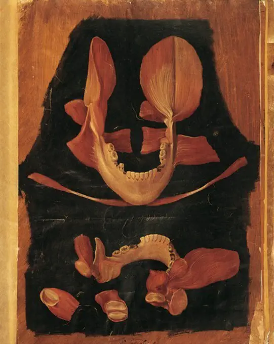
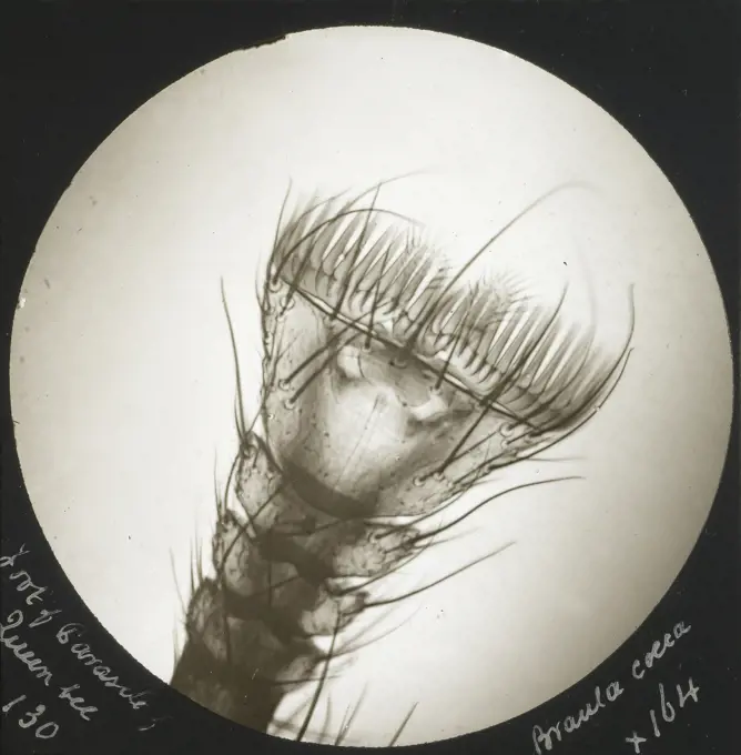
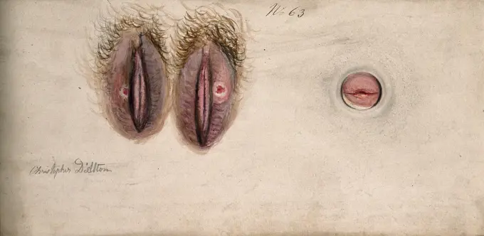
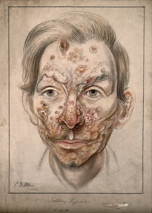
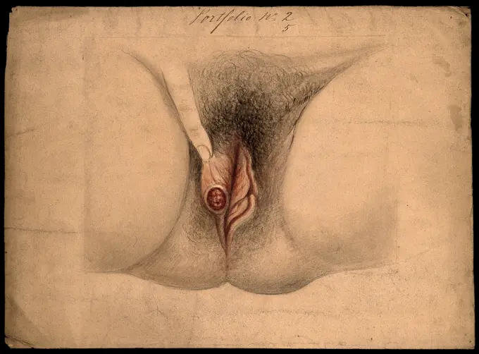
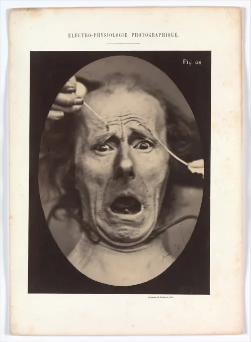
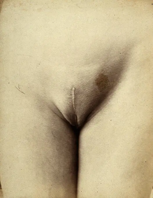
Intricate drawings depicting human and animal anatomy, showcasing muscles, organs, and structures in a vintage artistic style with muted colors.

Dissection made after the removal of a pregnant uterus, showing the parts lying immediately behind. Copperplate engraving by C. Grignion after I.V. Rymsdyk, 1774, reprinted 1851.
166 assets in this story
6145-29271787
Female genitalia showing severely diseased tissue. Watercolour by Christopher D' Alton, 1865.
1788-39873
Hieronymus Fabricius ab Aquapendente (1537-1619), Anatomy of the mouth, anatomical plate.
6145-29265850
Bones of the torso: two figures showing the shoulders, ribcage, spine and pelvis, with a cartoon-style face. Pencil drawing, by J.C. Whishaw, 1852/1854.
6145-52738358
X-ray recording of a woman's gloved hand holding a bracelet and a bouquet of flowers, captured with a four-minute exposure by Walter König, preserved on silver gelatin bromaryt paper in 1896.
4443-21971112
Foot of Parasite of Queen Bee. Braula Coeca; Frederick H. Evans (British, 1853 - 1943); about 1883; Lantern slide; 7.1 x 7.1 cm (2 13,16 x 2 13,16 in.)
6145-29293730
Muscles of the trunk: back view. Pencil and chalk drawing by J. Mongrédien, ca. 1880.
6145-29251528
A diseased penis showing symptoms of gangrene. Watercolour by C. D'Alton, 1857.
6145-29270090
Anterior view of thigh (fig. 1) and posterior view of thigh (fig. 2), with superimposed paper flaps. Coloured and laminated lithograph by S.G. Tovey after E.W. Tuson, 1828.
5513-75516859
Walt Disney's Full Length Feat Fantasia In Multiplane Technicolor - The Music in "Fantasia" was recorded by RCA and reproduced by equipment especially designed and developed by that company in collaboration with the Walt Disney Studio. This new sound is called Fantasound. The ostrich ballerina who appears in the "Dance of the Hors" sequence in Walt Disney's musical feature, Fantasia. March 8, 1941. (Photo by Guy K. Austin).
6145-29284555
Diseased tissue on the feet of a woman, showing symptoms of sloughing phagedena. Watercolour by C. D'Alton, 1857.
6145-29295705
Anatomical fugitive sheets
6145-29286156
Muscles of the legs and feet: an écorché figure, shown from behind, kneels to reveal the sole of the left foot. Colour mezzotint by J. F. Gautier d'Agoty after himself, 1759.
6145-29253266
Sores on the female genitalia: three details. Watercolour by Christopher D' Alton, 1836.
4409-17281132
CAP I PEDRES - 1990/91 - 50X65 CM. Author: MIQUEL BARCELO.
6145-29289571
Diseased tissue on the toe and nail of a woman (), showing symptoms of syphilitic onychia. Watercolour by C. D'Alton, ca. 1870.
6145-29289360
A diseased penis showing symptoms of "diphtheritic phagedena". Watercolour by C. D'Alton, 18--.
1746-21105540
Anatomical illustration by Amé Bourdon, from 'Nouvelles tables anatomiques'; Chambray: 1678.
6145-29297746
Section of the head of a child with extreme congenital proptosis and microcephalus
6145-59061533
Study d They; Waliszewski, Zygmunt (1897-1936); around 1914 (1910-00-00-1920-00-00);anatomy, gift (provenance), right, hands
6145-29261516
Skull, seen from above: Hole in sagittal suture. Pencil and chalk drawing by C. Landseer(), or a contemporary, ca. 1815.
6145-29292493
Bones of the leg and thigh. Drawing, ca. 1800.
6145-29283087
A marine animal (coelenterate). Coloured etching.
6145-58843532
Anatomic photo of a female corpse: 'The form of the chest in relation to the skeleton' (Die Form der Brust in Ihren views Zum Skelette, 1908  Florence paper. photographic support. cardboard collotype  Florence paper. photographic support. cardboard collotype
6145-59141351
Syphiloderma squamosum circinatum and syphiloderma tuberculo-ulcerativum. Dr. George Henry Fox, photographer (American, 1846 - 1937) 1881 Two images side by side. The left image shows a close view of the palm of a right hand held up to show circular squamous, or scaly, plaques from secondary syphilis. The right image shows a close view of the bottom of a foot to show ulcerative lesions from secondary syphilis. (Recto, sheet) lower center, printed in brown ink: "SYPHILODERMA SQUAMOSUM CIRCINATUM. SYPHILODERMA TUBERCULO-ULCERATIVUM. / PLATE XXII." Lower left, printed in brown ink: "ARTOTYPE, HARROUN & BIERSTADT, N.Y."
6145-29256971
Typhoid ulceration of the small intestine, in an early stage of the disease
6145-29291768
The dissected neck, chest and shoulder of a man: the skin being held back with a surgical instrument to reveal the muscles beneath. Drawing.
6145-29283117
A tumour on the torso of a young woman. Watercolour by C. D'Alton, 1870.
6145-59190614
Psoriasis généralisé. Arthur de Montmeja, photographer (French, born 1841) negative about 1867, print 1882 View of a nude man from behind to show severe generalized psoriasis plaques covering the neck, back, buttocks, arms, and legs. (Recto, mount) upper center, printed in black ink: "DARTRES" Lower left, printed in black ink: "A. de Montmeja" Lower center, printed in black ink: "PSORIASIS GÉNÉRALISÉ" Lower right, printed in black ink: "Ad naturam fecit"
6145-29285164
A section of the body (torso) with a skin disease. Chromolithograph, c. 1888.
6145-29268511
Liver with depressions on its surface caused by beading of the ribs due to rickets
6145-29247453
Muscles of the pelvis and leg: two écorchés, rear view. Colour mezzotint by J. F. Gautier d'Agoty after himself, 1745/1746.
4220-21905060
 The HEART and the CHEST  ARTERIES of the human body according to the anatomist  Haller.       Date: Circa 1760
4443-73253683
Gautier, Jean-Baptiste, Essai d'anatomie en tableaux imprimés: Figure 16, Anatomical muscle model, print, prints, height 606 mm, width 460 mm, French, 1775 - 1825, print maker, printmaker, 1745 - 1745, second quarter 18th century, paper, mezzotint
5514-62181441
solarized bean pod
4048-6089
Fully developed human fetus in utero. Engraving from William Hunter's THE ANATOMY OF THE HUMAN GRAVID UTERUS, 1774. Hunter was appointed the physician extraordinary to Queen Charlotte in 1764, who gave birth to 15 children between 1762 and 1783.
4443-73253670
Gautier, Jean-Baptiste, Essai d'anatomy en tableaux imprimés: déuxième planche (nr.2), Two heads with visible veins., print, prints, height 440 mm, width 330 mm, French, 1775 - 1825, print maker, printmaker, 1745 - 1745, second quarter 18th century, paper, papier, mezzotint
6145-29258091
Anatomy: a head with the skull removed showing meninges. Photograph, ca. 1900.
6145-46026071
Prints of the Brain and the Heart. Around 1735 Jan lAdmiral printed several remarkable anatomical prints in colour, among others on commission for the celebrated physician Frederik Ruysch. For his illustrations of the cerebral membrane (the skull of an unborn child) and a human heart he experimented with ever different colours, and made various versions of them. The objective was to provide medical practitioners with the most precise and realistic images possible.
6145-29256550
Anatomical fugitive sheets
6176-67041147
Anatomical sketch of a human foetus in the womb, c1510 (1954). Artist: Leonardo da Vinci
6145-29290865
Female genitalia showing diseased tissue. Watercolour by Christopher D' Alton.
1899-48156249
Medicine, Anatomy, Two views of the head, in Myologie Complette en Couleur et Grandeur Naturelle, Historical, digitally restored reproduction from a 19th century original.
1838-16290228
Annurca Apple, Malus domestica, Watercolor Illustration by Bertha Heiges ,U.S. Department of Agriculture Pomological Watercolor Collection, 1904
1788-20761861
Medicine: Human anatomy, circulatory system. The structure of the artery. Drawing
6145-29282738
Two examples of a penis with a diseased glans and excreting pus. Chromolithograph, c. 1888.
6145-29280657
An Alaskan Inuit skull, showing the effects of syphilis. Photograph of x-ray, by A. Hrdlička, ca. 1910.
6145-29254880
Section of a testicle removed from a man suffering from tertiary syphilis. Watercolour by C. D'Alton, 1858.
4409-17332998
TOPOGRAPHISCH- ANATOMISCHER ATLAS, 1872.
6145-29265815
Head of a man with a syphilitic lesions affecting his face. Watercolour by Christopher D' Alton, 1874.
6176-67075719
'Dissection of a Bear's Foot to the Right', c1480 (1945). Artist: Leonardo da Vinci.
1788-39655
Medicine: Human anatomy, eye. Drawing
6145-59136725
Chancre. Arthur de Montmeja, photographer (French, born 1841) negative about 1867, print 1882 Close view of male genitalia with an open sore, called a chancre, from primary syphilis. (Recto, mount) upper center, printed in black ink: "SYPHILIS" Lower left, printed in black ink: "A. de Montmeja" Lower center, printed in black ink: "CHANCRE" Lower right, printed in black ink: "Ad naturam fecit"
442-21984407
England, London, The Wellcome Collection, Colour Lithograph of a Heart Transplant Operation
1746-21105466
Anatomical illustration from 'Vier Bücher von menschlicher Proportion' ' by Albrecht Dürer (1471-1528), Nuremberg, 1528. His collection of drawings were used to apply the science of human anatomical proportions to aesthetics
6145-29254885
Female genitalia held open by a finger to show an area of diseased tissue. Watercolour by Christopher D' Alton, 1866.
6145-29162951
Electro-Physiologie, Figure 64 1854-56, printed 1862 Adrien Tournachon French In compiling a scientific treatise to aid artists, the physiologist Duchenne de Boulogne used electrical stimulation of the facial muscles to elicit expressions of the principal emotions. Wanting his transcriptions to be exact, he collaborated with Adrien Tournachon (brother of the famous Nadar), a photographer who specialized in portraiture. From the negatives they made together in 1854, Adrien produced a single set of carefully crafted prints that the doctor mounted in a large album (now École des Beaux-Arts, Paris). Later, on his own, Duchenne copied and cropped the images to create illustrations for his book Mécanisme de la physionomie humaine; ou, Analyse électro-physiologique de lexpression des passions applicable à la pratique des arts plastiques (1862). In the volume, Duchenne wrote that the subject of this image seems terrified of the idea of imminent death or torture: “This expression must be that
6145-29267819
Anatomical fugitive sheet,female
6145-29255986
Cosmetic surgery: post-operative stage: a woman's pubes, showing scarring to the mons pubis. Photograph by Félix Méheux, 1903/1905.
4409-17406764
Electro-Physiologie, Figure 64. Artist: Guillaume-Benjamin-Amand Duchenne de Boulogne (French, 1806-1875); Adrien Tournachon (French, 1825-1903). Dimensions: Image: 29.8 x 22.3 cm (11 3/4 x 8 3/4 in.)Mount: 40.1 x 28.5 cm (15 13/16 x 11 1/4 in.). Date: 1854-56, printed 1862.In compiling a scientific treatise to aid artists, the physiologist Duchenne de Boulogne used electrical stimulation of the facial muscles to elicit expressions of the principal emotions. Wanting his transcriptions to be exact, he collaborated with Adrien Tournachon (brother of the famous Nadar), a photographer who specialized in portraiture. From the negatives they made together in 1854, Adrien produced a single set of carefully crafted prints that the doctor mounted in a large album (now École des Beaux-Arts, Paris). Later, on his own, Duchenne copied and cropped the images to create illustrations for his book Mécanisme de la physionomie humaine; ou, Analyse électro-physiologique de l'expression des passions appli
6145-29284346
Anatomy: écorché human back, showing cutaneous nerves and the first layer of muscle. Photograph, ca. 1900.
4220-20406019
A colour illustration showing two lovers kissing in the moonlight.     Date: 14th November 1923
6177-V53560728
1848-51501667
Plastinate, human abdomen, diaphragm, Body Worlds, Menschen Museum, Berlin, Germany, Europe
6145-29253679
A penis with a patch of skin disease on the shaft; and female genitalia with a skin disease around the labia and vulva. Chromolithograph, c. 1888.
1746-30002792
Professor Dart's version of the Taungs Ape-Man.
6145-48770891
Quartz. minerals
PREVIOUS
of 2
NEXT
2401 S. Ervay, Suite 206
Dallas, Texas 75215
United States
Get Started
Free ResearchMy BoardsMy Cart
For Creators
How To License Your ContentContributor PortalFrame of Mind
Resources
API accessPricing
Contact
+1 866 236 0087help@viewpictures.co.uk Contact form
©2026 View Pictures. All Rights Reserved. -A
This site is protected by reCAPTCHA and Google Privacy Policy and Terms of Service apply.