My Boards
View Pictures
  • My Boards

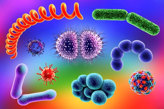
Bacterial Illustrations

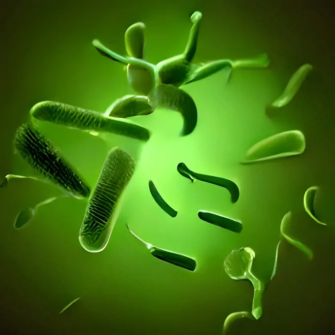
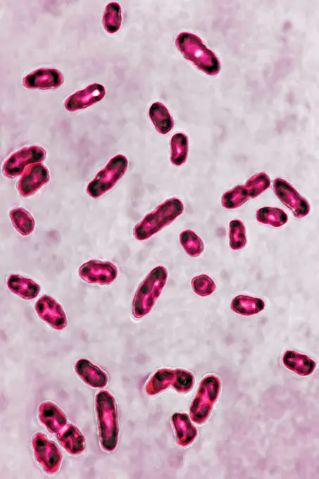
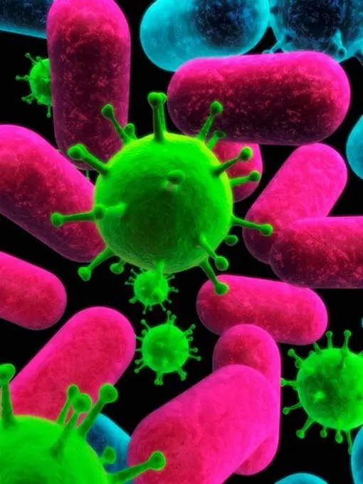
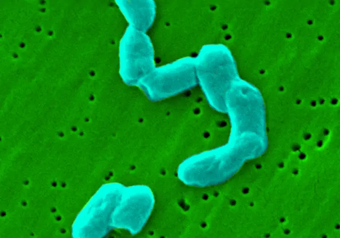
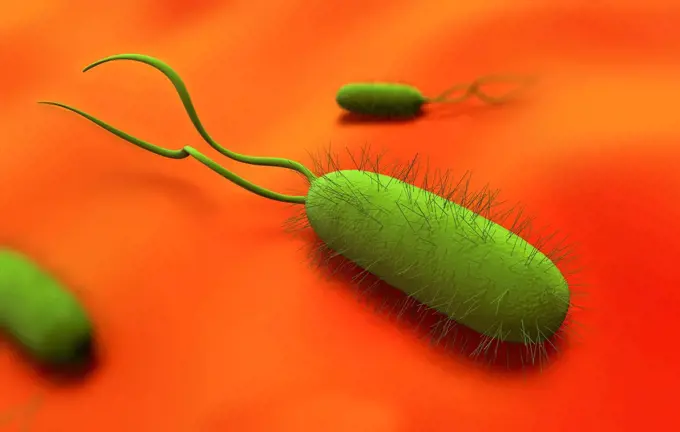
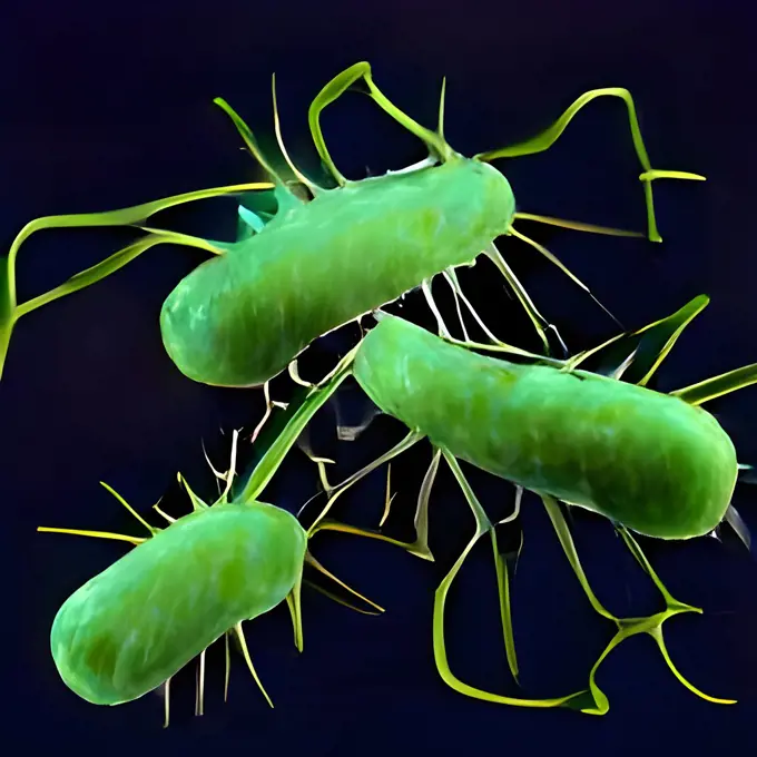
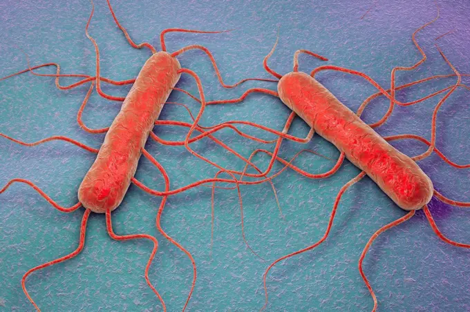
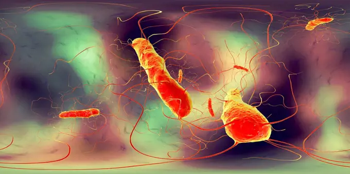
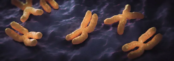
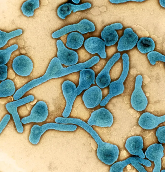
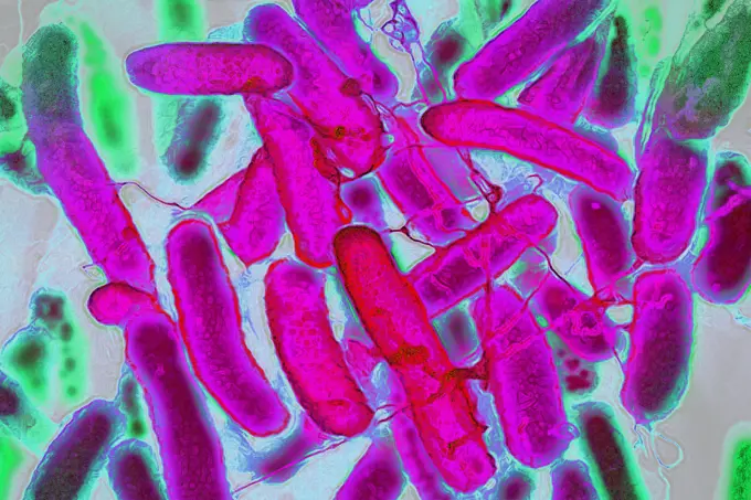
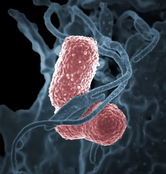
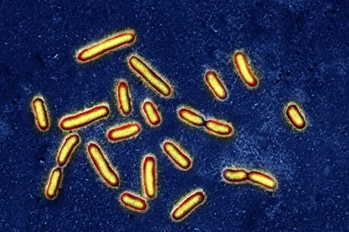
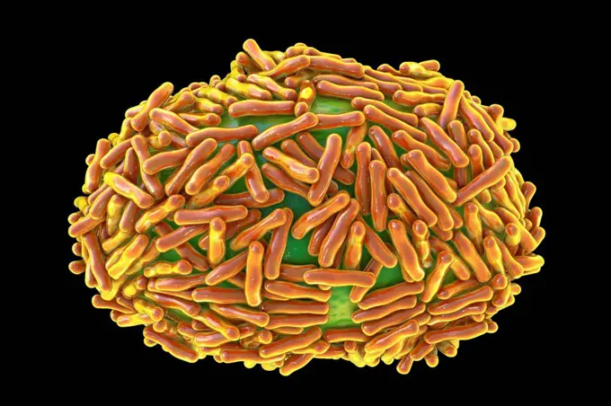
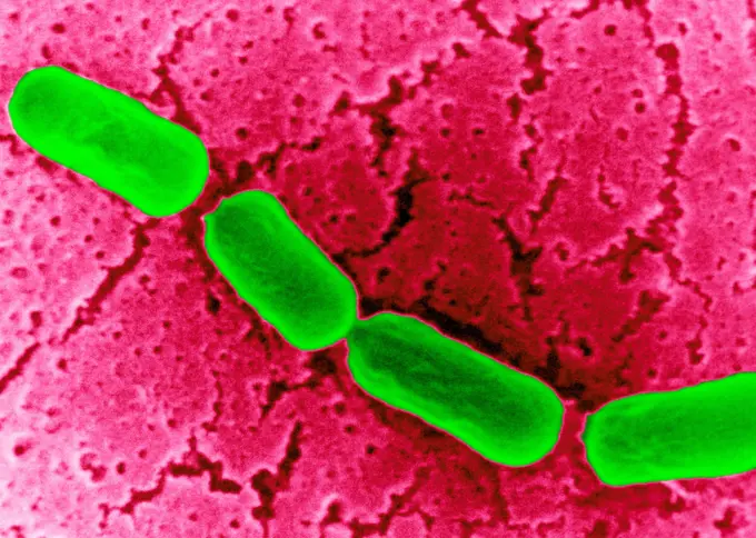
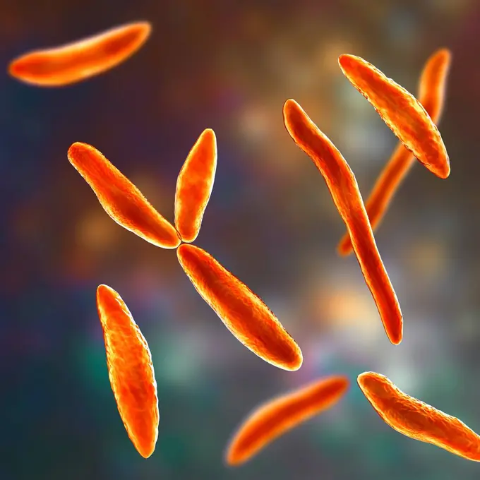
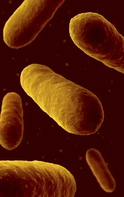
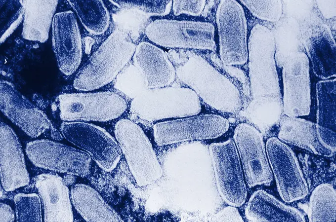
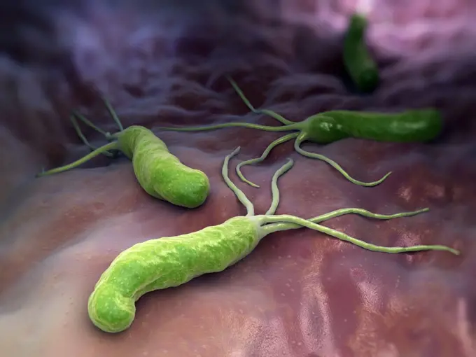
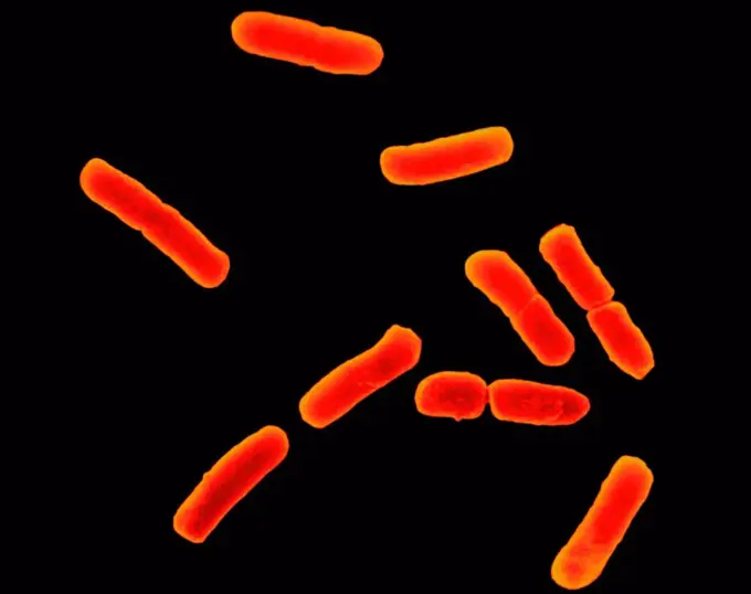
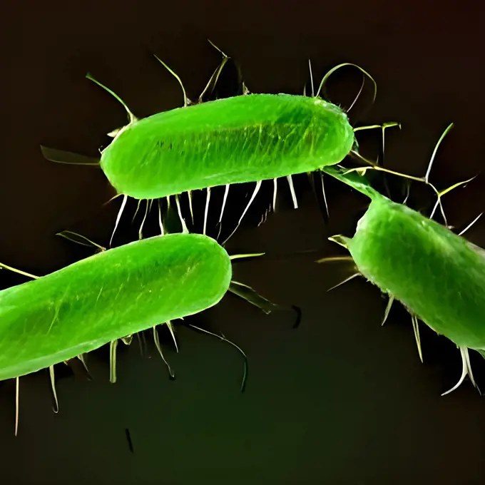
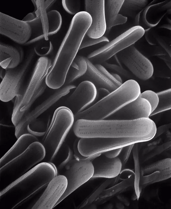
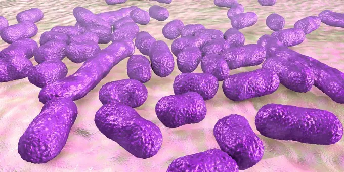
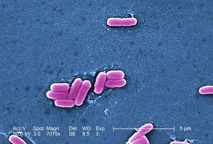
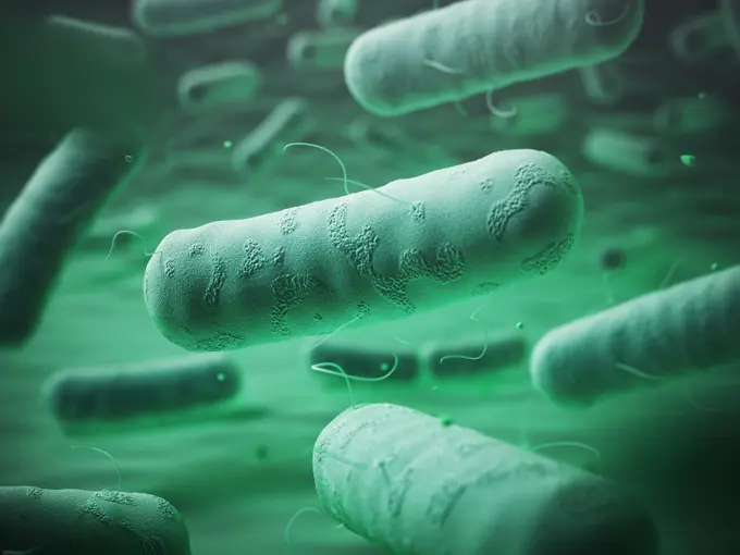
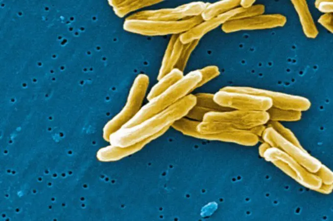
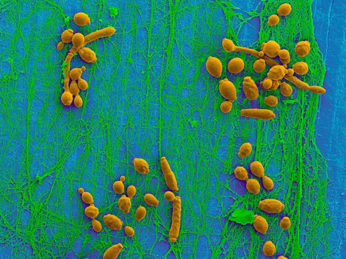
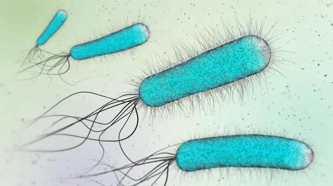
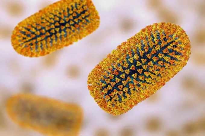
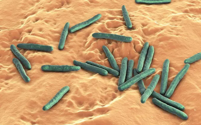
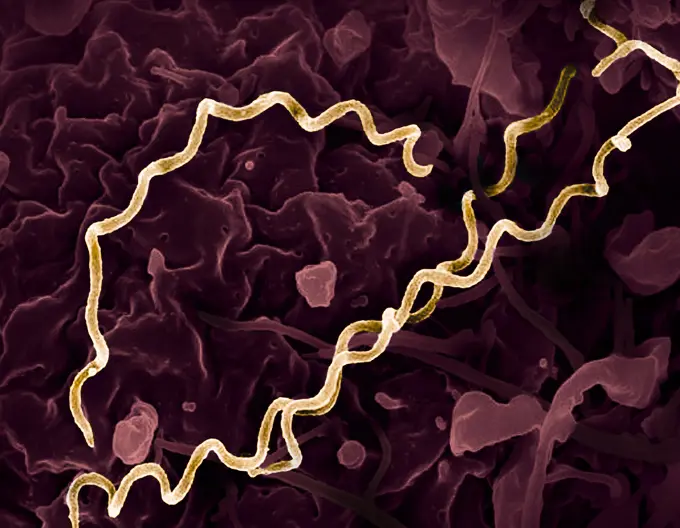
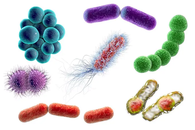
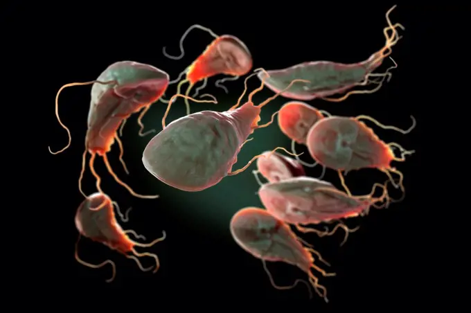
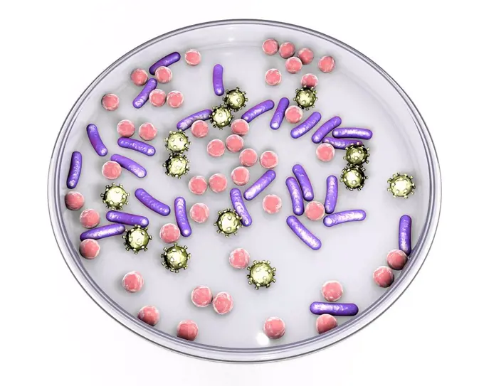
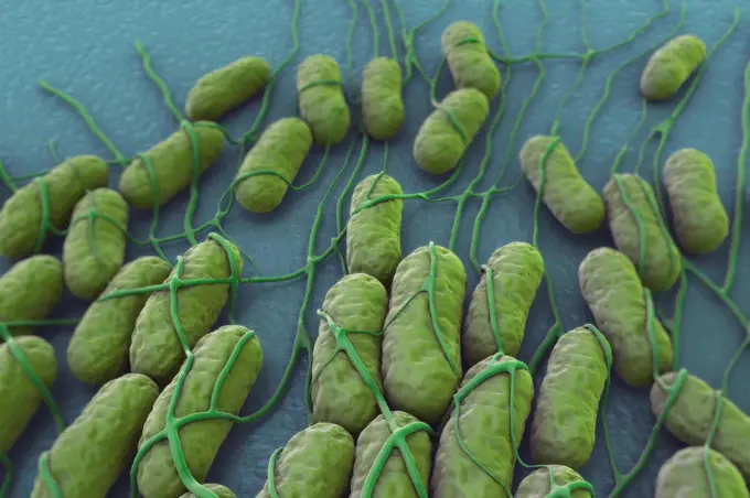
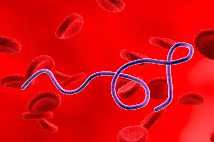
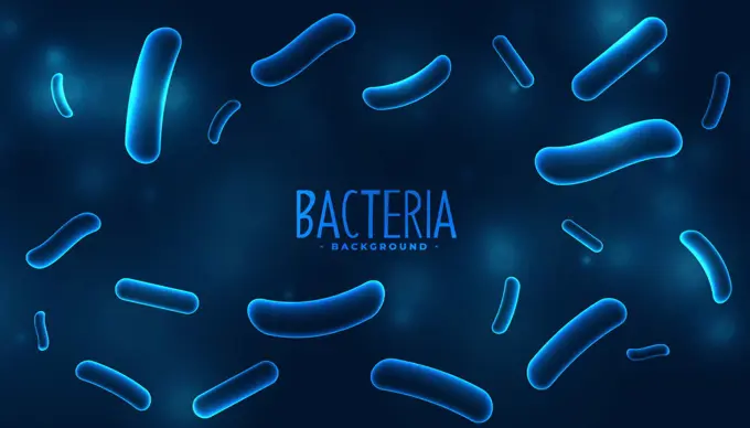
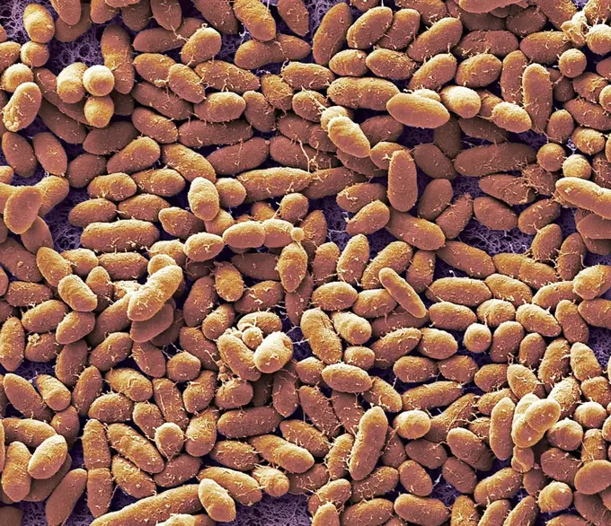
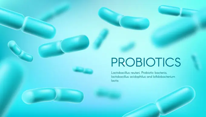
Computer-generated images of various bacteria and microorganisms, highlighting their shapes and characteristics in vibrant colors.

Medical, blue particles and germs or bacteria in science or clinical study and investigation. Healthcare, discovery and virus for research with innovation, future and experiment on microscope
278 assets in this story
1525-25996194
Helicobacter pylori bacteria green background on a dark background. Vector. Best for medical, scientific and commercial design.
6019-19982634
Microscopic view of bacteria. 3d illustration
4128R-15545784
Salmonella bacteria, illustration
824-63211612
Yersinia pestis (Pasteurella pestis). Bacteria responsible for the plague. Microscopy to optical microscopy X 1000.
1525R-176892
824-63128602
This scanning electron micrograph (SEM) depicts a number of highly magnified rod-shaped, motile, Gram-negative Salmonella infantis bacteria, some of which are attached, Magnification 18875x. Salmonellosis is an infection due to members of a genus of bacteria called Salmonella, of which S. infantis is a member. Most persons infected with Salmonella develop diarrhea, fever, and abdominal cramps 12 to 72 hours after infection. The illness usually lasts 4 to 7 days, and most persons recover without treatment. However, in some persons the diarrhea may be so severe that the patient needs to be hospitalized. In these patients, the Salmonella infection may spread from the intestines to the blood stream, and then to other body sites and can cause death unless the person is treated promptly with antibiotics. The elderly, infants, and those with impaired immune systems are more likely to have severe illness. See image n0830305 on the site.
4128-20044898
Tetanus bacteria, illustration
4128R-11310275
Helicobacter pylori bacteria, computer artwork.
6019-19982661
Bacteria outbreak and bacterial infection as a microscopic background
4128R-13242358
Listeria monocytogenes bacterium, computer illustration. L. monocytogenes is the causative agent of the human disease listeriosis. Listeriosis is cont...
4128-111582018
Clostridium difficile bacteria with peritrichous flagella, computer illustration. C. difficile is a Gram-positive, anaerobic, spore forming, rod-shape...
4378-1832
Genetic determining human chromosomes.
4128R-22308
Chromosome, computer artwork.
824-63223173
Colorized transmission electron micrograph of Marburg virus particles (blue) harvested from infected VERO E6 cell supernatant. Image captured and color-enhanced at the NIAID Integrated Research Facility in Fort Detrick, Maryland. Credit: NIAID.
4128R-10639
Ebola viruses, artwork
1525-26122184
Mitochondrion. Mitochondrion is a double membrane-bound organelle found in all eukaryotic organisms. 3D illustration
4128-20044919
Tetanus bacteria, illustration
824-29057231
Escherichia coli (intestinal bacteria that resides in the digestive tract of humans and warm-blooded animals, it is the cause of food poisoning. Electron microscopy.
824-63128595
This scanning electron micrograph (SEM) depicts a grouping of Vibrio vulnificus bacteria, Mag. 13184x. Vibrio vulnificus is a bacterium in the same family as those that cause cholera. It normally lives in warm seawater and is part of a group of vibrios that are called halophilic because they require salt. V. vulnificus can cause disease in those who eat contaminated seafood or have an open wound that is exposed to contaminated seawater. Among healthy people, ingestion of V. vulnificus can cause vomiting, diarrhea, and abdominal pain. In immunocompromised persons, particularly those with chronic liver disease, V. vulnificus can infect the bloodstream, causing a severe and life-threatening illness characterized by fever and chills, decreased blood pressure (septic shock), and blistering skin lesions. V. vulnificus bloodstream infections are fatal about 50% of the time. See image n0831605 on the site.
4128R-13711036
Illustration of bacteria.
824-57660039
Pseudomonas aeruginosa or pyocyanin bacillus, pathogenic bacteria, very resistant and often responsible for nosocomial infections. It is one of the most difficult bacteria to eliminate clinically. The infections it causes concern: the eyes, burns, surgical wounds, urine, lungs, stomach, intestines, meninges, blood. Image taken from an X 1000 optical microscopy.
824-63227252
A human neutrophil interacts with Klebsiella pneumoniae (pink), a multidrug-resistant bacterium that causes serious hospital infections.
1899-61461221
Pseudomonas aeruginosa or pyocyanin bacillus, pathogenic bacteria, very resistant and often responsible for nosocomial infections. It is one of the most difficult bacteria to eliminate clinically. The infections it causes concern: the eyes, burns, surgical wounds, urine, lungs, stomach, intestines, meninges, blood. Image taken from an X 1000 optical microscopy
4128-19489546
Monkeypox virus particles, illustration
6188-66486216
Gelb gefärbte Escherichia coli-Bakterien, die im Darm und im Stuhl vorkommen - 3d Illustration Yellow colored rod-shaped Escherichia coli bacteria found in the intestine and in feces - 3d illustration Copyright: xZoonar.com/ChristophxBurgstedtx 22602714
824-63128354
SEM x 15,000.
824-63179011
Bordetella pertussis (bacillus of Bordet-Gengou or Haemophilus pertussis), is the agent of whooping cough. The bacterium settles in the trachea or bronchi, and causes cough and fever with an accumulation of mucus. Optical microscopy
4128-28767343
Corn smut fungus, illustration
4128R-14058639
Lactobacillus crispatus bacteria, illustration
4128R-13271923
Microbes. Computer illustration of a microbial mixture containing bacteria and viruses of different types.
4128-30422581
Elizabethkingia meningoseptica bacteria, illustration
4128R-10089
Tuberculosis bacteria, artwork
6188-64770635
Peanuts in the shell levitate on a white background Peanuts in the shell levitate on a white background. Copyright: xZoonar.com/AlekseyxButenkovx 15083104
4128-48285256
Chromosomes, illustration
4239R-20483559
Microscopic view of bacteria.
824-63127918
Causative agent of vesicular stomatitis in cattle, horses and pigs. This electron micrograph depicts the vesiculovirus responsible for vesicular stomatitis (VS) in horses, cattle and pigs. As a member of the Rhabdoviridae family of viruses, you'll note the morphologic similarity, ie bullet-shaped virion, between this vesicular stomatitis virus (VSV), and the rabies virus. See image n1302405 on the site.
1525-19806358
Helicobacter Pylori
1899-53512002
Escherichia coli (intestinal bacteria) that resides in the digestive tract of humans and warm-blooded animals, it is the cause of food poisoning. Electron microscopy.
4128R-13620069
Coloured scanning electron micrograph (SEM) of Escherichia coli is a Gram-negative, facultatively anaerobic, enteric, rod prokaryote. This bacterium w...
1525-25341432
3d rendered scientific illustration of some bacteria
4128-V58615275
4128-111494928
Escherichia coli bacterium, illustration.
4128-20040355
Mitochondria, illustration
4128-V58574329
6019-19982666
Bacteria outbreak and bacterial infection as a microscopic background
4201-66311
Diatom (Diadesmis contenta) group SEM close-up view at 2800x magnification
4128R-13763224
Multi-drug resistant Acinetobacter baumannii bacteria, computer illustration. A. baumannii is a Gram-negative, oxidase negative, aerobic, coccobacillu...
4378-5203
Legionella Pneumophila Bacteria
4128R-13244740
E. coli bacteria, illustration.
4297-1762
Colorized scanning electron micrograph of E.coli O157:H7 at a magnification of 7075x.Escherichia coli bacteria of the strain O157:H7.E.coli O157:H7 is one of hundreds of strains of this bacterium
6188-63343256
Enterobacteriaceas. Gram-negative bacterias escherichia coli, salmonella, klebsiella, legionella, mycobacterium tuberculosis, yersinia pestis, and shigella, proteus, enterobacter, serratia, and citrobacter. Enterobacteriaceas. Gram-negative bacterias escherichia coli, salmonella, klebsiella, legionella, mycobacterium tuberculosis, yersinia pestis, and shigella, proteus, enterobacter, serratia, and citrobacter. 3d illustration Copyright: xZoonar.com/MaksymxYemelyanovx 10359619
4384-301
Mycobacterium tuberculosis Bacteria
4128-20044908
Tetanus bacteria, illustration
4128R-13724248
Yersinia pestis bacteria, illustration.
824-63211081
Legionella pneumophila is the bacteria responsible for legionellosis (pneumonia known as Legionnaires' disease). Optical microscopy X 1000.
4128R-13620120
Coloured scanning electron micrograph (SEM) of Candida albicans infected medical catheter. A common yeast, C. albicans can colonize catheters resultin...
4128-18680851
Bacteria on a tooth, illustration
6188-66486071
3d-Illustration von blauen Bakterien oder einzelligen Mikroorganismen 3d illustration of blue bacteria or single cell microorganisms Copyright: xZoonar.com/ChristophxBurgstedtx 22601076
4128-20239402
Mokola virus, illustration
4128-V58574253
4128-20045016
Cholera bacteria in human intestine, illustration
4239-18641714
Conceptual image of fusobacteria.
4128R-14846583
Tuberculosis bacteria, illustration
824-65830676
Colorized electron micrograph of Treponema pallidum, the bacteria that cause syphilis. Several spiral-shaped bacteria have been highlighted in gold.
4128R-13271913
Bacteria. Computer illustration of bacteria of different shapes.
4378-5169
Multiple Giardia Trophozoites
4128R-13587540
Petri dish with microbes, computer illustration. Conceptual image of laboratory diagnosis of infectious diseases.
4378-3367
A culture of Salmonella bacteria on a plane surface. Salmonella is a genus of rod-shaped, Gram-negative bacteria.
4378-3817
A culture of Salmonella bacteria on a plane surface. Salmonella is a genus of rod-shaped, Gram-negative bacteria.
4128R-13048937
Ebola virus, illustration.
4128R-13325696
Bacteria, illustration.
4128R-13023012
Computer illustration of Borrelia bacteria in blood. Borrelia causes relapsing fever (the most common cause is Borrelia recurrentis) and Lyme diseases...
1525-75679728
Bacteria floating virus infection background
4128-111453069
Bacteria found on mobile phone, SEM
4128-20041789
Faecal bacteria, sem
4384-123
This colorized transmission electron micrograph (TEM) revealed some of the ultrastructural morphology displayed by an Ebola virus virion. The exact origin, locations, and natural habitat (known as the 'natural reservoir') of Ebola virus remain unknown. However, on the basis of available evidence and the nature of similar viruses, researchers believe that the virus is zoonotic (animal-borne) and is normally maintained in an animal host that is native to the African continent. A similar host is pr
1525-27109118
Microscopic Probiotics Live Bacteria Banner Vector. Medical Laboratory Research Microfloral Microorganism Bacteria. Microbiological Fermentation Organism Concept Layout Realistic 3d Illustration. Microscopic Probiotics Live Bacteria Banner Vector
1525-76101424
Probiotic bacteria, lactobacillus acidophilus and bifidobacterium, vector background. Probiotic bacteria prebiotics, healthy microorganism bacterium for gut health and digestion with bifidobacterium. Probiotic bacteria, lactobacillus acidophilus
PREVIOUS
of 3
NEXT
2401 S. Ervay, Suite 206
Dallas, Texas 75215
United States
Get Started
Free ResearchMy BoardsMy Cart
For Creators
How To License Your ContentContributor PortalFrame of Mind
Resources
API accessPricing
Contact
+1 866 236 0087help@viewpictures.co.uk Contact form
©2026 View Pictures. All Rights Reserved. -A
This site is protected by reCAPTCHA and Google Privacy Policy and Terms of Service apply.