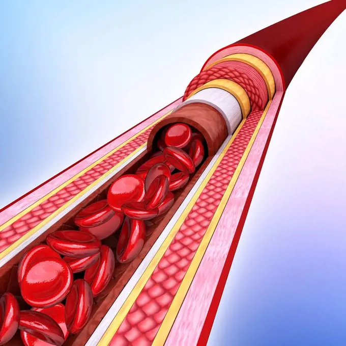
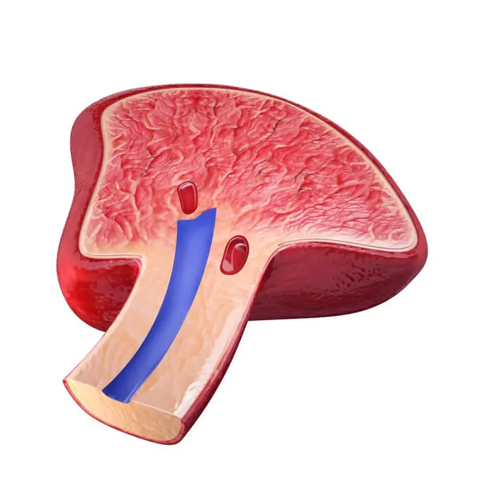
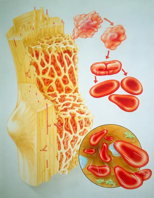
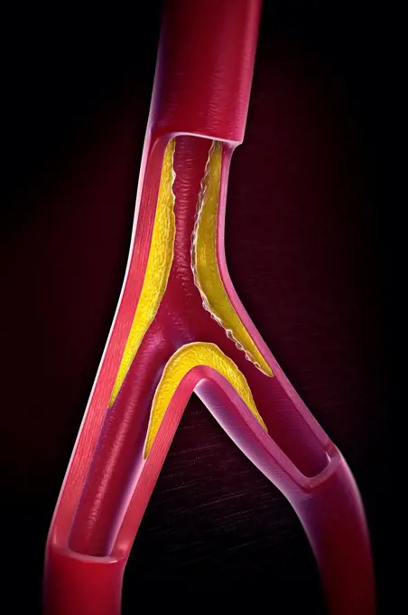
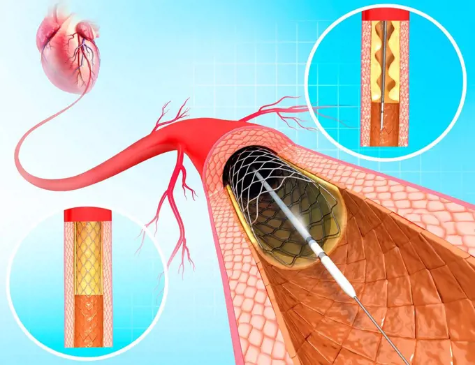
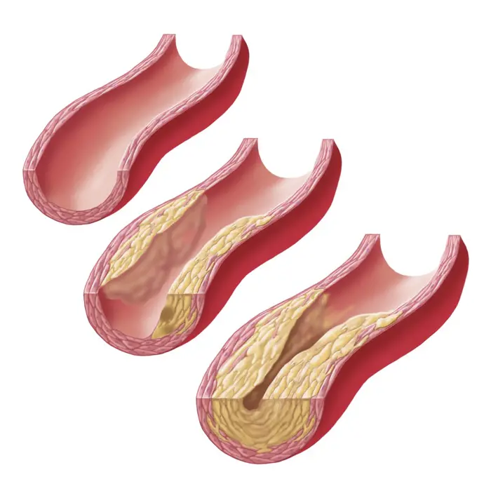
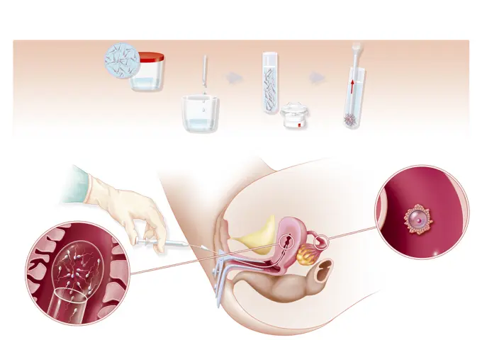
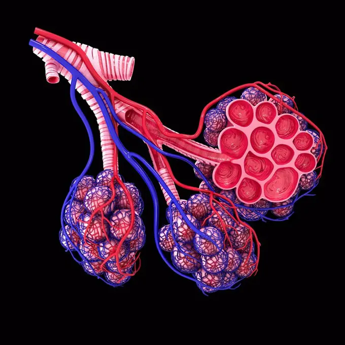
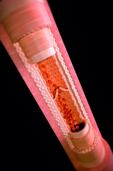
My Boards
View Pictures
  • My Boards

Blood Vessel Pathology Art

Computer-generated art showing human blood vessels, highlighting conditions like deep vein thrombosis and structural details of veins and arteries.

Human muscular structure, computer artwork.
175 assets in this story
824-63219042
Myocardial infarction, coronary artery with thrombus, atheromatous plaques, treatments. The atheromatous plaques (yellow) in the wall of the coronary artery cause coronary disease. In the zoom of the coronary vessel, a thrombus forms at the bottom right, in contact with the blood by platelet aggregation. Treatments act on platelets to prevent platelet aggregation, on the secretion of aldosterone by the kidneys and adrenals, but also upstream, on the liver by limiting the synthesis of cholesterol at the origin of atheromatous plaques.
4128R-11476350
Nerve cell, computer artwork.
4239-18641688
Medical illustration of an asthmatic bronchiole, with labels.
4128R-11315209
Human artery, cut-away computer artwork.
1525-26235643
intravenous intervention in brain and nerve diseases3D illustration. intravenous intervention in brain and nerve diseases
4128R-11312488
Human spleen, cut-away computer artwork.
4128R-13310
Genetics research, conceptual computer artwork.
1525-20586940
Blood clot and thrombosis medical illustration concept as a group of human blood cells clumped together by sticky platelets and fibrin creating a blockage in an artery or vein as a health disorder symbol for circulatory system danger.
4128R-11315768
Human lymph node, computer artwork.
1899-53508968
Female genitalia, ovarian cycle, ovulation, fertilization, embryo segmentation, implantation.
824-63206052
Illustration of an atheromatous plaque which obstructs the lumen of a large artery, such as coronary arteries, and reduces blood flow.
4128R-11313019
Human artery, cut-away computer artwork.
824-63219990
Chronic obstructive pulmonary disease COPD and its treatments. At the top left the lungs with the bronchial tree in transparency, from where a sub-segmental bronchus in zoom. A transected left bronchiole is normal. The transected right bronchiole shows bronchoconstriction with hypersecretion of mucus, inflammatory cells producing cytokines in the mucosa. Below the pulmonary lobules. On the left, the alveolar sacs are normal, on the right, an emphysema seen in section shows the distended alveoli.
4128R-15465536
Trichomoniasis, illustration
824-63219988
Chronic obstructive pulmonary disease COPD and its treatments. At the top left the lungs with the bronchial tree in transparency, from where a sub-segmental bronchus in zoom. A transected left bronchiole is normal. The transected right bronchiole shows bronchoconstriction with hypersecretion of mucus, inflammatory cells producing cytokines in the mucosa. Below the pulmonary lobules. On the left, the alveolar sacs are normal, on the right, an emphysema seen in section shows the distended alveoli.
4128R-11292826
Colon cancer, computer artwork.
1525-25144088
Illustration of bad cholesterol in vein section
4128R-12923234
Cross section of liver, illustration
824-63208817
Cross-section illustration of a blood vessel containing red blood corpuscles.
824-63167603
The bone marrow is where the blood cells are regenerated. Shown here, red blood corpuscles being produced by the bone marrow
1525-56996888
3D illustration. Fresh slices or meat. Marbling of pork or beef tenderloin for steak.
4128R-11291336
Human bone structure, computer artwork.
4128R-11313701
Human lymph node, computer artwork.
4128R-11289354
Human artery, cut-away computer artwork.
4128R-13244780
Infected human bronchus of the lungs, illustration.
4378-4259
Gallstones
1899-53513225
Renal glomerulus. At the extremity of each nephron there is the renal glomerulus, filtration unit of the kidney. It is constituted of the Bowman capsule (in violet) that enfolds the capillary leading the blood to filter (afferent glomerular arteriole). The Bowman capsule ends with the proximal convoluted tubule (at the bottom), by which the urine transits and the one by which blood was filtered goes back towards the body (efferent glomerular arteriole). The afferent and efferent glomerular arterioles are covered with a layer of cells, the podocytes, represented for the left part of the image (in beige).
4128R-29271
Healthy artery, computer artwork.
4128R-24439
Atherosclerosis. Computer artwork of a narrowed artery, due to a cholesterol plaque.
4128-19248150
Mitochondria, illustration.
6188-58187792
Animal cell structure isolated on white background. 3D illustration Animal cell structure isolated on white background. 3D illustration. Copyright: xZoonar.com/CigdemxSimsekx 21429803
4128R-14324148
Prostate cancer, illustration
1848-77390817
3D Illustration Concept of Human Internal Organs Pancreas with Gallbladder Anatomy
4128R-14057544
Ovarian follicles, micrograph and illustration
4128R-18114
Breast anatomy, computer artwork.
1525-24977246
Poster of medical illustration of the heart.
1525-24345969
Vector illustration of pancreas
4378-5423
Atherosclerosis
824-63167624
Hematopoiesis occurs in three successive cell compartments. Stem cells are pluripotent cells guaranteeing permanent production of blood cells. Precursor cells (or determined cells) are only capable of a limited number of divisions. Maturing cells constitute a transition between the preceding compartments and the bloodstream, cells in the bloodstream have identifiable morphologies.
824-63191587
Illustration of the Hepatitis C virus that infects the liver. It is transmitted through the bloodstream, reproduces and spreads in the hepatic lobules, causing inflammation, then necrosis of the hepatic lobular tissue.
824-63180639
Illustration of the chain reaction involved in muscle cramp.
824-63163768
Healthy dorsal vertebrae with osteoporosis. (zooms on the spongy bone).
4128R-12916217
Small intestine, illustration
4128R-23895
Angioplasty. Computer artwork of a stent being placed in a narrowed blood vessel.
4239-20481096
Build-up of atherosclerotic plaque in an artery.
824-63214084
Illustration of inter-uterine insemination (IUI). This technique is used in the case of reduced quality sperm, insufficient sperm, abnormal cervical mucus or in the case of anti-spermatozoa antibodies or unexplained infertility. The top of the illustration shows sperm preparation. The lower part shows insemination, the doctor slowly places the sperm in the uterus during ovulation.
824-63195464
Representation of the transmission of nerve impulses from one neuron (purple) to a 2nd neuron (red). The electrical signal is transformed into a chemical signal, vesicles release glutamate (blue) which binds to the receptors (yellow) which open the sodium channels and allow Na+ ions to penetrate the dendritic button, thus creating an electric current.
4128R-11315763
Human alveoli, cut-away computer artwork.
1899-53508976
Atheroma, lesion of the inner surface of the arteries caused by a deposit of cholesterol.
4128R-13723757
Illustration of female reproductive system.
4128-30420494
Microsporum canis infection of the foot, illustration
5507-46943614
Cartoon illustration of muddy blood vessels
4378-20393151
Anatomy of a Vein
4128-18800964
Clogged milk duct and thrush comparison, illustration
824-63189060
Illustration of the ovarian cycle in the ovary : the follicle matures (pink) which contains the ovocyte (dark purple), then the egg is released into the Fallopian tube and finally, the formation of corpus luteum (yellow) then disintegration of the egg isnt fertilised.
4128R-14060111
Normal and infected bronchi, illustration
1525-75955909
coronary arteries with accumulated fat in the body
4128-19249262
Peripheral artery disease, illustration.
4128R-13446805
Illustration of a child's alveoli.
4128R-29324
Atherosclerosis. Computer artwork of a narrowed artery, due to a cholesterol plaque.
6188-67437837
3D Isometrische flache konzeptionelle Illustration des Häm-Stoffwechsels, Bildungsdiagrammbr3D-Isometrische flache konzeptionelle Illustration des Häm-Katabolismus, Lehrdiagramm 3D Isometric Flat Conceptual Illustration of Heme Catabolism, Educational Diagram 3D Isometric Flat Conceptual Illustration of Heme Catabolism, Educational Diagram Copyright: xZoonar.com/AnastasiiaxTorianykx 22973181
4128-19358517
Female breast cancer, illustration.
4128R-11313185
Human pancreas, computer artwork.
5507-45991448
Levels of cholesterol plaque in vessels
4128R-13267664
Lymph node of a child, illustration.
1525-24012682
Organ bioprinting icon isometric vector. Medical printer. Bio science. Organ bioprinting icon isometric vector. Medical printer
1525-27988795
Levels of cholesterol plaque in vessels vector illustration
4128R-13410319
Human liver, illustration
4128R-14060539
Toxoplasma gondii parasites, illustration
4128-19249212
Ischaemic and haemorrhagic strokes, illustration.
4128-30420497
Athlete's foot, illustration
4378-20014146
Rat Brain Anatomy
5507-43151555
chronic iron deficiency yellow skin
1525-20790325
Blood vessel of  healthy person with normal leukocytes. Bloody Artery Boxer. Blood cells red boxing gloves. Dead man  blood- lymph cells skull. Anatomical human blood vein

4409-28579794
Infographic that reviews the main achievements of nanomedicine, a branch that tries to diagnose, prevent and cure at a molecular level with nanobots and nano structures. QuarkXPress (.qxp); Adobe InDesign (.indd); QuarkXPress (.qxd); 4960x3188.
PREVIOUS
of 2
NEXT
2401 S. Ervay, Suite 206
Dallas, Texas 75215
United States
Get Started
Free ResearchMy BoardsMy Cart
For Creators
How To License Your ContentContributor PortalFrame of Mind
Resources
API accessPricing
Contact
+1 866 236 0087help@viewpictures.co.uk Contact form
©2026 View Pictures. All Rights Reserved. -B
This site is protected by reCAPTCHA and Google Privacy Policy and Terms of Service apply.