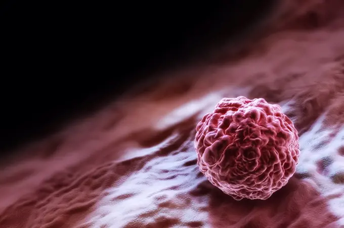
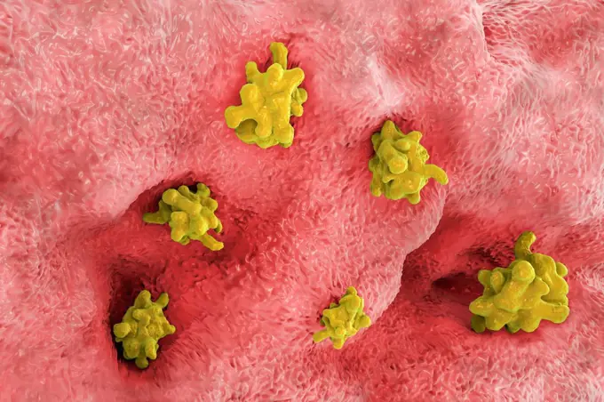
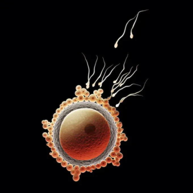
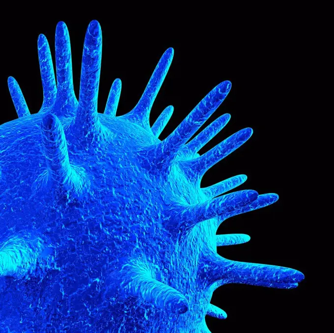
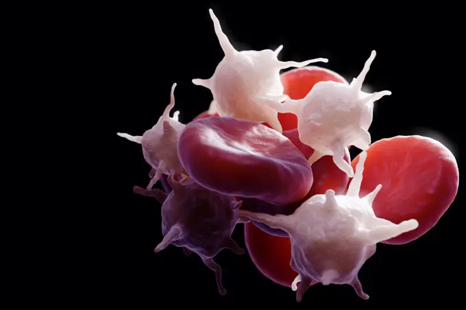
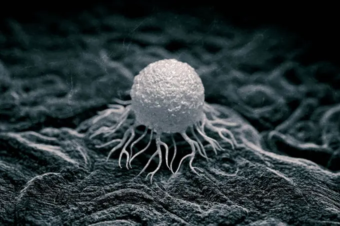
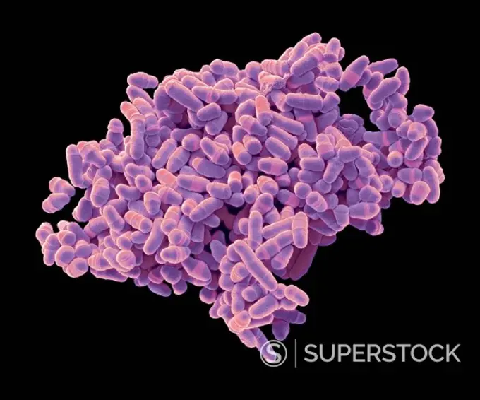
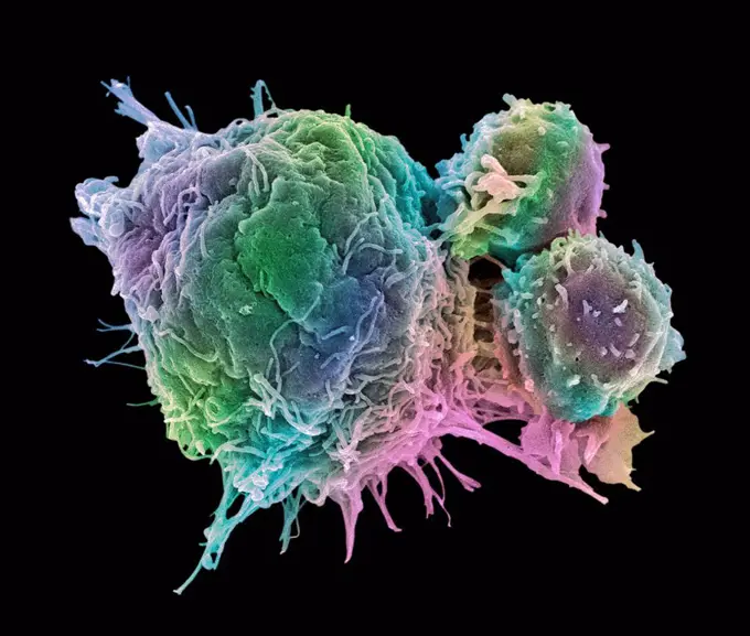
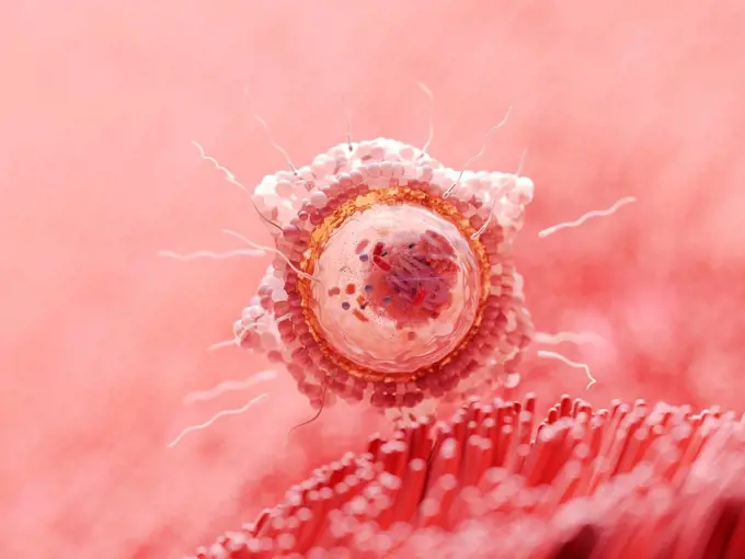
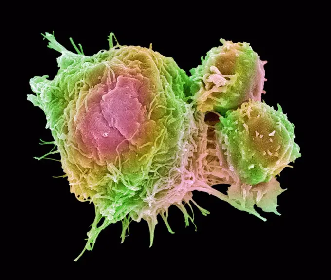
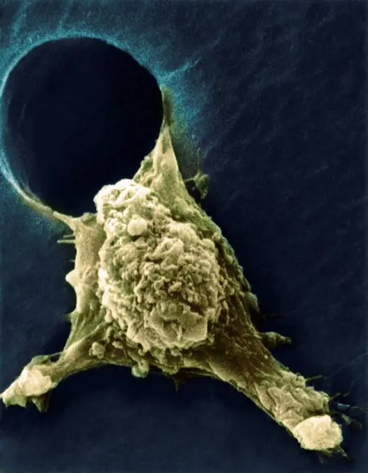
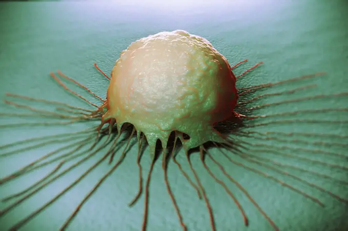
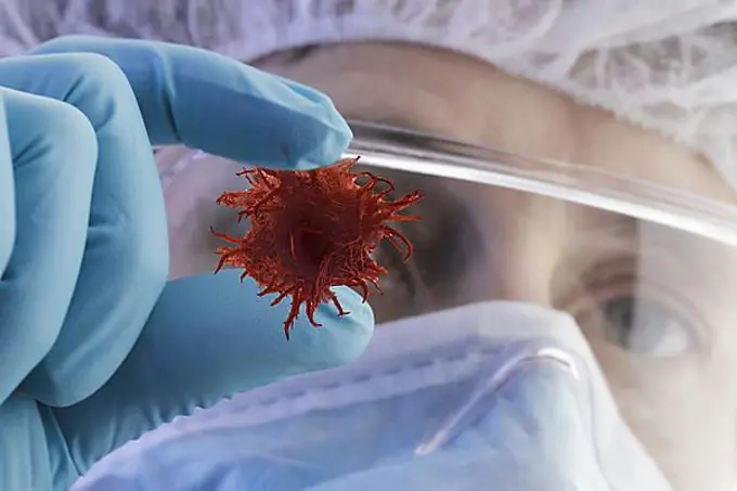
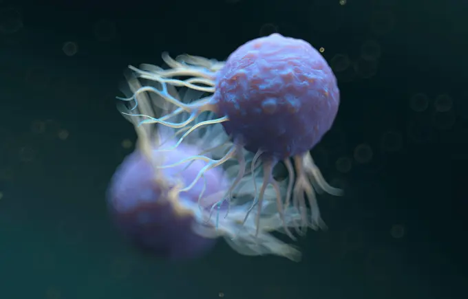
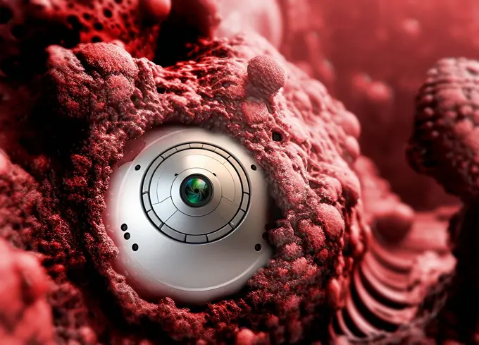
My Boards
View Pictures
  • My Boards

Cell Biology Illustrations

Vibrant 3D renderings of cancer cells, white blood cells attacking them, sperm and ovum dynamics, and embryonic stem cells, showcasing cellular structures and interactions.

Embryonic Stem Cell
Embryonic Stem Cell
233 assets in this story
4128R-13928467
Parasitic amoeba (Entamoeba histolytica) invading the intestine, computer illustration. This single-celled organism causes amoebic dysentery and ulcer...
4378-1782
Fertilization between egg and sperm.
4128R-20392
Medical nanoparticles, conceptual image
4378-4439
Blood Cells
4128-48286028
Cancer cell, illustration
4128R-14057788
Cancer cell and T cell, illustration
4413-43447
Spores of Rose rust
4128R-11473200
Coloured scanning electron micrograph (SEM) of Schizosaccharomyces pombe yeast. S. pombe is a single-celled fungus that is studied widely as a model o...
4128R-13047385
T lymphocytes and cancer cell. Coloured scanning electron micrograph (SEM) of T lymphocyte cells (smaller round cells) attached to a cancer cell. T ly...
4128R-15516388
Illustration of fertilisation
4128R-12571798
Blood cells. Coloured scanning electron micrograph (SEM) of human blood showing a red and white cell ( lymphocyte). Red blood cells (erythrocytes) hav...
1525R-177074
4378-4628
Birch Tree Pollen
4378-20013766
Colon Cancer Cells
1525-23843278
blood- brain barrier and central nervous system 3d illustration. blood- brain barrier and central nervous system
4128R-13844351
Illustration of a fat cell (adipocyte).
4269-6802
Scanning electron micrograph (SEM) of the bronchus wall, consisting in ciliated cells. At center, a gobelet cell expulsing a drop of mucus. Magnification x3000.
4128R-13047355
T lymphocytes and cancer cell. Coloured scanning electron micrograph (SEM) of T lymphocyte cells (smaller round cells) attached to a cancer cell. T ly...
4128R-11474475
Neurone. Scanning electron micrograph (SEM) of a PC12 neurone in culture.The PC12 cell line, developed from a pheochromocytoma tumor of the rat adrena...
4128R-1787
Metastasis of a cancerous cell, SEM
4378-1780
A breast cancer cell.
1899-65662338
Colorized scanning electron micrograph of a T lymphocyte (also known as a T cell) (yellow).
824-65830645
Colorized scanning electron micrograph of a T lymphocyte (also known as a T cell) (yellow)
4128R-11312731
Human lymph node, computer artwork.
1848-54695954
Scientist and tumor, illustration
4269-27696
Malignant cell. Scanning electron micrograph (SEM) of a cancerous cell showing pseudopodia ( pseudopod ). This 'arms' are generated by a protein named autocrine motility factor (AMF) enabling them to migrate to other parts of the body. Locomotion is integral to the entire process of metastasis.
4128-48286022
Cancer cell, illustration
4128-V58562151
4128-48285283
Neuroblastoma cancer cells, illustration
4378-5416
Colon Cancer Cells
4128-48285720
Engineered bacteria, conceptual illustration
4128R-31105
White blood cells. Coloured scanning electron micrograph (SEM) of white blood cells (leucocytes) and a platelet (thrombocyte, orange). Magnification: ...
4128-19358370
Bladder cancer stages, illustration.
PREVIOUS
of 3
NEXT
2401 S. Ervay, Suite 206
Dallas, Texas 75215
United States
Get Started
Free ResearchMy BoardsMy Cart
For Creators
How To License Your ContentContributor PortalFrame of Mind
Resources
API accessPricing
Contact
+1 866 236 0087help@viewpictures.co.uk Contact form
©2026 View Pictures. All Rights Reserved. -A
This site is protected by reCAPTCHA and Google Privacy Policy and Terms of Service apply.