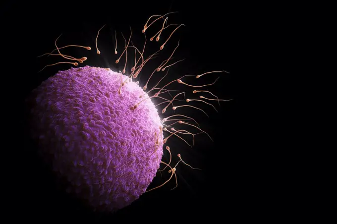
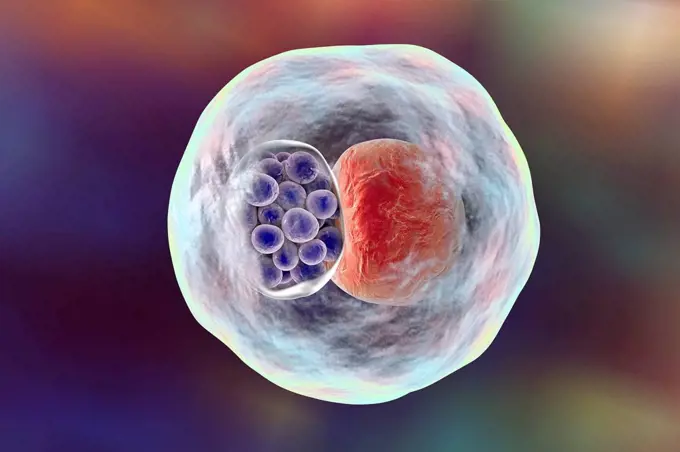
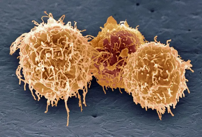
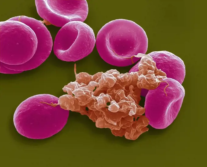
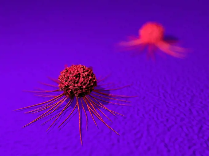
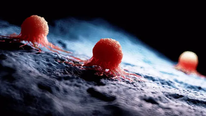
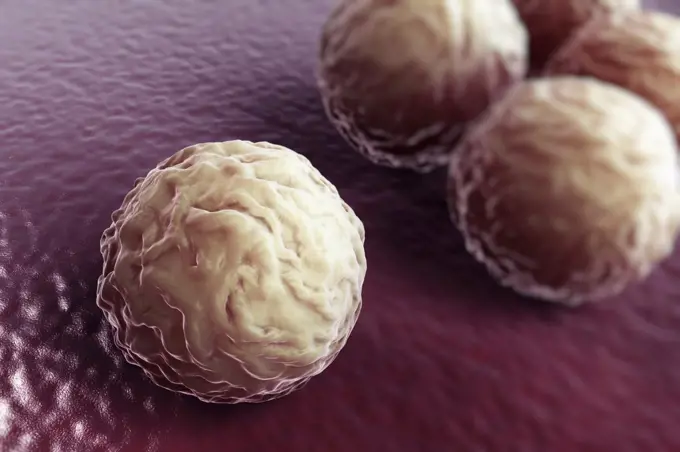
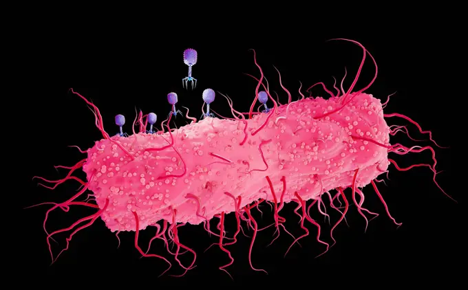
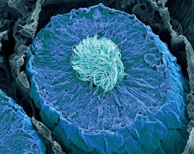
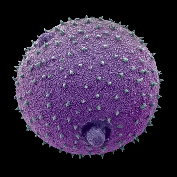
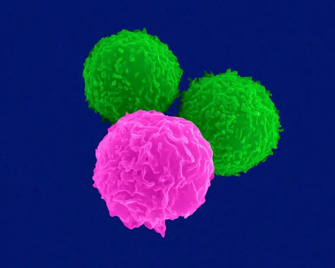
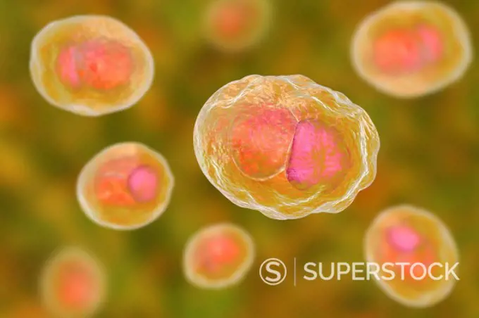
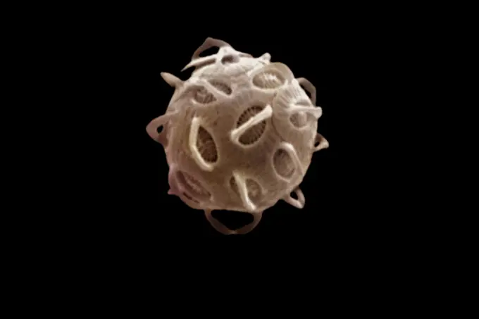
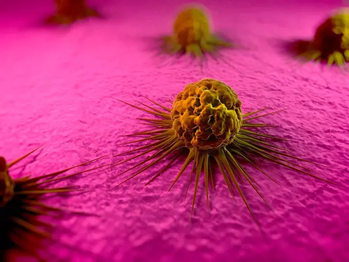
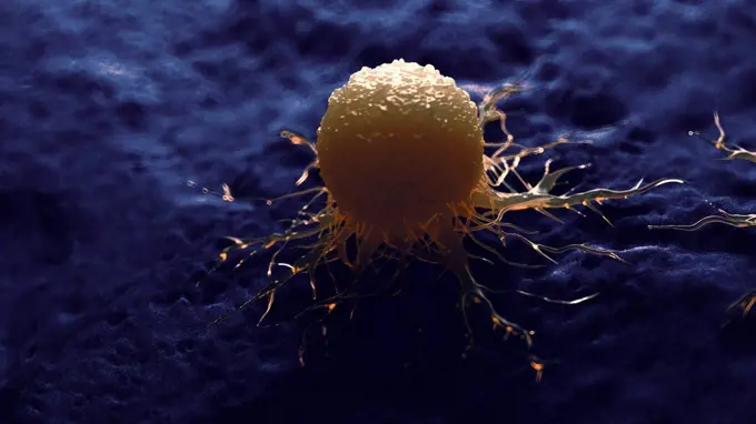
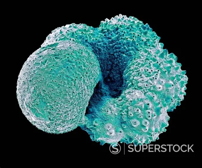
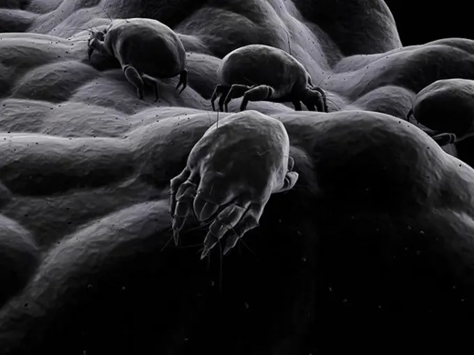
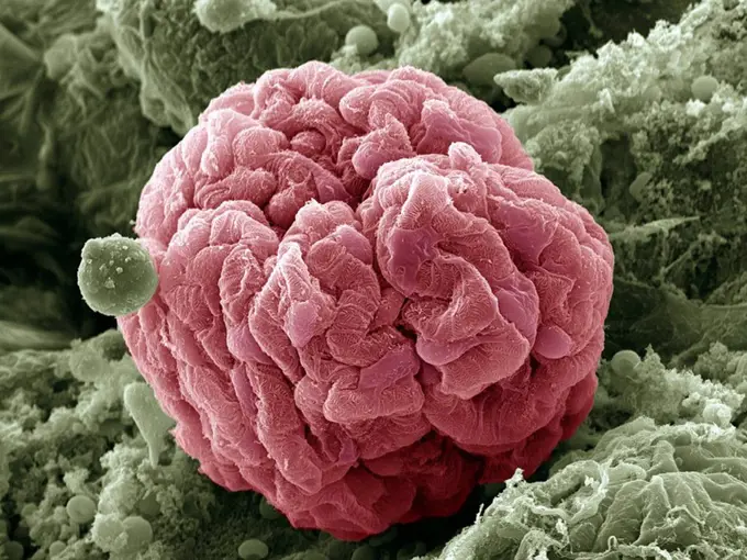
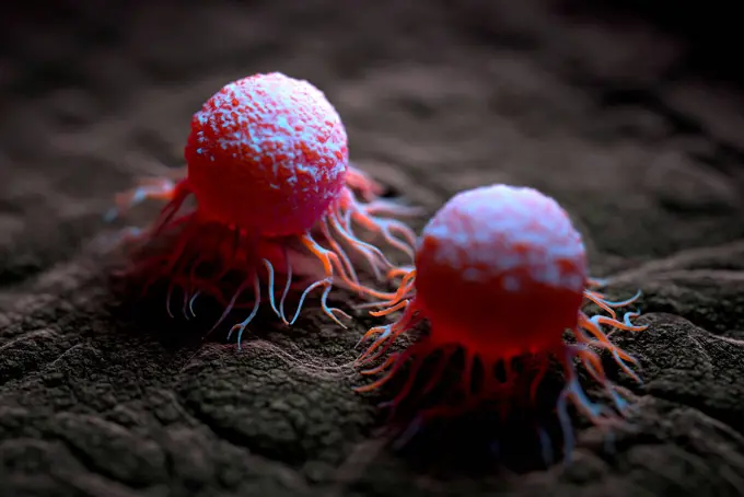
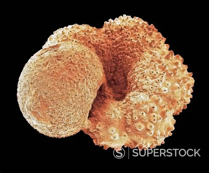
My Boards
View Pictures
  • My Boards

Cells and Viruses

Microscopic illustrations of human cells, viruses, and bacteria, highlighting scientific and medical themes with vibrant colors and detailed imagery.

Human sperm surrounding an unfertilized ovum prior to conception.
246 assets in this story
4269-27732
Hartmanella vermiformis. Color enhanced scanning electron micrograph of cysts of the protozoan Hartmanella vermiformis on a laboratory-grown potable water biofilm. Aquatic bacteria were grown as biofilm on steel for one week. Hartmanella vermiformis protozoans were then added, and phagocytized the bacteria that multiplied within vesicles that became cysts in which the bacteria will live until they rupture.
4128R-13282650
Chlamydia bacteria in a cell. Computer illustration showing an inclusion composed of a group of chlamydia (violet) near the nucleus (red) of a cell.
824-63225828
A cluster of CCL-81 cells infected with SARS-CoV-2 virus particles, which are the small spherical structures found on the surface of the cells. The protrusions extending from the cells are cell projections or pseudopodium. Image captured at the NIAID Integrated Research Facility (IRF) in Fort Detrick, Maryland.
4128R-2540
White blood cells, SEM
4128R-12870
Blood cells. Coloured scanning electron micrograph SEM of red blood cells pink and an aggregate of platelets red. Magnification: x4000 when printed at...
4128-20041797
Mesenchymal stem cell, SEM
1848-49387900
Nerves The cells transmit signals
4128R-13619767
Blood vessel with blood cells, computer illustration. Red blood cells and white blood cells inside a blood vessel.
4128-20041257
Athlete's foot fungus, illustration
824-63191232
Scanning electron micrograph of an apoptotic HeLa cell. Zeiss Merlin HR-SEM.
4128-111493991
Computer illustration of a blood clot inside a vein, known as a venous thrombus.
4128R-14057784
Cancer cell and T cell, illustration
824-57657676
Colorized scanning electron micrograph of monkeypox virus (green) on the surface of infected VERO E6 cells (red).
4128R-14649625
Cancer cells, illustration
4128R-15516236
Illustration of a cancer cell
4378-2471
Group of chlamydia bacteria.
824-65830618
Colorized scanning electron micrograph of Marburg virus particles (blue) both budding and attached to the surface of an infected VERO E6 cell (gold).
824-63223191
Colorized scanning electron micrograph of Middle East Respiratory Syndrome virus particles (yellow) attached to the surface of an infected VERO E6 cell (blue). Image captured and color-enhanced at the NIAID Integrated Research Facility in Fort Detrick, Maryland. Credit: NIAID.
4128-V58557856
4128-111493984
Genetic engineering, illustration. Synthetic small-ribonucleic acid (sRNA) being injected (needle tip at upper right) into the cell cytoplasm as part ...
4128-28515257
Bacteriophages infecting E. coli bacterium, illustration
4128-15751128
Seminiferous tubules in the testis, SEM
4070-48287210
Harebell (Campanula rotundifolia) pollen grain,  false-coloured Scanning Electron Micrograph. Pollen grain 0.025mm across, image x4000 magnification when printed at 10cm wide.
4128R-13619969
T lymphocytes (pre-T cells) and granulocyte (neutrophil), coloured scanning electron micrograph (SEM). Lymphocytes are involved in the specific immune...
4128R-15405902
Chlamydia trachomatis bacteria, illustration
4128-V58557900
4220-21334514
Scanning Electron Micrograph (SEM): Coccolithophore (Calcareous Nannoplankton), Gephyrocapsa ericsonii.     Date:
1525-27967505
Red blood cells and activated platelets or thrombocytes. 3D illustration. Red blood cells and activated platelets or thrombocytes
4128R-14425
Cancer cells, computer artwork. The uneven surface and cytoplasmic projections are characteristic of cancer cells.
4128R-15516227
Illustration of a cancer cell
4128R-11310472
Globigerina foraminiferan. Coloured scanning electron micrograph (SEM) of the shell (test) of a Globigerina sp. planktonic foraminiferan. Foraminifera...
1525R-178824
4391-241
A scanning electron microsope's view of blood cells. The round non-nucleated doughnut shaped cells are red blood cells, which transport oxygen and remove carbon dioxide. The spherical cells with a rough texture are white blood cells, which produce antibodies and fight infection. The smallest peices are platelets, tiny cells needed for blood clotting. Photo by Bruce Wetzel and Harry Schaefer.
4128R-187
Kidney glomerulus, SEM
4128-V58569707
4128-48286030
Cancer cells, illustration
4128R-11310470
Globigerina foraminiferan. Coloured scanning electron micrograph (SEM) of the shell (test) of a Globigerina sp. planktonic foraminiferan. Foraminifera...
4413-1788
Thalli of the calcareous corraligenous alga Pseudolithophyllum
4128-V58574924
4128R-9405
Bay tree anther, SEM
4128R-15516226
Illustration of a cancer cell
4128R-13376670
Human embryo destruction, illustration
4128R-11313165
Human lymph node, computer artwork.
4128R-15290694
Lyme disease tick, illustration
4128R-15516269
Illustration of a cancer cell being attacked by white blood
4239-18641687
Medical comparison of healthy alveoli and an unhealthy emphysema alveoli.
PREVIOUS
of 3
NEXT
2401 S. Ervay, Suite 206
Dallas, Texas 75215
United States
Get Started
Free ResearchMy BoardsMy Cart
For Creators
How To License Your ContentContributor PortalFrame of Mind
Resources
API accessPricing
Contact
+1 866 236 0087help@viewpictures.co.uk Contact form
©2026 View Pictures. All Rights Reserved. -A
This site is protected by reCAPTCHA and Google Privacy Policy and Terms of Service apply.