My Boards
View Pictures
  • My Boards

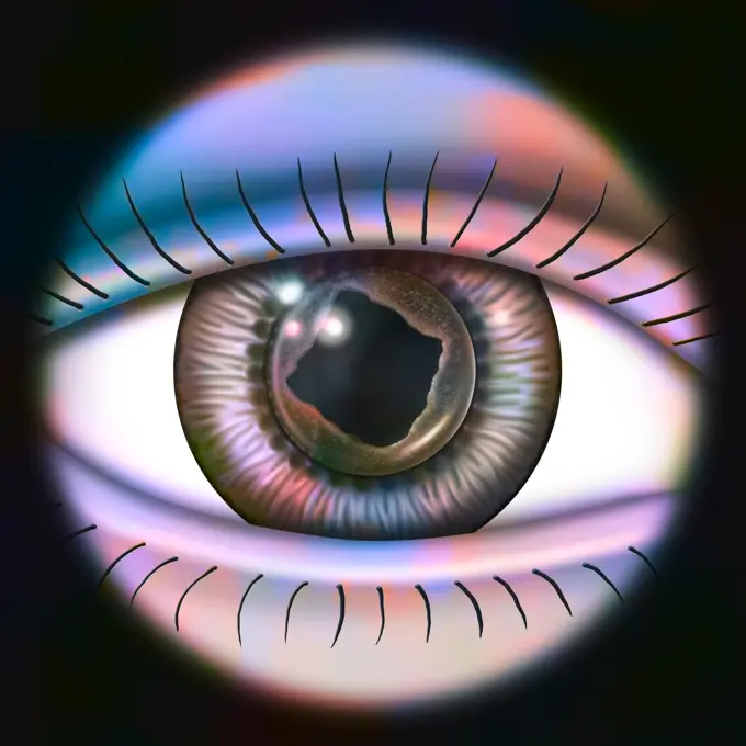
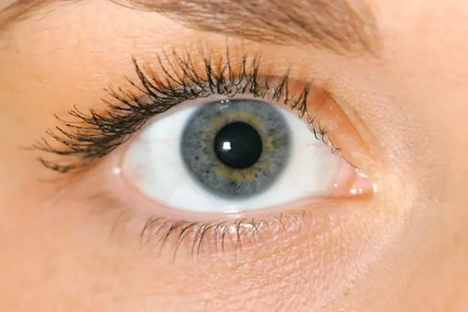
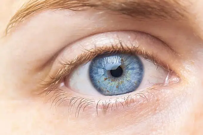
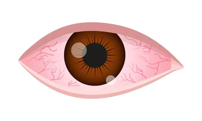
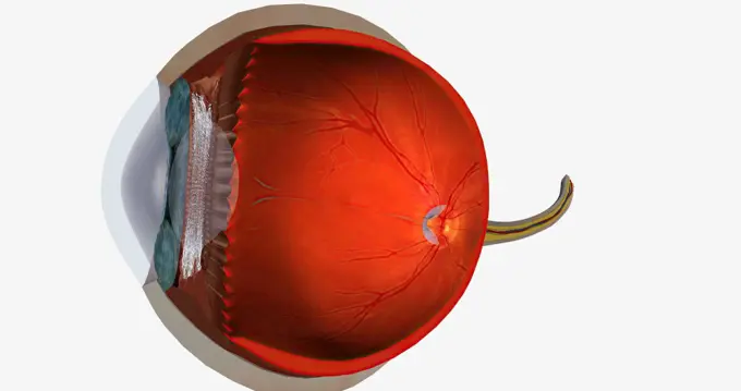
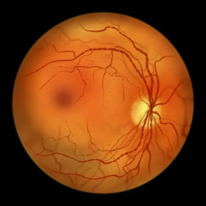
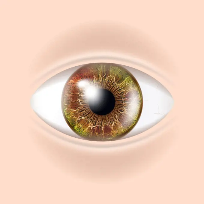
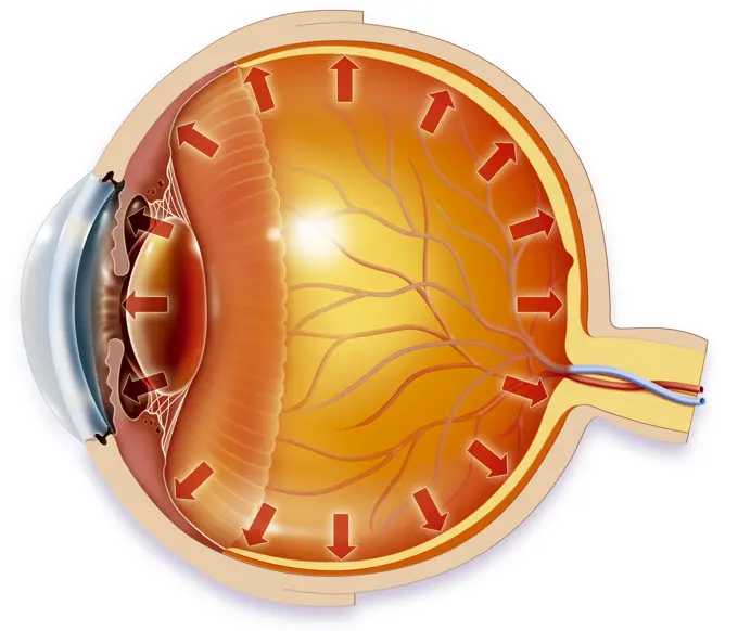
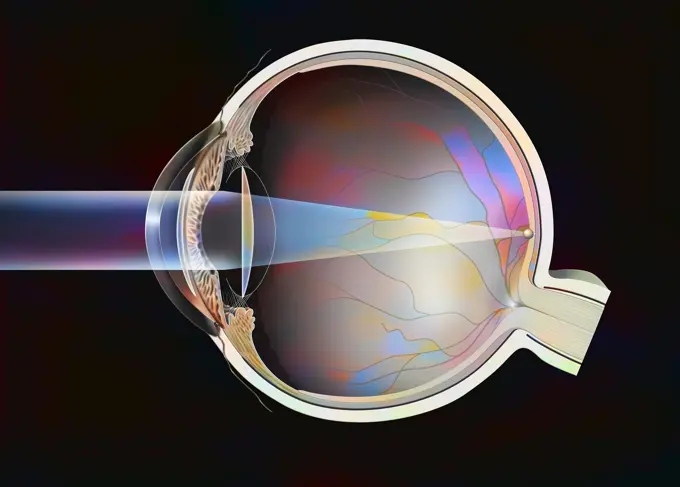
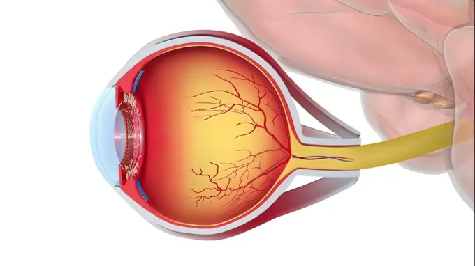
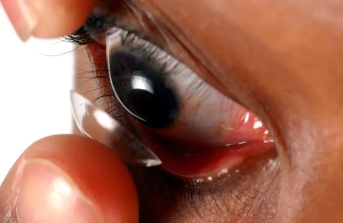
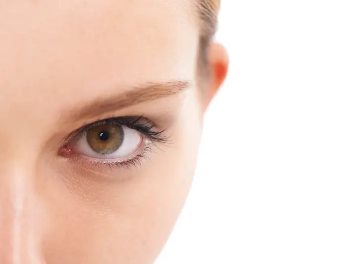
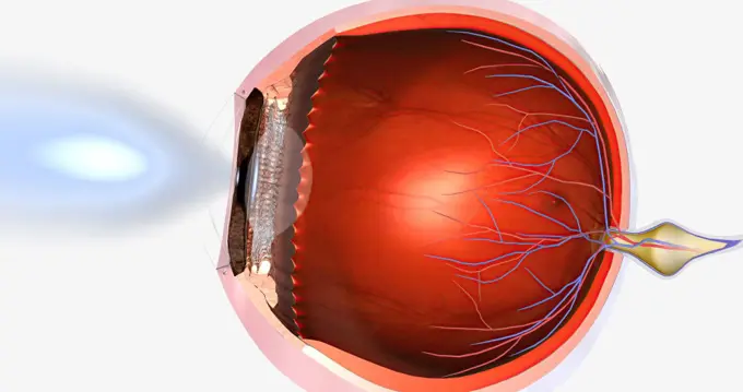
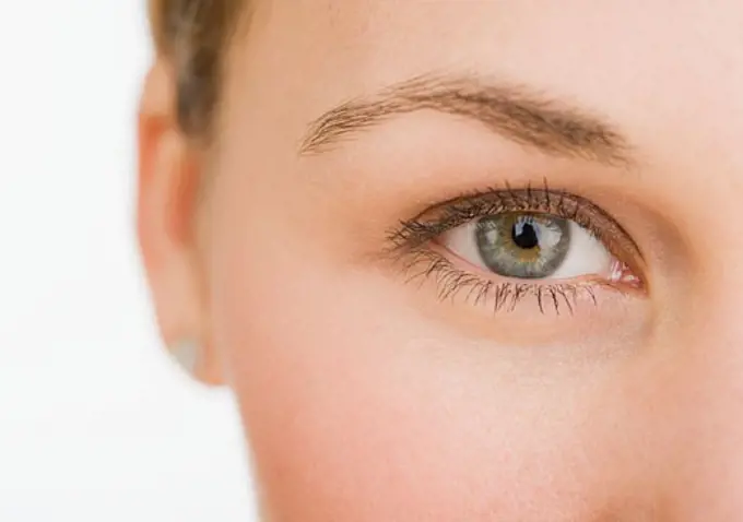
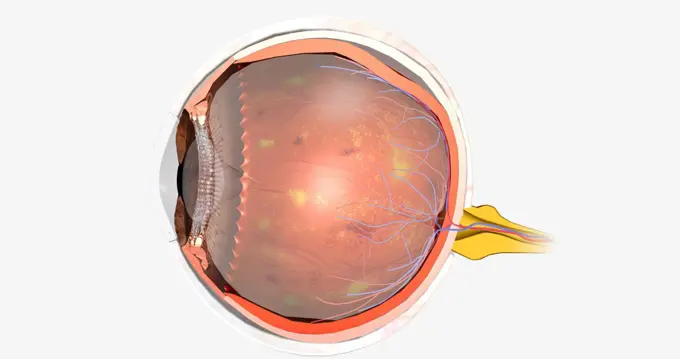
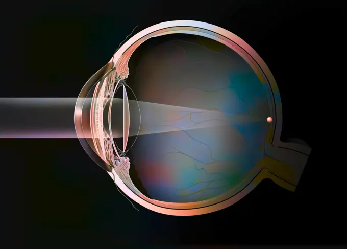
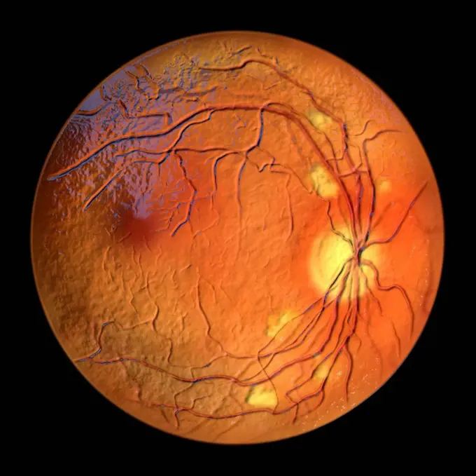
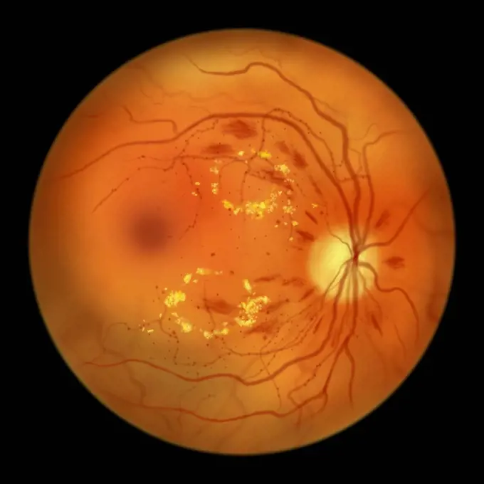
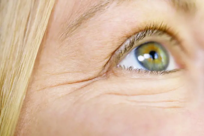
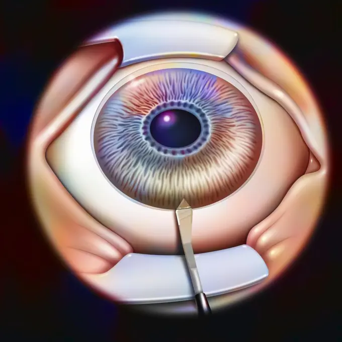
Close-Up Eye Details

Detailed close-up images of human eyes showing various colors and conditions, highlighting the uniqueness of eye features.

close up of a brown eye
327 assets in this story
1525-21748242
green eye
1525-23035412
Red eye of a woman
4286-65896
Eye
1525-23035409
Red eye of a woman
1889-48996150
Woman's Eye
1525-22442676
left blue eye of child with long eyelashes close up
1525-20704497
Close up of woman eye
1889R-53587
close up of a brown eye
1525-24650420
Red eye of a woman
4292-59561
Woman's eye
1899-53513472
Optical and surgical treatments of refractive disorders. Refractive disorders are caused by myopia, hyperopia, astigmatism, anisometropia, presbyopia, aphakia. Optical treatments consist of the use of lenses in front of the cornea. While the surgical treatment consists of correcting the curvature of the cornea either by surgery or by laser.
1848-55997670
Beautiful blue color woman eye close up
1525-56722707
Higher than normal intraocular pressure in the absence of ocular hypertension, optic nerve damage, or visual field loss 3D rendering. Higher than normal intraocular pressure in the absence of ocular hypertension, optic nerve damage, or visual field loss
5507-51526011
Dry eye syndrome. Inflamed bloodshot eyeball with swelling, irritation and red conjunctiva. Symptoms of infection, allergy, keratitis, blepharitis, conjunctivitis, uveitis
824-63165268
Sectional eye with secondary cataract treated with YAG laser.
6188-63295809
Injection at spa salon. Doctor hands in gloves. Closeup. Pretty female patient Injection at spa salon. Doctor hands in gloves. Closeup. Pretty female patient. Beauty treatment. Healthy skin procedure. Young women face. Plasmolifting rejuvenation ,model released, Symbolfoto Copyright: xZoonar.com/ElenaxSafonovax 22197638 ,model released, Symbolfoto ,property released
4269-1746
Woman putting a colored contact lens into her eye.
1525-56854519
Higher than normal intraocular pressure in the absence of ocular hypertension, optic nerve damage, or visual field loss 3D rendering. Higher than normal intraocular pressure in the absence of ocular hypertension, optic nerve damage, or visual field loss
1899-53513471
Optical and surgical treatments of refractive disorders. Refractive disorders are caused by myopia, hyperopia, astigmatism, anisometropia, presbyopia, aphakia. Optical treatments consist of the use of lenses in front of the cornea while the surgical treatment consists of correcting the curvature of the cornea either by surgery or by laser.
824-63165296
Eye, cataract: intraocular implants in anterior and posterior chamber.
6145-29259946
Trachoma grading: trachomatous inflammation - follicular
824-63195471
Illustration of combined surgery for cataracts and glaucoma. An intraoccular implant is placed in the eye to correct the cataract which is combined with glaucoma.
1525-56722640
Higher than normal intraocular pressure in the absence of ocular hypertension, optic nerve damage, or visual field loss 3D rendering. Higher than normal intraocular pressure in the absence of ocular hypertension, optic nerve damage, or visual field loss
4128-20041567
Retina damage from diabetes, illustration
1525-25669552
Human Eye Vector. Visual Examination. Body Check. Realistic Anatomy Illustration. Human Eye Vector. Sight, Eyesight. Body Care. Realistic Detail Vision Illustration
824-63180708
Illustration of the anatomy of the eye showing the trabecular meshwork located at the base of the cornea. This natural filter allows the aqueous humour to drain and maintain constant and equal pressure on the interior of the eye. In glaucoma, the blocked trabecular meshwork prevents the aqueous humour from draining correctly, which leads to intraocular pressure that can damage the optic disc and nerve.
4128-20041421
Retina damage from diabetes, illustration
1899-53509138
Eye (cataract), intraocular implants in anterior and posterior chamber.
824-63164015
Eye (cataract): intraocular implants in anterior and posterior chamber.
824-63190442
Illustration of a close-angle glaucoma : trabeculums are blocked due to the pupil which blocks the entrance. The aqueous humour does not drain normally. This leads to increased intraocular pressure, which deforms the optic disc and progressively destroys the optic nerve.
4128-30420931
Best vitelliform macular dystrophy, illustration
1899-30608852
Different intraocular implants, phakic implant (anterior, posterior chamber), iris, aphakic implant.
1525-26220630
Glaucoma is a group of eye conditions that damage the optic nerve, the health of which is vital for good vision. 3D illustration. Glaucoma is an eye condition that damages the optic nerve, which is vital for good vision.
1899-66224
Close up of young man putting contact lens onto eye
4197-63525872
Beauty model, portrait or eye makeup cosmetics with eyeshadow mascara in skincare vision, innovation or ideas. Zoom, macro or woman face with lashes on white background studio in microblading mock up
1525-56854560
Myopia, is a common visual condition in which the eye s ability to focus on distant objects is impaired. 3D rendering. Myopia, is a common visual condition in which the eye s ability to focus on distant objects is impaired.
1899-30608858
Different possible corrections of a hyperopic eye, spectacle lenses - external lenses.
1795R-13787
Extreme close up of woman's eye
824-63165229
Eye, glaucoma: examination of a fundus with the result.
4128-20041539
Retina damage from diabetes, illustration
824-63180614
Illustration of laser eye surgery, used in various pathologies such a short-sightedness, presbyopia etc.
1525-56722711
Diabetic retinopathy is a complication of diabetes that affects the eyes. 3D rendering. Diabetic retinopathy is a complication of diabetes that affects the eyes.
824-63165144
Different intraocular implants: phakic implant (anterior, posterior chamber), iris, aphakic implant.
1525-23843192
Low-tension or normal-pressure glaucoma. In normal-tension glaucoma the optic nerve is damaged even though the pressure in the eye is not very high. 3d illustration. 3D Medical Animated Normal Tension Glaucoma on Blue Background
1525-26220627
Glaucoma is a group of eye conditions that damage the optic nerve, the health of which is vital for good vision. 3D illustration. Glaucoma is an eye condition that damages the optic nerve, which is vital for good vision.
4197-V65364052
4128-30420934
Best vitelliform macular dystrophy, illustration
1525-26182457
Glaucoma eye disease 3D illustration medical .. Glaucoma eye disease 3D illustration medical
824-63190441
Illustration of open-angle glaucoma : the trabeculums are blocked and dont allow the aqueous humour to drain normally. This leads to increased intraocular pressure, which deforms the optic disc and progressively destroys the optic nerve.
1525-56854507
Higher than normal intraocular pressure in the absence of ocular hypertension, optic nerve damage, or visual field loss 3D rendering. Higher than normal intraocular pressure in the absence of ocular hypertension, optic nerve damage, or visual field loss
1525-22623125
Eye Iris Realistic Vector. Anatomy Concept Illustration. Eye Iris Vector. Vision Medical Concept Illustration
4128-20041414
Retina damage from diabetes, illustration
4128-30420928
Best vitelliform macular dystrophy, illustration
1525-26220629
Glaucoma is a group of eye conditions that damage the optic nerve, the health of which is vital for good vision. 3D illustration. Glaucoma is an eye condition that damages the optic nerve, which is vital for good vision.
1525-22623222
Human Eye Iris Close Up Vector. Healthy Medical Concept Illustration. Human Eye Iris Vector. 3D Realistic Eyeball Illustration
1525-56854457
Higher than normal intraocular pressure in the absence of ocular hypertension, optic nerve damage, or visual field loss 3D rendering. Higher than normal intraocular pressure in the absence of ocular hypertension, optic nerve damage, or visual field loss
4269-25471
ARMD. Eye angiography of an Age-Related Macular Degeneration (ARMD).
824-63208864
Illustration of glaucoma treatment using ultrasound. The ultrasound beams enable the ciliary body, the gland which produces aqueous humour, to coagulate, reducing the secretion long-term. The optic nerve is no longer under pressure, sight is maintained.
824-63165198
Open-angle glaucoma: the trabeculae are blocked and no longer allow the aqueous humor to drain normally.
1525-56854375
Diabetic retinopathy is a complication of diabetes that affects the eyes. 3D rendering. Diabetic retinopathy is a complication of diabetes that affects the eyes.
1525-56854253
Keratoconus is an eye disease that affects the structure of the cornea. 3D rendering. Keratoconus is an eye disease that affects the structure of the cornea.
4269-6022
Corneal graft. Stitches are seen on the transplanted cornea.
824-63193773
Illustration of a healthy eye (left) and an eye affected by diabetic retinopathy (right).
1899-30609097
Eye (secondary cataract), clouding of the posterior lens capsule.
6145-29257696
Trachoma WHO grade: corneal opacity (CO)
4128-20041490
Retina damage from diabetes, illustration
4128-20041441
Retina damage from diabetes, illustration
4128-20041361
Retina damage from diabetes, illustration
1899-30609089
Eye, glaucoma, examination of a fundus with the result.
4128-20041378
Retina damage from diabetes, illustration
4269-27056
Diabetic retinopathy. Fundus camera image (ophthalmoscope) of the retina showing acute non-proliferative diabetic retinopathy.
824-63165230
Eye, glaucoma: examination of a fundus with the result.
824-63165249
Eye (secondary cataract): clouding of the posterior lens capsule.
4128-20041549
Retina damage from diabetes, illustration
1525-23035410
Red eye of a woman
824-63163812
Eye: position and size of soft lenses.
1525-25223088
beautiful human eye and contact lens isolated
4128-20041559
Retina damage from diabetes, illustration
1899-30609107
Eye, clouding of the lens in case of posterior subcapsular cataract.
1525-56854447
Higher than normal intraocular pressure in the absence of ocular hypertension, optic nerve damage, or visual field loss 3D rendering. Higher than normal intraocular pressure in the absence of ocular hypertension, optic nerve damage, or visual field loss
4128-20041351
Retina damage from diabetes, illustration
824-63180705
Illustration of the anatomy of the eye showing the trabecular meshwork located at the base of the cornea. This natural filter allows the aqueous humour to drain and maintain constant and equal pressure on the interior of the eye. In glaucoma, the blocked trabecular meshwork prevents the aqueous humour from draining correctly, which leads to intraocular pressure that can damage the optic disc and nerve.
4128-20041304
Retina damage from diabetes, illustration
4269-27057
Diabetic retinopathy. Fundus camera image (ophthalmoscope) of the retina showing acute proliferative diabetic retinopathy.
4128-20041505
Retina damage from diabetes, illustration
824-63178731
Angiography exam of the left eye. Small irritation (here clear area) situated just in the middle of the macula (Here dark area) creating for the patient a discomfort in reading. This irritation can be due to big stress and should disappear in some weeks
4128-20041374
Retina damage from diabetes, illustration
1795-16462591
Close up of woman's eye
4128-20041807
Healthy retina,
1525-23239200
Close up old women blue eye
1899-30608866
Intraocular implant, step 1, a small incision is made in the cornea.
1848-61083207
Woman's eye injured due to rupture of capillary, causing hematoma or bruising. It could also be conjunctivitis or other allergic eye inflammation
824-63164169
Eye, intraocular implant, step 1: small incision in the cornea.
4128-20041507
Retina damage from diabetes, illustration
4128-20041475
Retina damage from diabetes, illustration
4239-18641897
Astigmatism and correction with corrective lens.
4239-18641723
Medical illustration of conjunctivitis in the human eye.
824-63165278
Eye: clouding of the lens in case of posterior subcapsular cataract.
1899-53508243
Fundus of a patient with macular edema.
824-63165254
Eye with secondary cataract treated with YAG laser.
PREVIOUS
of 4
NEXT
2401 S. Ervay, Suite 206
Dallas, Texas 75215
United States
Get Started
Free ResearchMy BoardsMy Cart
For Creators
How To License Your ContentContributor PortalFrame of Mind
Resources
API accessPricing
Contact
+1 866 236 0087help@viewpictures.co.uk Contact form
©2026 View Pictures. All Rights Reserved. -B
This site is protected by reCAPTCHA and Google Privacy Policy and Terms of Service apply.