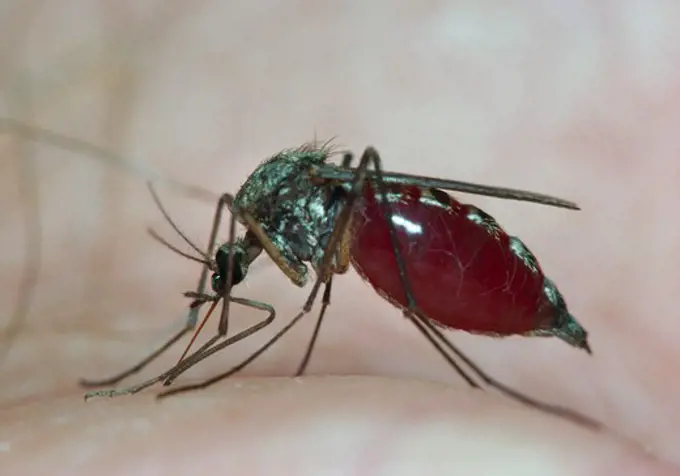
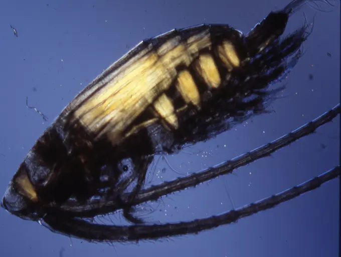
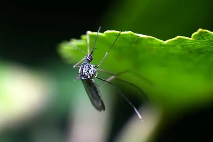
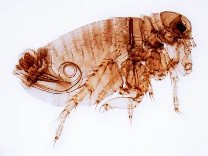
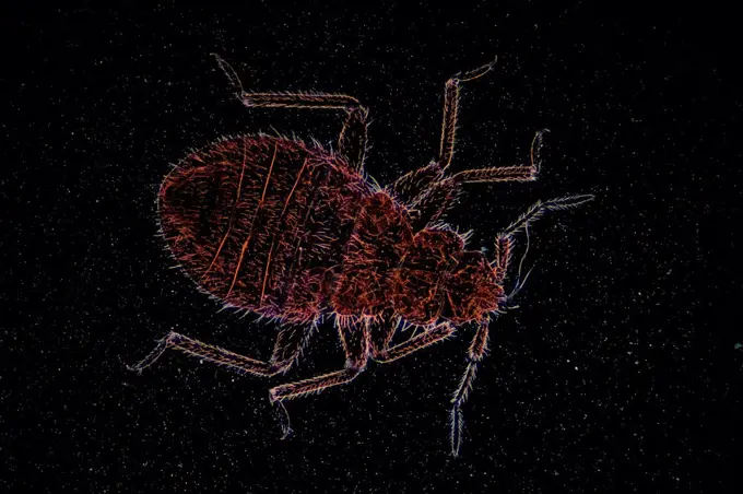
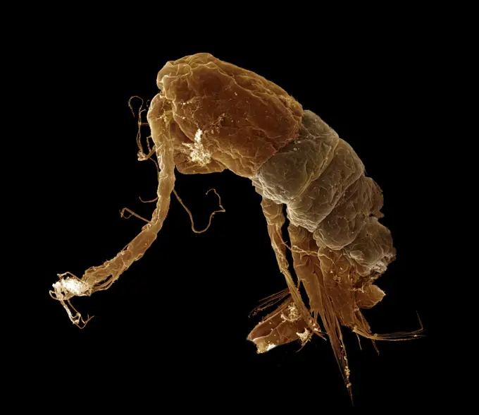
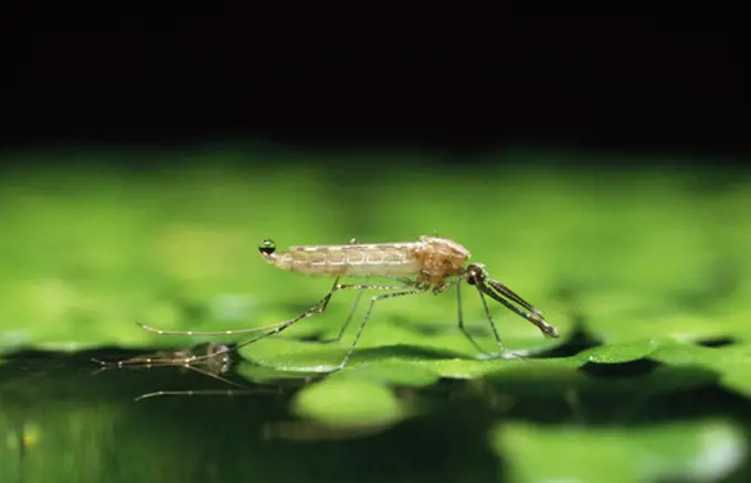
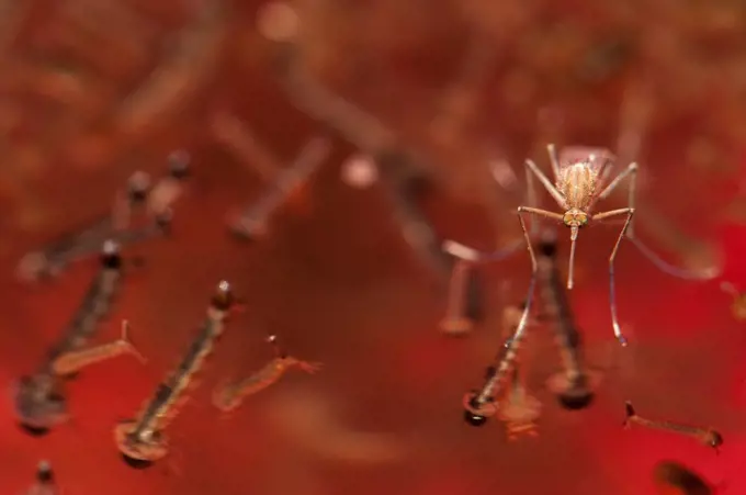
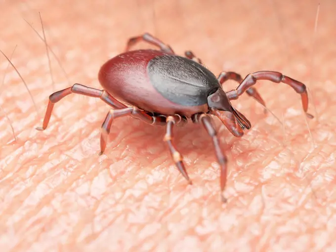
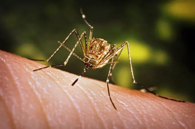
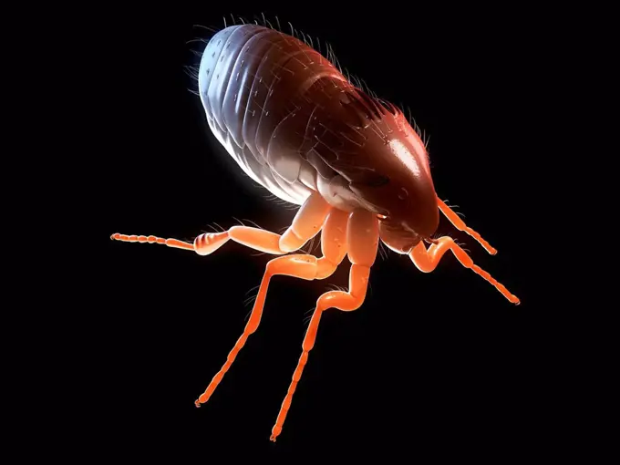
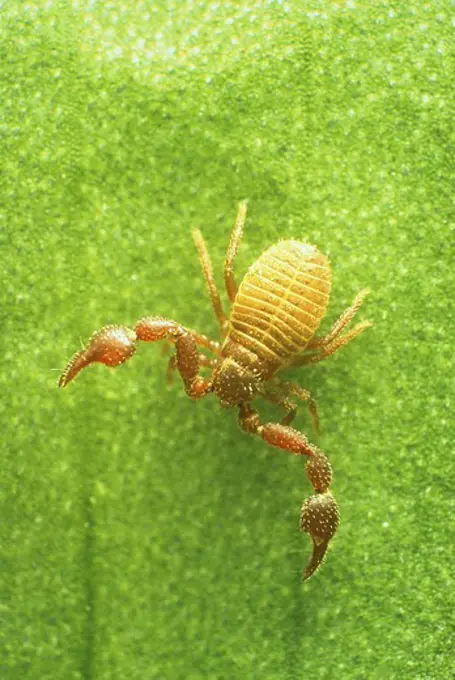
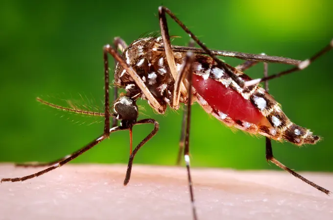
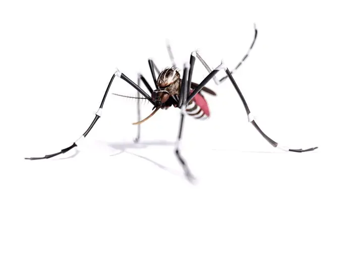
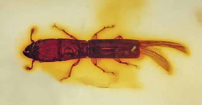
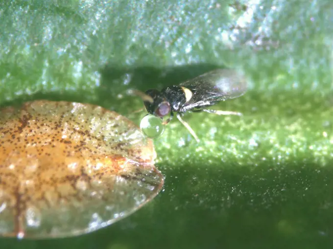
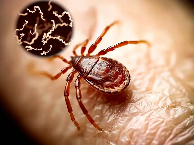
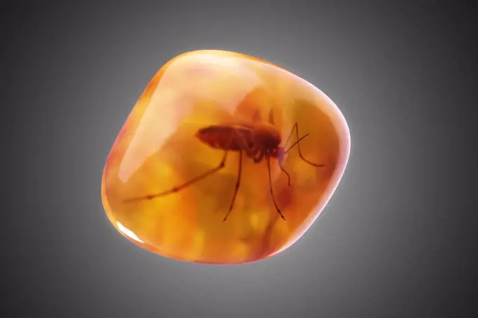
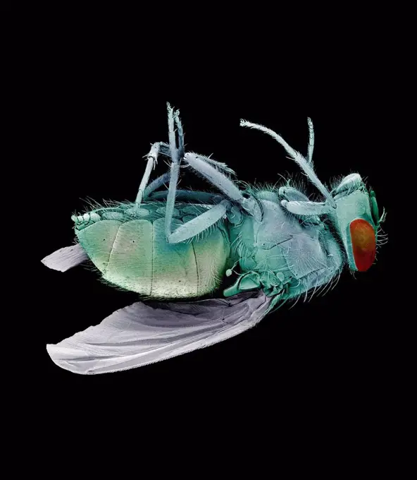
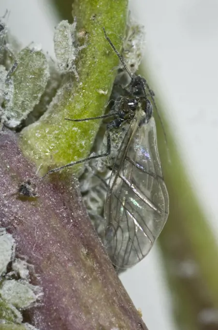
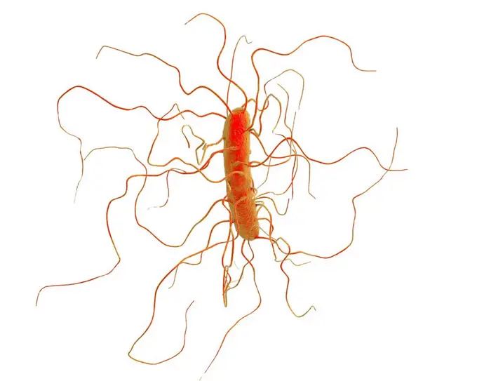
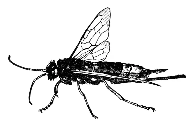
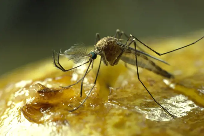
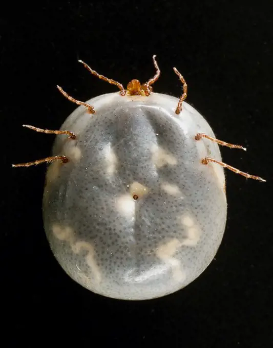
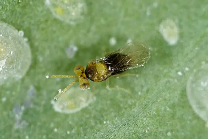
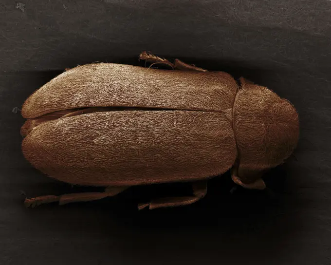
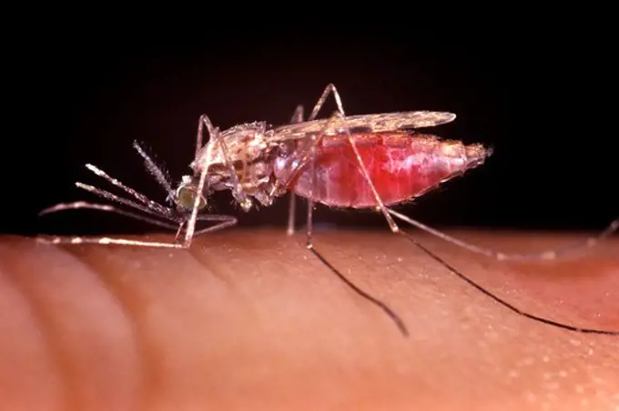
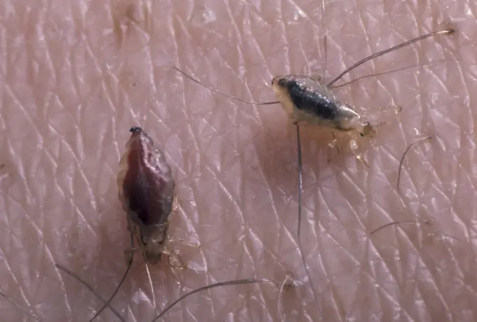
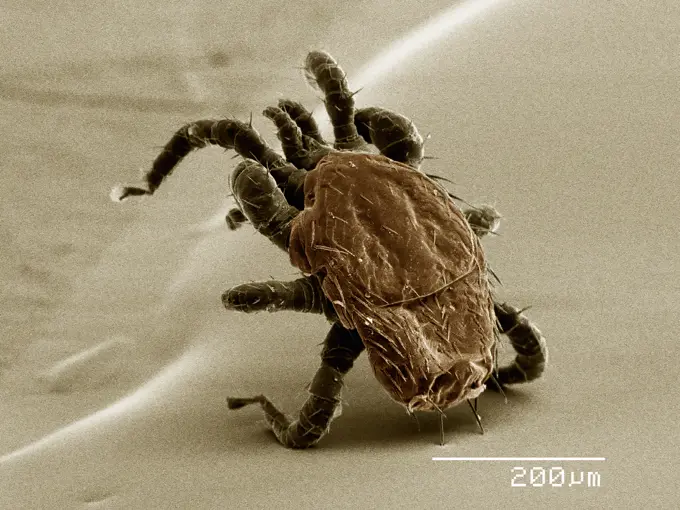
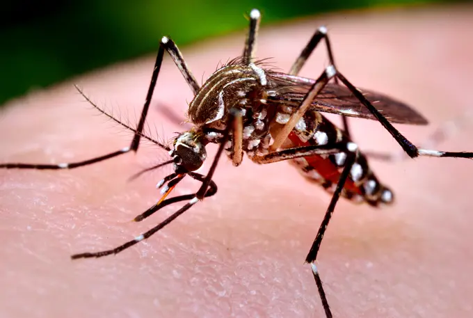
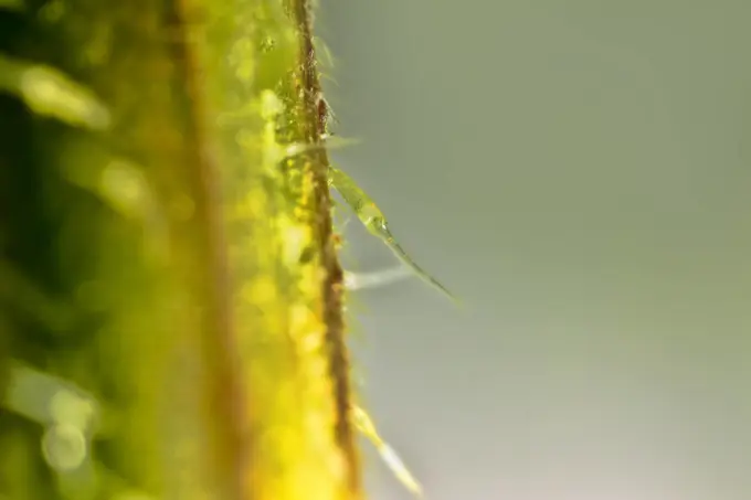
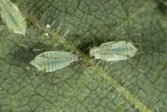
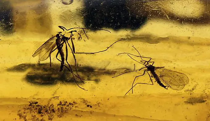
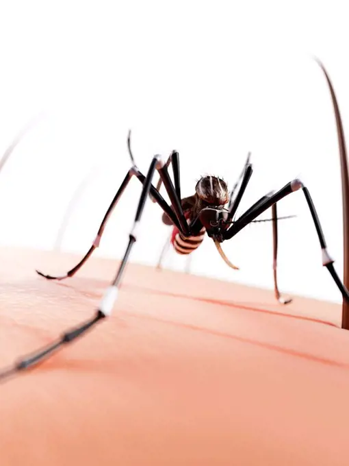
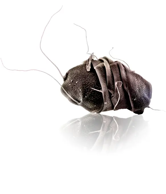
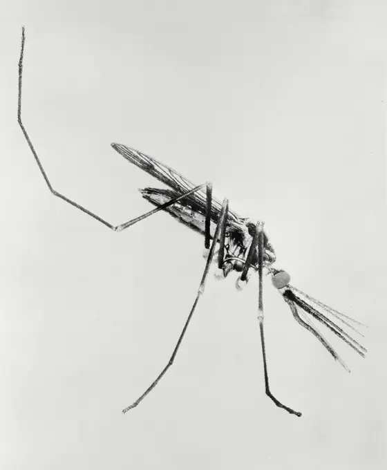
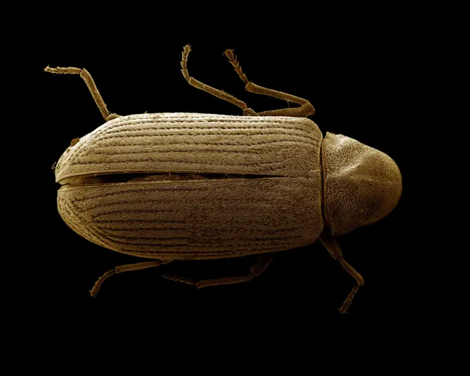
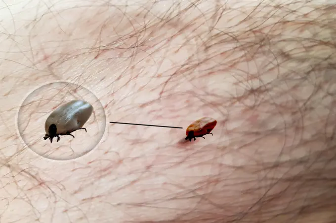
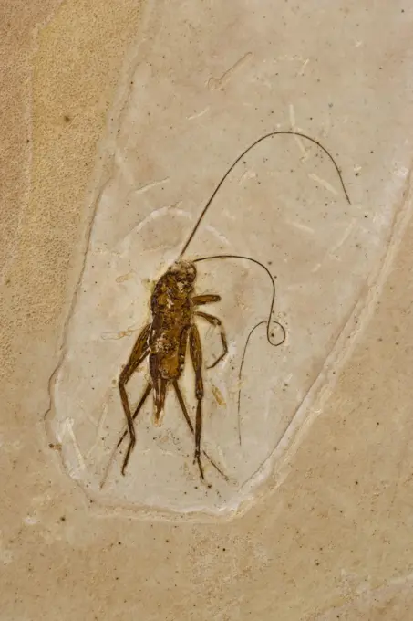
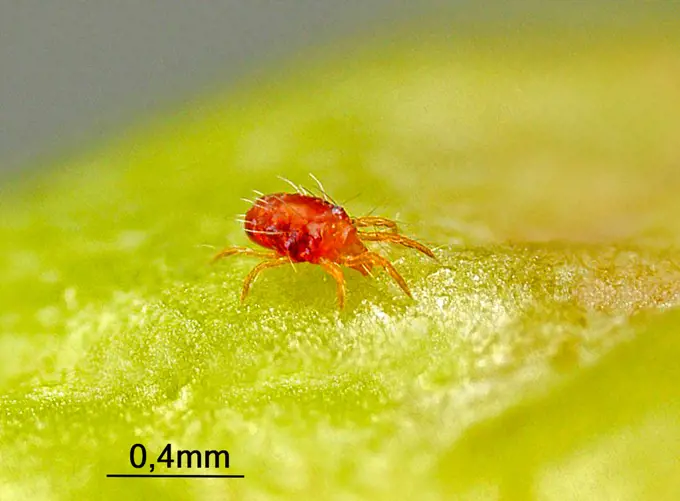
My Boards
View Pictures
  • My Boards

Close-ups of Insects and Vectors

Detailed images of various insects under magnification, including mosquitoes and bedbugs, showcasing their anatomy and significance in research.

A close up view of the Cimex Lectularius or bedbug.
120 assets in this story
4413-28224
Biting Midge vector of the Blue tongue disease France
1439-57949074
Extreme closeup of an insect under electron microscope
4378-1217
A close up view of the Cimex Lectularius or bedbug.
1848-49594338
Scuttle Fly, Phoridae in Amber, Colombia 10 MYO Pliocene
4297-1580
An Aedes aegypti female mosquito with a blood meal visible through her abdomen. After having filled with blood, the abdomen becomes distended, stretching the exterior exoskeletal surface, causing it to become transparent
4141-50140
gall gnat fossil length 1.5mm, between 27 & 38 million years old, in, baltic amber also preserved in amber, are patterns caused by wings as gnat >
1899-18906650
Male of Tachinaephagus zealandicus ASHMEAD (Hymenoptera Encyrtidae) under intense magnification.
4128R-15515358
Illustration of a tick on skin
4141-6796
mosquito biting human culex sp. michigan, usa.
6204-56614285
Rose Aphid mummified by parasitic Chalcid Wasp, Oxon UK
4141-49875
fruit fly male, drosophila melanogaster, genetic variation ebony body
4141-16176
bedbug view under microscope showing head, mouthparts, legs & abdominal segementation
6188-55645592
Small cancer in salt water Small cancer in salt water
1848-69294605
Blood-sucking mosquito (Culicidae), epizoic parasite
4128R-13447202
Human flea. Light micrograph (LM) of a male human flea (Pulex irratans). Fleas are wingless and flattened from side to side, which makes them difficul...
1525-24460464
Scanning electron micrograph of a tree hopper
4413-89073
Common Bed bugin light microscopy
4220-21334537
Scanning Electron Micrograph (SEM): Calanus sp..     Date:
4141-50066
mosquito newly emerged, anopheles stephansi, (note mecomium droplet) sequence
4413-113045
Freshly emerged from pupa newborn female mosquito
4413-9718
Planktonique stage of a Lobster larva
4201-66112
Fruit Fly (Drosophila melanogaster) caught by Cape Sundew (Drosera capensis), native to South Africa
6188-55645467
Schweinelaus as a parasite under the microscope 50x Schweinelaus as a parasite under the microscope 50x copyright: xzoonar.com/DR.XNORBERBERTXLANGEX 13865923
1525R-176916
4128R-15515182
Illustration of a dust mite
4128R-15537296
Illustration of a tick crawling on human skin
4384-195
This close-up view of a Culex tarsalis mosquito resting on human skin, shows the dark-scaled proboscis which has a broad median white band.  Other identifying characteristics include white scales around the joints of its tarsi, a line of white scales along the tibia and femur, and two silver dots on its scutum. The epidemiologic importance of C. tarsalis lies in its ability to spread Western Equine Encephalitis (WEE), St. Louis Encephalitis (SLE), and California Encephalitis, and is currently th
4269-5514
Photograph of a body louse Pediculus humanus.
4128R-15701
Cat flea Ctenocephalides felis, computer artwork.
1848-68972704
Varroa mite - on dead honey bees (Varroa destructor)
824-63185947
This 2006 photograph depicted a dorsal view of a male body louse, Pediculus humanus var. corporis. Some of the external morphologic features displayed by members of the genus Pediculus include an elongated abdominal region without any processes, and three pairs of legs, all equal in length and width. The distal tip of the males abdomen is rounded, whereas, the females is concave. Body lice are parasitic insects that live on the body, and in the clothing or bedding of infested humans. Infestation is common, found worldwide, and affects people of all races. Body lice infestations spread rapidly under crowded conditions where hygiene is poor, and there is frequent contact among people. Note the sensorial setae, or hairs that cover the louses body, which pick up, and transmit information to the insect about changes in its environment such as temperature, and chemical queues. The dark mass inside the abdomen is a previously ingested blood meal.
4413-18972325
Abyssal shrimp (Rimicaris exoculata). Ifremer. The animal lives on the Mid-Atlantic Ridge, at a depth of 3500 m, near smokers, underwater volcanoes that provide the fauna with minerals and energy.
4413-98628
Syrphid in studio
4128-111581579
Head louse, computer illustration.
4409-50098898
Madrid 10-25-2007 report for science on the Exploitation and study of the Ambar site by the Members of the Museum of Natural Sciences of Vitoria Jesus Alonso and Rafael Lopez photo Jaime Garcia Archdc in the image Insects Found in the Ambar Hematophagous mosquito ( New species).
4179-17493
Pseudoscorpion aka False Scorpion aka Book Scorpion, appr. 4x magnified
1916-111278727
Tick seen through a microscope for research into Deer tick and Lyme disease at College Park, Maryland, USA
4141-49887
fruit fly male, drosophila melanogaster, genetic var. purple cinnamon eye
4128R-13620143
Coloured scanning electron micrograph (SEM) of House fly (Musca domestica). The house fly, Musca domestica, is a well-known cosmopolitan pest of both ...
824-63194395
This photograph depicts a female Aedes aegypti mosquito while she was in the process of acquiring a blood meal from her human host, who in this instance, was actually the biomedical photographer, James Gathany, here at the Centers for Disease Control. The feeding apparatus consisted of a sharp, orange-colored fascicle that was covered in a soft, folding sheath called the labellum while not feeding. The labellum was shown here retracted as the sharp stylets contained within pierced the host's skin surface, thereby, allowing the insect to obtain its blood meal. The orange color of the fascicle was due to the red color of the blood as it migrated up the thin, sharp translucent tube. Note the distended abdominal exoskeleton, which being translucent, allowed the color of the ingested blood meal to be visible.
4128R-11283573
Mosquito, computer artwork.
1848-49594332
Bark Beetle, Colydium sp. in Amber (1/4 " long), Pliocene, Colombia 10 MYO
4413-43369
Parasitoid wasp beside a scale
4413-193476
Male parasitoid hatched gall on Fennel
4070-19450
Wild Common fruit fly (Drosophila melanogaster) Vienna Drosophila RNAi Center, Institute for Molecular Pathology, Austria  focus stacking
6188-55454269
sketch of spider on white paper training drawing in suibokuga style with watercolor paints - sketch of spider on white paper ,property released Copyright: xZoonar.com/ValeryxVoennyyx 11038825 ,model released, Symbolfoto ,property released
4269-26303
Anophele. Artwork of an Anophele, a kind of mosquito known as vector of the Plasmodium sp parasite, causing agent of malaria.
1439-57949065
Closeup of an ant under a scanning electron microscope
824-63189039
Illustration of a mosquito.
824-66066030
An Aedes mosquito and 3D renderings of several viruses this species can transmit. Credit: NIAID See related August 20, 2024 NIAID Now article, &quotWorld Mosquito Day 2024 The Metabolic Mysteries of Mosquito Metabolism,&quot at.
4128-30420850
Tick transmitting Lyme disease, illustration
4378-20013503
Mosquito in Amber
4128R-15537146
Illustration of a spider
4128R-12882
Dead bluebottle fly Calliphora vomitoria, coloured scanning electron micrograph SEM. Magnification: x10 when printed at 10 centimetres wide.
4128-16374059
Beetle, X-ray
4421-21543931
Mealy cabbage aphid, Brevicoryne brassicae, alate on a brassica stem, June
4128R-13818024
Clostridium difficile bacterium with peritrichous flagella, computer illustration. C. difficile is Gram-positive, anaerobic, spore forming, rod-shaped...
6188-55550619
Sirex gigas, vintage engraving. Sirex gigas, vintage engraved illustration. Copyright: xZoonar.com/PatrickxGuenettex 10648124
1525-27730604
Sirex gigas, vintage engraved illustration.
4201-81272
Mosquito (Aedes sp) male, feeding from pear
1848-51606067
Tetranichid spider mites (Tetranichidae) on the underside of a sycamore leaf
4409-20837023
MICROCOSMOS (1996) -Original title: MICROCOSMOS: LE PEUPLE DE L'HERBE-, directed by CLAUDE NURIDSANY and MARIE PERENNOU.
4297-1479
Ventral view of an engorged female 'lone star tick' Amblyomma americanum. An Ixodes or 'hard' tick, A. americanum is found through the southeast and south-central states, and has been shown to transmit the spirochete, Borrelia lonestari
1848-51606055
An adult parasitoid wasp, Encarsia tricolor, with larval scales of the cabbage whitefly, Aleyrodes proletella
4220-21334661
Scanning Electron Micrograph (SEM): Death Watch Beetle.     Date:
4297-1467
Female Anopheles gambiae mosquito feeding
1848-53966731
Spider imago, transparent, visible blood vessels and nerves
4491-111311792
Closeup of magnified grey head of flying insect with round convex green eyes
6056-18184281
Mosquito - 3D Illustration
4413-19309060
Head louse (Pediculus humanus) stinging a human
4220-20142227
Velvet Mite (Dinothrombium)
1916-111284730
Portrait of a Grasshoper; it has a very unusual nuclear symbol like vertex
5507-39047360
Drosophila melanogaster(Fruit Fly)
1439-57952388
A closeup sepiatoned microscopic image of a dust mite
824-63194394
This photograph depicts a female Aedes aegypti mosquito while she was in the process of acquiring a blood meal from her human host, who in this instance, was actually the biomedical photographer, James Gathany, here at the Centers for Disease Control. The feeding apparatus consisted of a sharp, orange-colored fascicle that was covered in a soft, folding sheath called the labellum while not feeding. The labellum was shown here retracted as the sharp stylets contained within pierced the host's skin surface, thereby, allowing the insect to obtain its blood meal. The orange color of the fascicle was due to the red color of the blood as it migrated up the thin, sharp translucent tube. When viewed in cross-section, the larger of the two needle-sharp stylets, known as the labrum, takes on the shape of an inverted V, and acts as a gutter, which directs the ingested host blood towards the insect's mouth.
1525-22874152
3D illustration of the liver and gallbladder with the word cancer presented for quick understanding. For medical, educational, advertising, scientific and industrial use.
6188-56037129
Stem and stinging needles of nettle Urticating hair stimulus, cnida of nettle. Visible glandular trichomes with burning acetylcholine. Nidle breaks off, fluid from retort-shaped cell enters skin. Stinging nettle Urtica dioica macro Copyright: xZoonar.com/Max5128x 20829254
1848-51649127
Stinging nettle aphid, Microlophium carnosum, infestation of a stinging nettle leaf
1848-49594347
40 MYO Male & Female Mosquitoes in Amber, Dominican Republic, Central America
4128R-11283547
Mosquito on skin, computer artwork.
4220-21334724
Scanning Electron Micrograph (SEM): Tardigrade or Water Bear.     Date:
6056-18184717
Mosquito - 3D Illustration
255-32323
Vintage Photograph. Close up of mosquito on white background
1848-50941716
Pyrrhocoridae ( Pyrrhocoridae ) in Venus flytrap ( Dionaea muscipula )
4128-16224672
Beetle, X-ray
4269-27262
Anopheles gambiae. Female Anopheles gambiae mosquito feeding on human blood. This mosquito is a vector of the parasite Plasmodium, the agent of malaria.
1525-27730607
Sirex juvencus, vintage engraved illustration.
1848-50408271
Model of a house fly (Musca domestica) magnified 50 times, Museum fuer Naturkunde, Berlin, Germany, Europe
1525-57009693
3d illustration. Halloween spiders and cobwebs.
4128R-15537294
Illustration of a tick crawling on human skin
4220-21334654
Scanning Electron Micrograph (SEM): Powder Post Beetle.     Date:
1439-57949069
Monochrome micrograph of an insect in flight
4179-15668
Brazil, Sao Paulo State, breeding of Silkworms (Bombyx mori) newly hatched
4220-21334655
Scanning Electron Micrograph (SEM): Common Furniture Beetle.     Date:
6188-55551194
Myrmeleon, vintage engraving. Myrmeleon, vintage engraved illustration. Copyright: xZoonar.com/PatrickxGuenettex 10648715
4413-193424
Caterpillar of Citrus leaf miner on Lemonleaf
6188-56030764
Tick crawls on the skin Tick crawls on the skin. Walking over the human body. The tick transmits encephalitis, Lyme disease, babesiosis and ehrlichiosis. Copyright: xZoonar.com/JFsxPicxFactory xThielemannx 12697315
4220-20060500
Cricket Fossil . Ceara - Brazil - from Santana Formation - Lower Cretaceous.
4413-20031209
Twospotted Spider Mite (Tetranychus urticae) in motion on an apple
6188-67661659
Dickfühlerweichwanze Heterotoma planicornis Dickfühlerweichwanze Heterotoma planicornis Copyright: xZoonar.com/Falkex 23083878
PREVIOUS
of 2
NEXT
2401 S. Ervay, Suite 206
Dallas, Texas 75215
United States
Get Started
Free ResearchMy BoardsMy Cart
For Creators
How To License Your ContentContributor PortalFrame of Mind
Resources
API accessPricing
Contact
+1 866 236 0087help@viewpictures.co.uk Contact form
©2026 View Pictures. All Rights Reserved. -A
This site is protected by reCAPTCHA and Google Privacy Policy and Terms of Service apply.