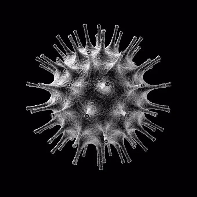
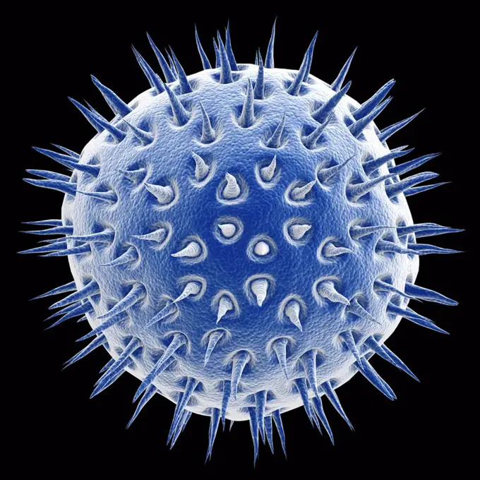
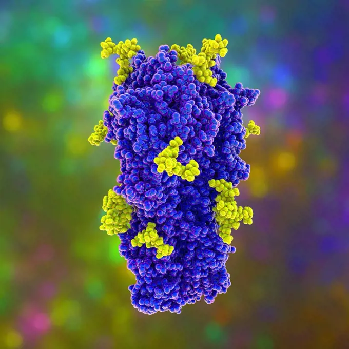
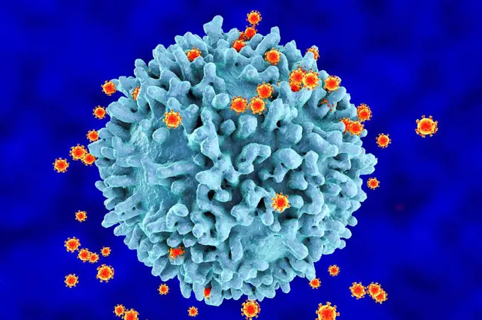
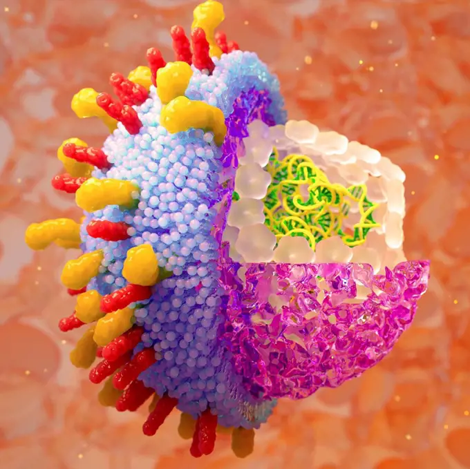
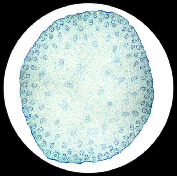
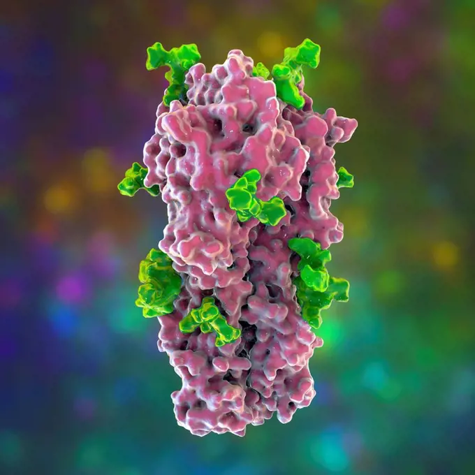
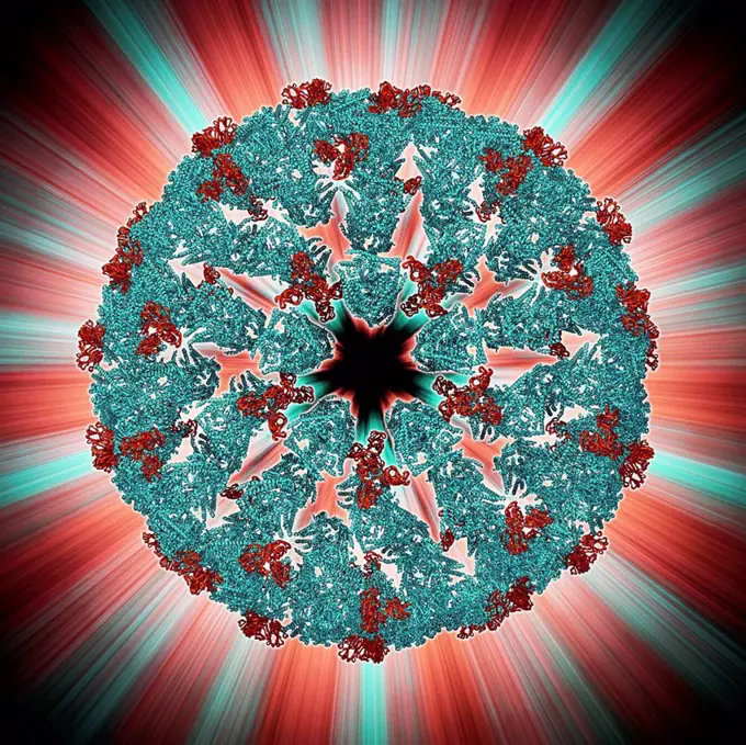
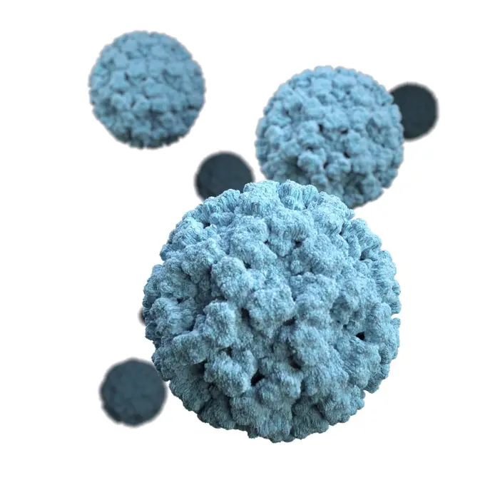
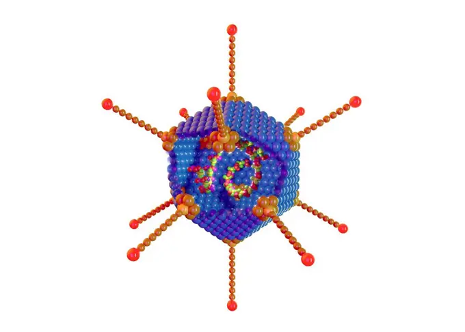
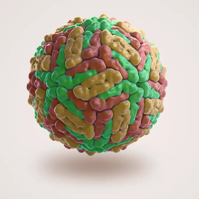
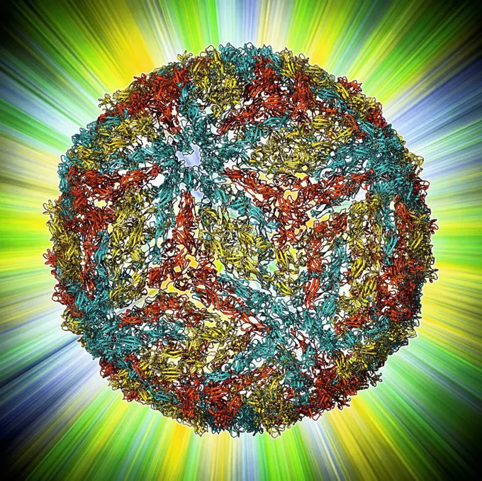
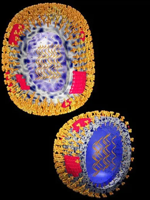
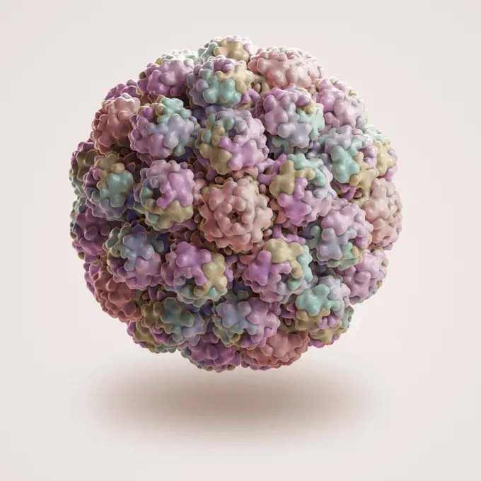
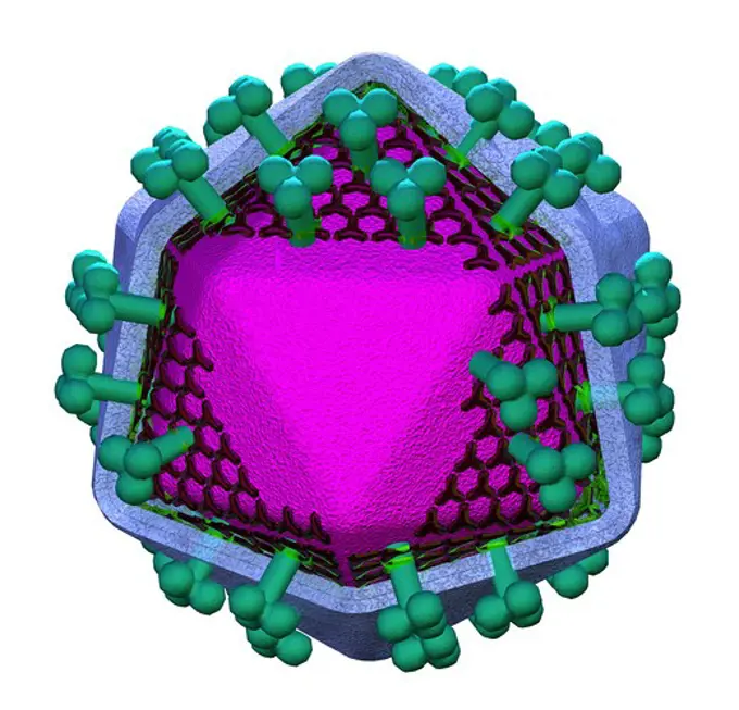
My Boards
View Pictures
  • My Boards

Colorful Virus Illustrations

High-resolution digital renderings of viruses in vibrant blues and greens, showcasing detailed structures against a black background.

Sindbis virus particle
261 assets in this story
4128R-595
Avian influenza virus, computer artwork
4128R-22341
Virus particle, computer artwork.
1899-54028001
Influenza virus (from the Orthomyxoviridae family and the Influenzavirus genus). Image made from a transmission electron microscopy view.
4378-3195
Structure of Rice Yellow Mottle Virus (PDB 1F2N). From the genus Sobemovirus RYMV is a major pathogen that dramatically reduces rice production in many African countries.
4128R-11474044
Ichnovirus, computer artwork.
824-63194377
Colorized scanning electron micrograph of filamentous Ebola virus particles (blue) budding from a chronically infected VERO E6 cell (yellow-green). Produced by the National Institute of Allergy and Infectious Diseases (NIAID), this digitally-colorized scanning electron micrograph (SEM) depicts numerous filamentous Ebola virus particles (blue) budding from a chronically-infected VERO E6 cell (yellow-green). Ebola hemorrhagic fever (Ebola HF) is one of numerous Viral Hemorrhagic Fevers. It is a severe, often fatal disease in humans and nonhuman primates (such as monkeys, gorillas, and chimpanzees). Ebola HF is caused by infection with a virus of the family Filoviridae, genus Ebolavirus. When infection occurs, symptoms usually begin abruptly. The first Ebolavirus species was discovered in 1976 in what is now the Democratic Republic of the Congo near the Ebola River. Since then, outbreaks have appeared sporadically.
4128-V58569726
4128R-21047
Virus, artwork
1815-16117096
Three dimensional render of single coronavirus cell
4128-16007392
Virus, illustration
6188-56055948
3d render of flu or virus for epidemic disease 3d render of flu or virus for epidemic disease Copyright: xZoonar.com/mtxKANGx 21159941
4128R-11543544
Virus, conceptual illustration.
4128R-11474182
Rice dwarf virus (RDF), computer artwork.
4128R-11312873
HIV virus, computer artwork.
824-63188685
Illustration of the seasonal flu virus. There are 3 types of virus that all have a nearly identical morphology : Type A and B viruses have 2 types of envelope glycoproteins or viral surface proteins (Neuraminidase et hemagglutinin). Type C virus doesnt have surface Neuraminidase. The lipid bilayer (purple) is visible, then the internal part of the envelope made up of M proteins (purple). In the centre, in green, is the ribonucleoprotein (RNP).
4128R-11284839
Monocyte white blood cell, computer artwork. Monocytes are the largest white blood cell. They engulf and digest invading bacteria and cell debris.
1525-24120859
Photorealistic model of coronavirus covid-19 mutating into new viruses isolated on white background, pandemic epidemic concept
4128-19054263
Haemagglutinin from H3N2 1968 influenza virus, illustration
4128R-11474638
Human papillomavirus (HPV), computer artwork.
1525-22197402
hi virus
4128R-15302855
Nipah virus particles, illustration
4197-63595688
Virus, bacteria and molecule structure of disease closeup in series for medical investigation or research. Covid, particle and healthcare with a microscope view of living cell samples for biology
4378-3292
The atomic structure of the Bluetongue Virus (PDB 2BTV) responsible for Bluetongue Disease or Catarrhal Fever.
4128R-13818088
MRSA bacteria. Computer artwork of methicillin-resistant Staphylococcus aureus (MRSA) bacteria.
4128-V58570349
4128R-13372947
HIV viruses infecting T-lymphocytes, computer illustration. The surface of the T-cell has a lumpy appearance with large irregular surface protrusions....
4128-111494006
Computer illustration of a varicella zoster (chickenpox) virus. The virus consists of a lipid membrane envelope (blue) with glycoproteins (red and yel...
1525-19842901
Stalk of a cereal cross-section under the microscope (Corn Stem C.S.), 40x
4128-19054261
Haemagglutinin from H3N2 1968 influenza virus, illustration
4269-21219148
A representation of the surface of the Zika virus is shown. A team led by Purdue University researchers is the first to determine the structure of the Zika virus, which reveals insights critical to the development of effective antiviral treatments and vac
4128R-11472024
Bluetongue virus capsid, molecular model. This virus, transmitted by midges, causes bluetongue disease in ruminants (commonly sheep, and less frequent...
4239-18641836
3D illustration of norovirus virions set against a white background. This artistic recreation is based upon scanning electron microscopic (SEM) imagery.
4128-111493725
Adenovirus structure, illustration. Adenovirus is a virus that can cause respiratory tract illnesses such as the common cold and bronchitis.
4378-3929
Structure of Sindbis Virus (PDB 1LD4). SINV is a member of the Togaviridae family, in the alphavirus subfamily. In humans the virus is transmitted from mosquito bites and causes sindbis fever.
4128R-13986
Mandelbulb fractal. Computer_generated image of a three_dimensional analogue derived form a Mandelbrot Set.
4128R-11473758
Authentic providence virus, computer artwork.
4128R-11472141
Flock house virus capsid, molecular model. The flock house virus is a member of the Nodaviridae family. It kills the New Zealand grass grub insect.
4128R-14846719
Egg fertilisation, illustration
4378-3538
Structure of the Dengue virus (DENV) (PDB 1K4R) the cause of dengue fever. It is a mosquito-borne single positive-stranded RNA virus of the family Flaviviridae and genus Flavivirus.
1848-50784785
Love in a mist (Nigella damascena), seed stand immature, Mediterranean region, Germany, Europe
4128-28767024
Rotavirus structure, illustration
4128R-11312841
Adenovirus, computer artwork.
4128-28575140
Mpox virus, illustration
4128R-13681991
Lymphoma cancer cell, illustration.
4128R-11471994
Bacterial nanocompartment. Molecular molecule of a nanocompartment from the bacterium Thermotoga maritima. This is a shell formed from encapsulin prot...
4128R-11471446
Dengue virus capsid, molecular model. This virus, transmitted by mosquito bites, causes the tropical disease dengue fever in humans. In viruses, the c...
824-63194382
Colorized scanning electron micrograph of filamentous Ebola virus particles (green) attached to and budding from a chronically infected VERO E6 cell (blue) (25,000x magnification). Produced by the National Institute of Allergy and Infectious Diseases (NIAID), under a magnification of 25,000X, this digitally-colorized scanning electron micrograph (SEM) depicts numerous filamentous Ebola virus particles (blue) budding from a chronically-infected VERO E6 cell ( yellow-green). Ebola hemorrhagic fever (Ebola HF) is one of numerous Viral Hemorrhagic Fevers. It is a severe, often fatal disease in humans and nonhuman primates (such as monkeys, gorillas, and chimpanzees). Ebola HF is caused by infection with a virus of the family Filoviridae, genus Ebolavirus. When infection occurs, symptoms usually begin abruptly. The first Ebolavirus species was discovered in 1976 in what is now the Democratic Republic of the Congo near the Ebola River. Since then, outbreaks have appeared sporadically.
4297R-1992
Illustration of the structure of a typical human virus
4128R-11313669
Human coxsackievirus, computer artwork.
4378-3295
Structure of the Murine Polyomavirus (PDB 1SID). Polyomaviruses have been extensively studied as tumor viruses leading to fundamental insights into carcinogenesis, DNA replication and protein processing in humans and animals.
4297-1561
Model of an HIV virion showing its major structural features, including gp120 Docking Glycoprotein, gp41 Transmembrane Glycoprotein and the Lipid Membrane
4128R-11474042
Ichnovirus, computer artwork.
4128R-13622327
Ragweed pollen (Ambrosia psilostachya), coloured scanning electron micrograph (SEM). This pollen is an allergen. Ragweed is a main cause of weed aller...
4417-16039622
The world is covered in a virus focused on North America infected.
4048-16067047
Novel Coronavirus SARS-CoV-2, causes 'coronavirus disease-2019', abbreviated as COVID-19. Scientific representation created in the form of a 3-D print. The virion surface capsid (blue) is covered with spike proteins (red) that enable the virus to enter and infect human cells. The spikes on the surface of coronaviruses give this virus family its name, 'corona', which is Latin for 'crown'. Model created at National Institute of Health, NIH  (BSLOC_2020_2_252)
1849-66221408
Close up of a radiolarian
6145-29276162
Sickle cell disease - mol. model haemoglobin
1848-50875489
Earth as a coronavirus
4128-38524184
Bacteriophage T4 structure, illustration
824-63167654
Bacteriophage. A bacteriophage is constituted of a proteic envelope (called capsid), containing its nucleic acid (DNA or RNA), and a tail. The tail includes a collar (covered with contractile proteins for the most elaborated bacteriophages, such as the T2 and T4 phages) and ending by tail fibers enabling it to attach to the bacteries it infects.
1525-27128141
Colorful diagram of Herpes simplex virus particle structure with annotations on white background. Vector illustration.. Diagram of Herpes simplex virus particle structure
PREVIOUS
of 3
NEXT
2401 S. Ervay, Suite 206
Dallas, Texas 75215
United States
Get Started
Free ResearchMy BoardsMy Cart
For Creators
How To License Your ContentContributor PortalFrame of Mind
Resources
API accessPricing
Contact
+1 866 236 0087help@viewpictures.co.uk Contact form
©2026 View Pictures. All Rights Reserved. -A
This site is protected by reCAPTCHA and Google Privacy Policy and Terms of Service apply.