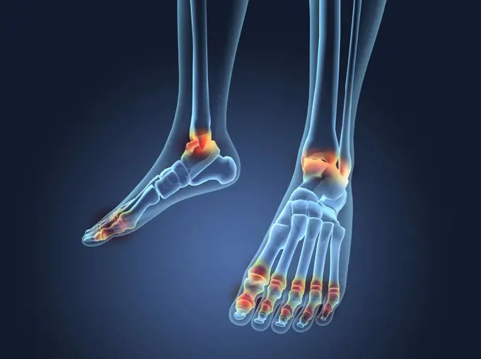
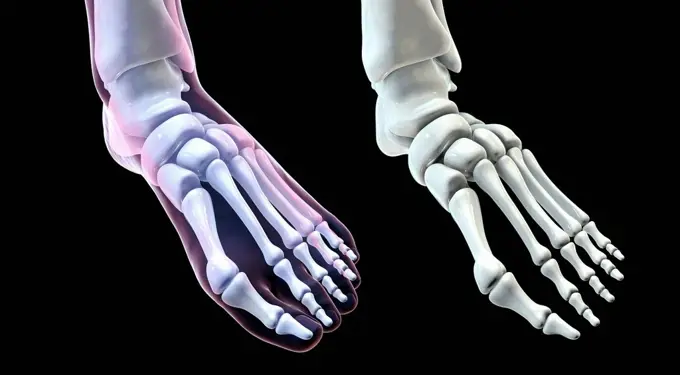
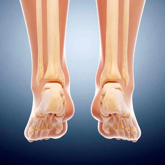
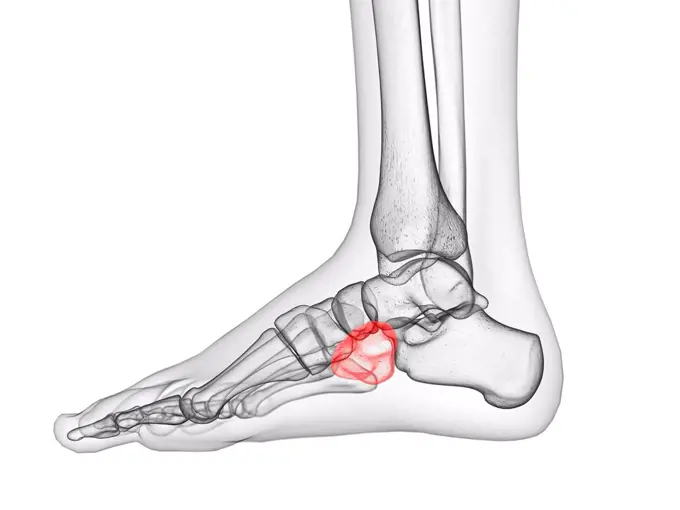
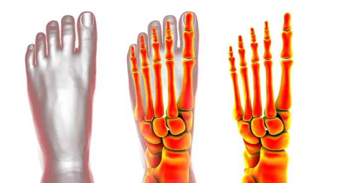
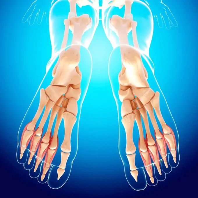
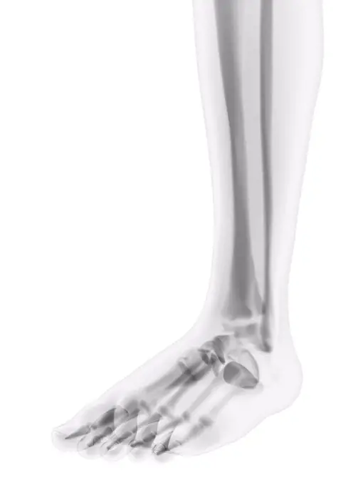
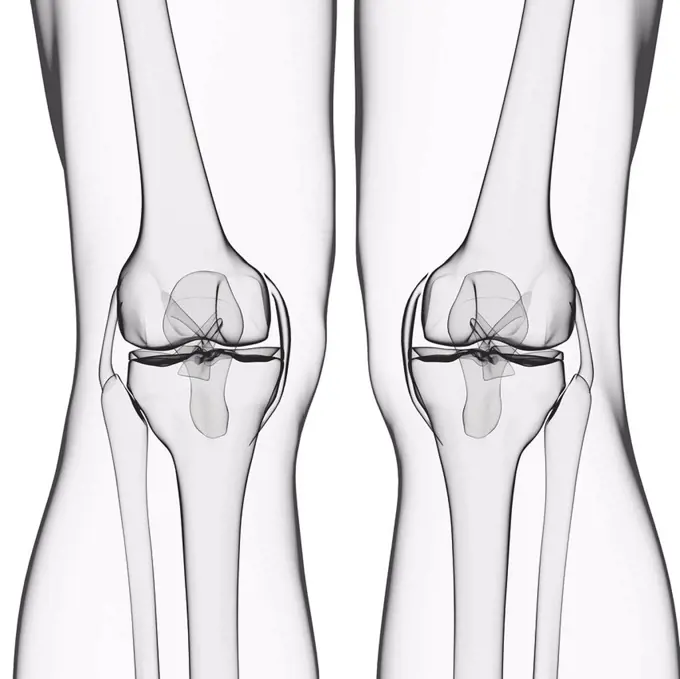
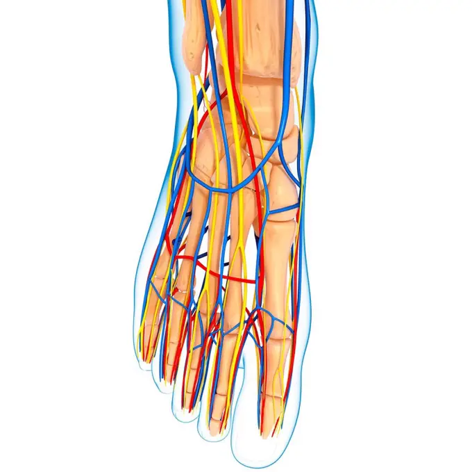
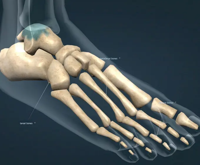
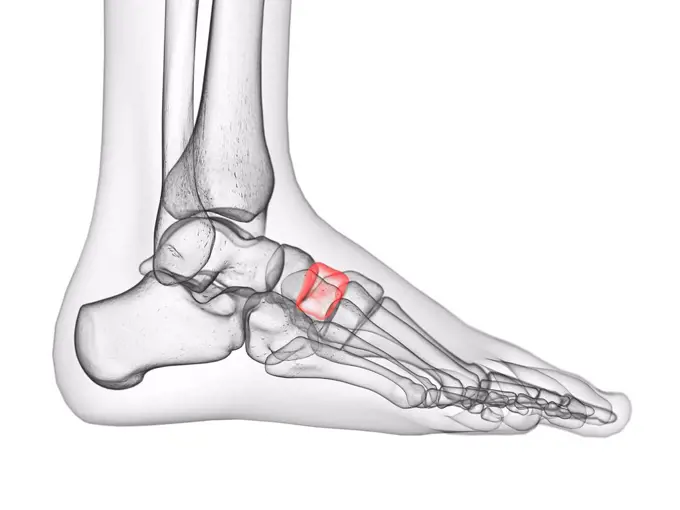
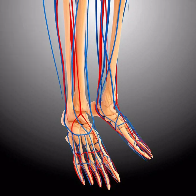
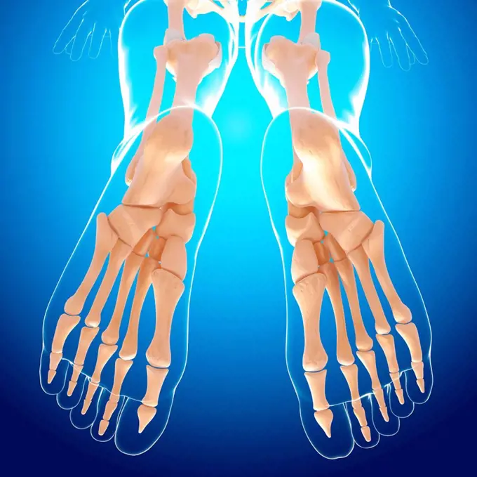
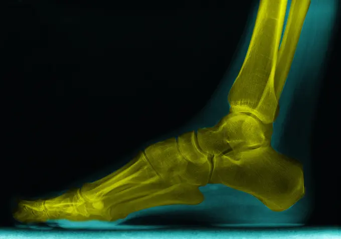
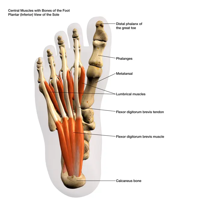
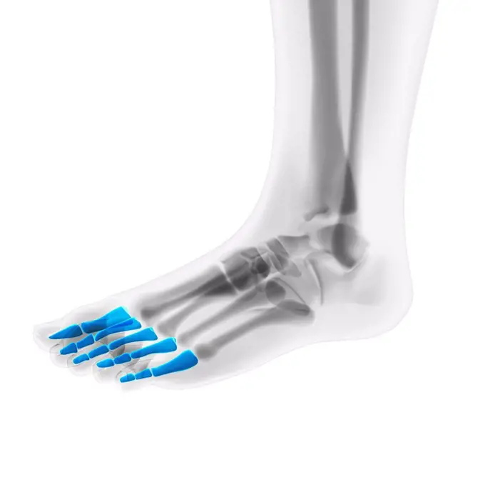
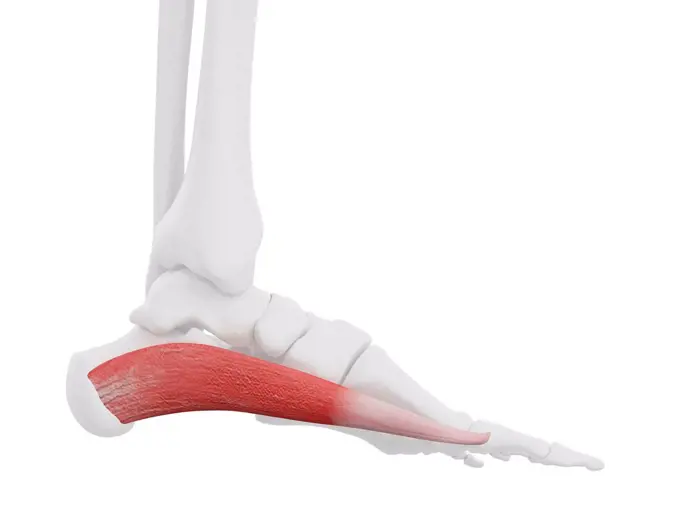
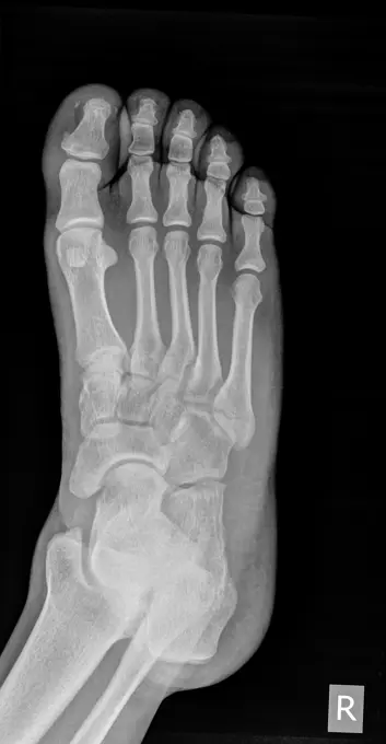
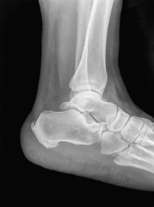
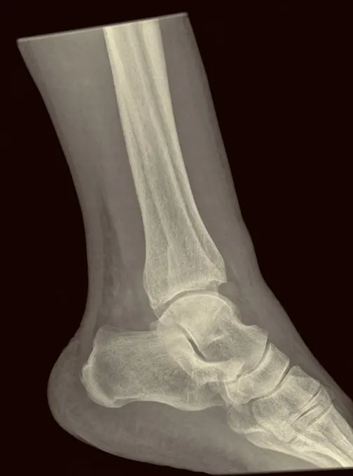
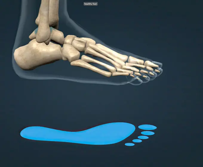
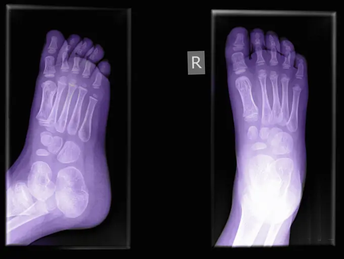
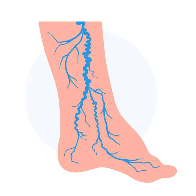
My Boards
View Pictures
  • My Boards

Foot Anatomy Illustrations

Detailed computer artworks depicting human foot bones, cardiovascular systems, and x-ray views, showcasing anatomical features and conditions.

X-ray view of inflamed foot bones
158 assets in this story
4128R-13372383
Human foot anatomy, illustration.
4128R-13385072
Human foot anatomy, illustration.
4128-15666107
Calcaneus bone, illustration
1525-23843092
Nerve endings in the toes 3d illustration. Nerve endings in the toes
4128-30418351
Lymphatics of the foot, illustration
4128R-32284
Human foot bones, computer artwork.
4128-30421491
Skeletal anatomy of the foot, illustration
4128-20041976
Bunion, illustration
4128R-12580262
Human nervous system, computer illustration.
4128R-31866
Human foot bones, computer artwork.
4128-15666137
Lateral cuneiform bone, illustration
4128-20045478
Navicular bone, illustration
4128-15666131
Cuboid bone, illustration
4128R-35040
Human foot musculature, computer artwork.
4128R-13574692
Illustration of the interosseous plantar muscle.
4128-20041908
Human foot anatomy, illustration.
6188-58135805
human bones radiography scan image human bones radiography scan image Copyright: xZoonar.com/OxanaxRishnyakx 8008131
6188-58114057
human radiography scan human radiography scan made in 3D Copyright: xZoonar.com/OxanaxRishnyakx 4219400
4128R-11316307
Human leg bones, computer artwork.
4239R-8498
Conceptual image of human foot with nervous system.
4128R-26169
Foot bones, computer artwork.
4128R-21597
Male musculature, computer artwork.
4128R-36889
Human leg bones, computer artwork.
4128R-34936
Human leg bones, computer artwork.
4128R-28240
Foot anatomy, computer artwork.
4128R-11542215
Human foot anatomy, computer illustration.
4128R-13587244
Human foot bones, illustration.
4128R-13572579
Illustration of the tibeonavicular ligament.
1525-56184101
The foot is the terminal portion of the lower limb, whose primary function is to bear weight and facilitate locomotion 3D illustration. The foot is conventionally divided into three zones. hindfoot, midfoot, and forefoot
4128R-33198
Human hand musculature, computer artwork.
4128-15666020
Intermediate cuneiform bone, illustration
4128R-23923
Lower leg anatomy, computer artwork.
4128-30422907
Bones of the foot, illustration
4128R-11476677
Human foot bones, computer artwork.
4128R-36148
Human foot musculature, computer artwork.
4128-30416923
Brain and nerves, illustration
4128R-13572633
Illustration of the middle phalanx bones.
4128-15666008
Medial cuneiform bone, illustration
4128R-13372310
Human foot anatomy, illustration.
4128R-11287718
Knitting needle through arm, coloured X-ray.
824-63126823
This color enhanced x-ray demonstrates the normal appearance of the foot. All of the bones visualized are intact. See image n8686606 on the site.
1428-67304563
Central Muscles and Bones of the Foot Labeled, 3D Rendering on white background
4128-30417335
Anatomy of the left foot, illustration
4128R-26191
Foot bones. Computer artwork showing the phalange bones.
4128-111577866
Abductor hallucis muscle, computer illustration.
1439-57940363
Xray image of a human foot revealing the intricate bone structure inside
1439-57940614
Xray image of a human ankle showing bones
4128-30421480
Blood vessels of the foot, illustration
1439-57940795
Xray of a human leg showing bones in detail
4128-30421482
Male foot, illustration
1525-56184094
Healthy foot skeleton and blue footprint 3D illustration. Healthy foot skeleton and blue footprint
1439-57939920
Xray image of a human right foot seen from the bottom
1899-66324
X-ray image of female foot
1525-26243976
Foot bones with ligaments and blood vessels 3D illustration. Foot bones with ligaments and blood vessels
1525-22347108
Close-up of a human foot
4128-20043838
Varicose veins, illustration
4128-20041924
Bunion, illustration
1899-54028625
Illustration of the bone renewal cycle. On the left, the loss of bone mass due to bone-dissolving cells (osteoclasts), and on the right the reconstruction of bone mass due to bone-building cells (osteoblasts). This cycle last 3 to 4 weeks in an adult. (Photo by: BSIP/UIG via Getty Images)
PREVIOUS
of 2
NEXT
2401 S. Ervay, Suite 206
Dallas, Texas 75215
United States
Get Started
Free ResearchMy BoardsMy Cart
For Creators
How To License Your ContentContributor PortalFrame of Mind
Resources
API accessPricing
Contact
+1 866 236 0087help@viewpictures.co.uk Contact form
©2026 View Pictures. All Rights Reserved. -A
This site is protected by reCAPTCHA and Google Privacy Policy and Terms of Service apply.