My Boards
View Pictures
  • My Boards

Histopathology Samples

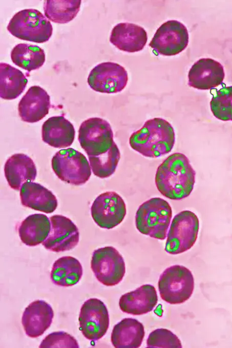
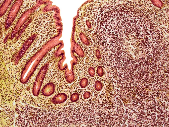
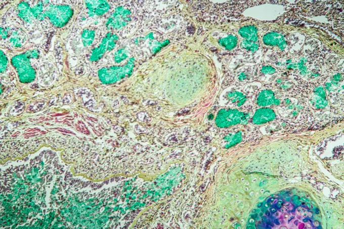
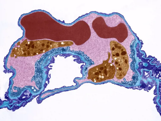
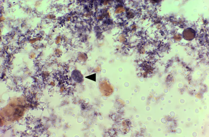
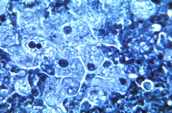
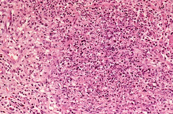
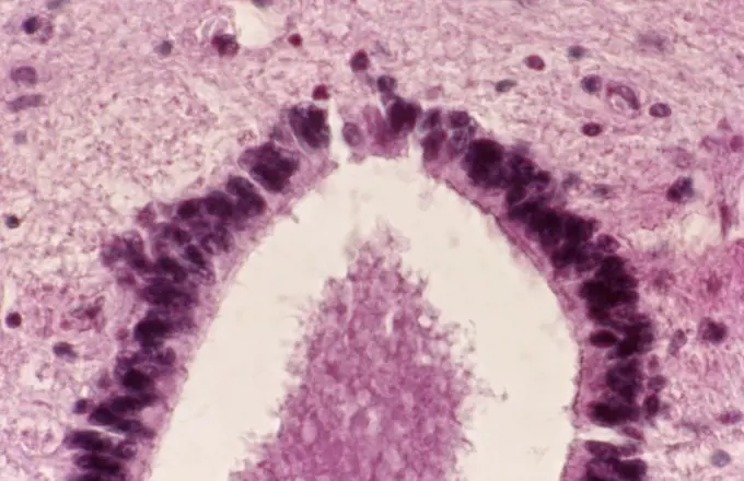
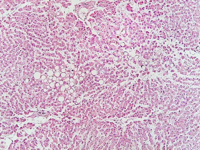
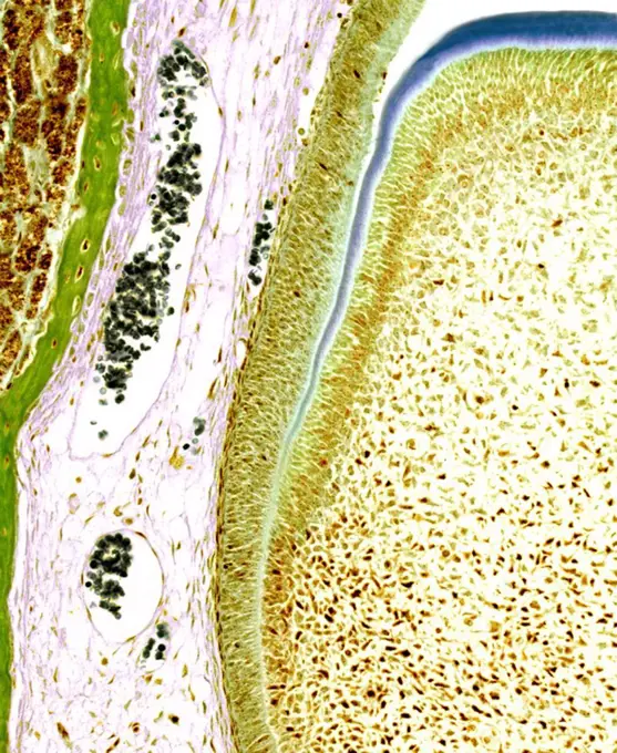
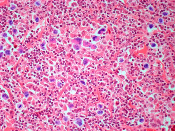
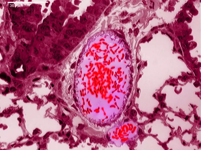
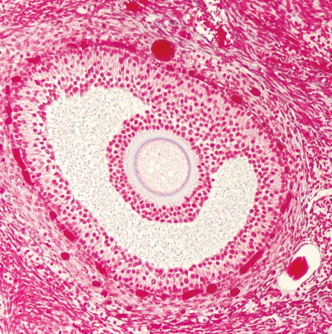
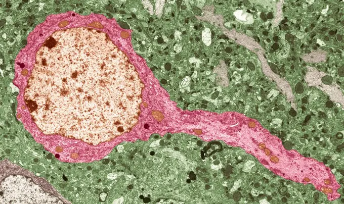
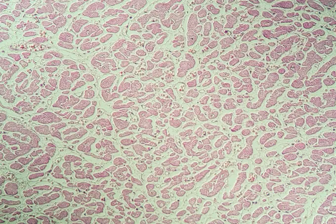
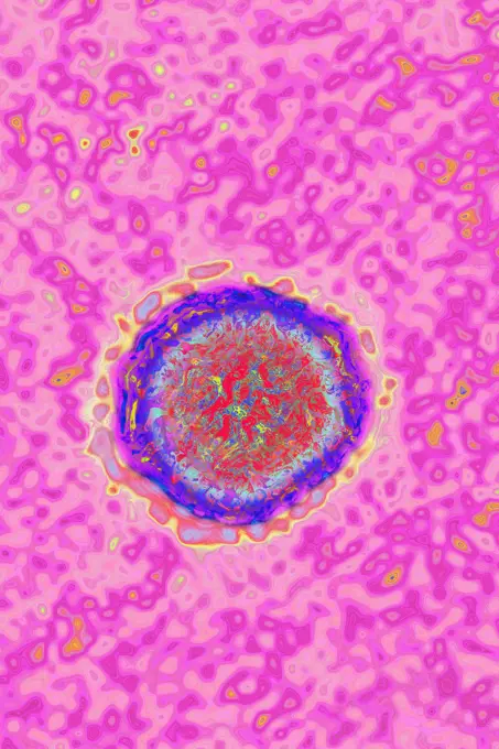
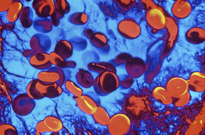
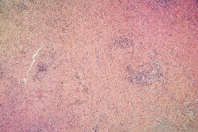
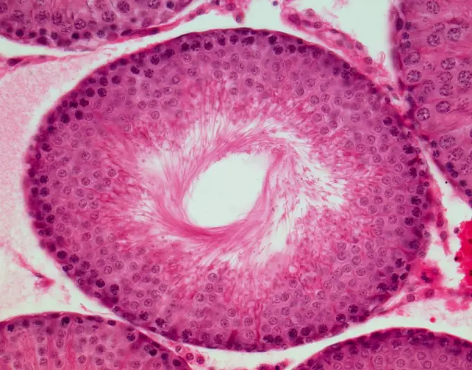
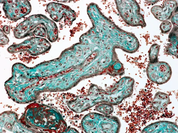
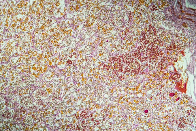
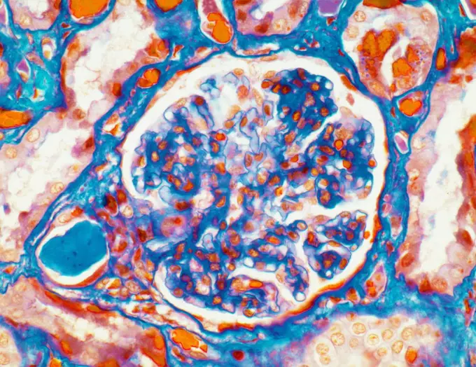
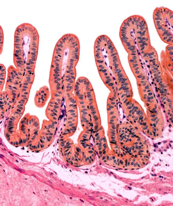
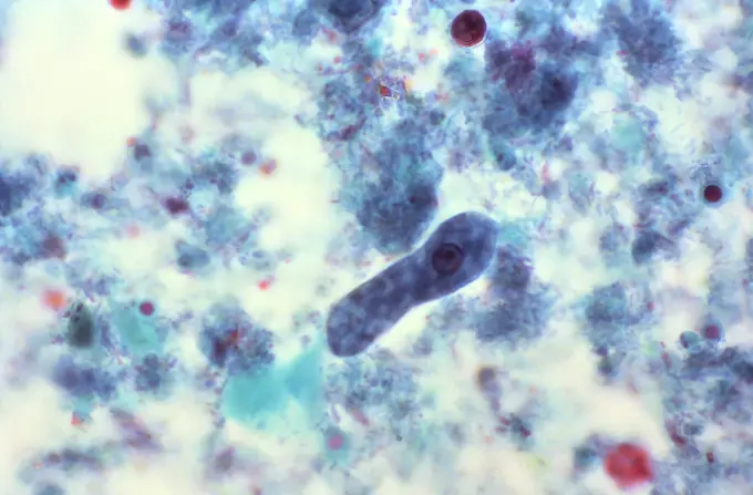
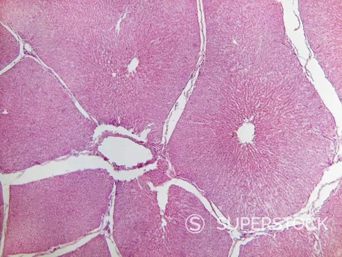
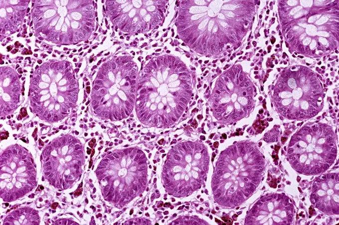
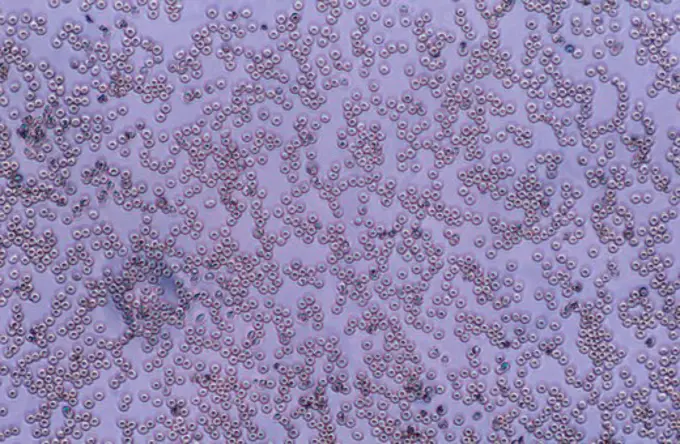
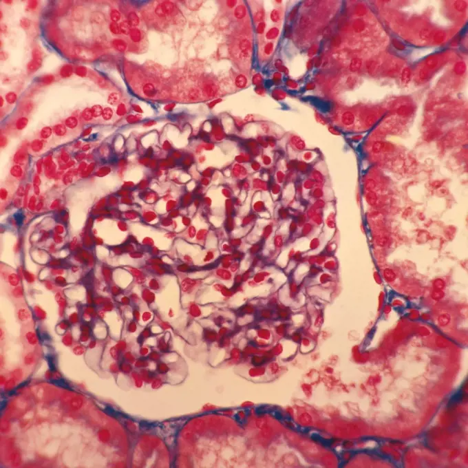
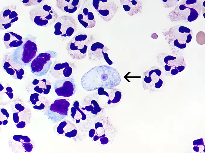
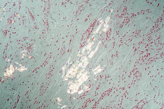
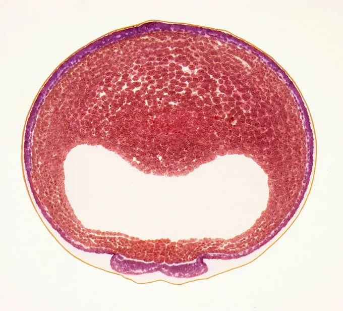
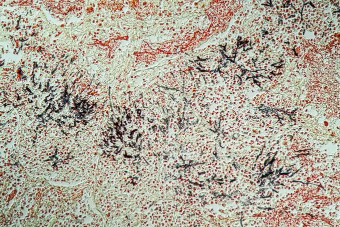
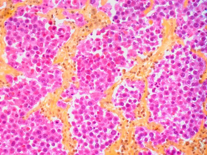
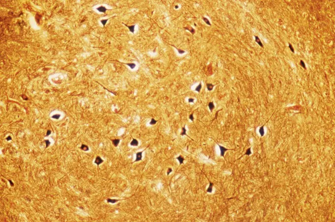
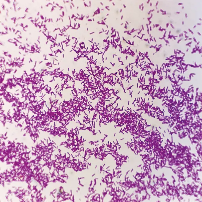
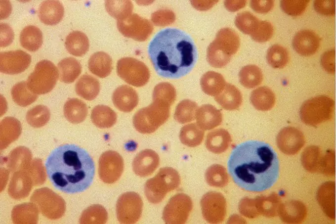
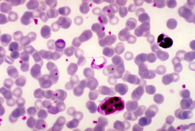
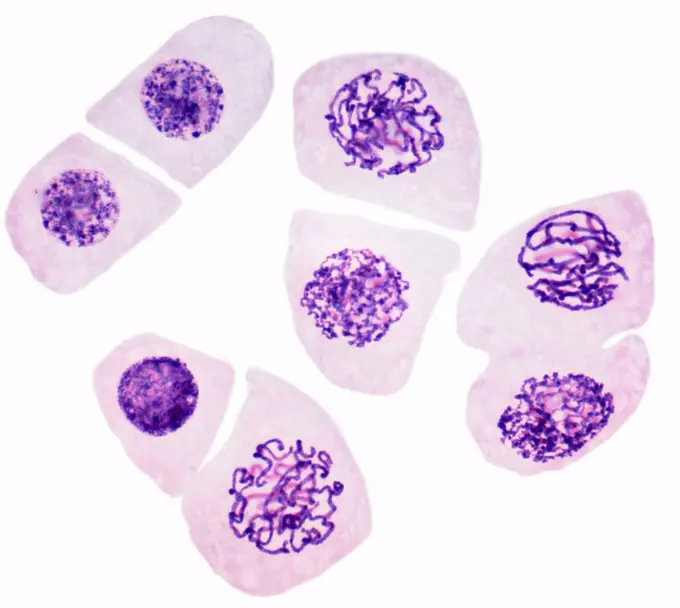
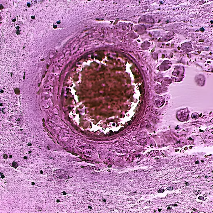
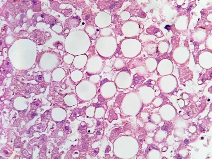
Microscopic images of tissue samples illustrating various diseases, showcasing cellular structures and pathological changes from infections and conditions.

Micrograph of human liver tissue with characteristic vacuolization resulting from fat deposits.  Magnification 400x
Micrograph of human liver tissue with characteristic vacuolization resulting from fat deposits. Magnification 400x
266 assets in this story
1788-111175563
Section of human intestinal villi seen under a microscope.
1899-53511538
Plasmodium falciparum is a malaria-causing parasite or malaria, transmitted by the bite of female anopheles which releases merozoites into the blood which enter red blood cells. Optical microscope view.
1788-111174431
Microphotograph of a section of human salivary gland (exocrine), where the epithelium is evident, with histological staining, at x220 magnification.
6188-55644266
Lung dog lung dog healthy lung tissue 200x copyright: xzoonar.com/DR.XNORBERBERTXLANGEX 13706789
4128R-8802
Appendicitis, light micrograph
6188-55644283
Tongue of the cat tongue of the cat tissue with flavor buds cross 200x copyright: xzoonar.com/dr.xnorberbertxlangex 13706815
4269-27394
Rabies virus. Transmission electron micrograph (TEM) of rabies viruses seen as numerous dark, bullet-shaped particles. This RNA virus is a Lyssavirus (Rhabdoviridae).
6188-55644501
Asthma Asthma Lungengewebe unter dem Mikroskop 100x Copyright: xZoonar.com/Dr.xNorbertxLangex 13728239
4128-17928151
Lung capillary, TEM
1899-61460870
This photomicrograph of an iron-hematoxylin stained specimen revealed the presence of an Entamoeba hartmanni trophozoite (arrow). E. hartmanni is classified as a nonpathogenic intestinal protozoan organism. CDC/Dr Mae Melvin 1977
824-57659460
Under 900X magnification, this photomicrograph revealed the presence of a number of trophozoites of the parasitic organism, Entamoeba histolytica, strain J93, in this tissue sample which was harvested from a rat and prepared using Kohn's staining method. CDC/Dr Healy 1964
6145-29280475
Lung: nocardiosis with HIV
6145-29293909
Leprosy: histopathology of skin
6188-55644625
Mozi infection of the heart Candida in the heart muscle of an AIDS patient, 200x copyright: xzoonar.com/dr.xnorbertxlangex 13747161
1788-111173440
Microphotograph of a cross section of a portion of the neural canal from the human spinal cord.
1788-111173844
Microphotograph of section of an invasive malignant tumor, carcinoma, human connective tissue.
4298-1064
Micrograph of human liver tissue with characteristic vacuolization resulting from fat deposits.  Magnification 100x
4128R-2257
Developing tooth, light micrograph. At centre are odontoblasts yellow overlaying the dental pulp. A layer of dentine blue surrounds the pulp. The oute...
6188-55643847
Blood sacrifice Red and Weoße Blood cells 400x copyright: xzoonar.com/dr.xnorberbertxlangex 13660597
1525-20843597
Stone Marble 17
4128R-23736
Hodgkin´s lymphoma. Light micrograph of a section through a lymph node affected by Hodgkin´s lymphoma. Several Reed_Sternberg cells are present, these...
1773-97837
Photomicrograph of bronchiolar epithelium tissue
1899-85800
Avian Influenza Virus TEM
1788-111177840
Corona radiata of a cat's oocyte, at x75 magnification.
4128R-8494
Nerve cell, TEM
6188-55644493
Heart attack Heart attack sick tissue under the microscope 200x copyright: xzoonar.com/DR.XNORBERBERTXLANGEX 13728228
824-63218207
Flavivirus (family Flaviviridae), these viruses are responsible for yellow fever, dengue fever, Japanese encephalitis, Zika virus, West Nile encephalitis. They are transmitted by mosquitoes or ticks. image produced from transmission electron microscopy (viral diameter approximately 40 to 60 nm).
1439R-995034
Red blood cells
6237-V70988060
6188-55643968
Heart calluses in the heart muscle 100x copyright: xzoonar.com/DR.XNORBERBERTXLANGEX 13667530
4128-18929735
Testis, LM
4128R-4236
Placenta. Light micrograph of a section through a full term placenta showing numerous chorionic villi green. Their connective tissue carries branches ...
6188-55644903
Hypophyse Hypophyse Gewebeschnitt unter dem Mikroskop 100x Copyright: xZoonar.com/Dr.xNorbertxLangex 13772968
4128-18929694
Glomerulus, LM
4128R-10347
Gall bladder lining, light micrograph
824-57659436
This photomicrograph of a trichrome-stained specimen revealed the presence of a typical Entamoeba coli trophozoite, which contained a characteristically large single nucleus with its peripherally scattered chromatin and an eccentric karyosome. Note the overall dirty appearance of the granulated cytoplasm of the amoebas. CDC/Dr Mae Melvin 1977
4128-17928266
Blood cells, light micrograph
4298-20535940
Dog liver tissue showing venules, central vein, and other hepatic structures.  Magnification 40X
1439R-1081395
Colon
1788-111174270
Microphotograph of neisseria meningitidis or meningococcus, bacteria which causes cerebro-spinal meningitis.
4141-49895
human erythrocytes, (red blood cells), displayed under microscope
4128R-22536
Uterine cancer. Light micrograph of a section through an adenocarcinoma cancer of the endometrium womb lining. Magnification: x300 when printed at 10 ...
1525-26234284
purple water painted with powder
1788-111177770
Section of renal glomerulus, at x230 magnification.
4413-19312472
Surra ; Transmitted by biting flies such as Tabanidae and Stomoxys species, as well as by vampire bats including Desmodus rotondus
1899-61460529
A fixed cerebrospinal fluid cytospin showing a Giemsa-Wright stained Naegleria fowleri trophozoite (arrow) amid polymorphonuclear leukocytes and a few lymphocytes. In the trophozoite, the nucleus and the nucleolus can be seen. Magnification: 1000x
6188-55644427
Kidney tissue with gout kidney fabric with gout tissue under the microscope 200x copyright: xzoonar.com/dr.xnorberbertxlangeex 13722693
4128R-13573039
Developing frog egg. Light micrograph of a transverse section through a developing egg laid by a common frog (Rana temporaria). The egg has reached th...
6188-55644557
Lungs of an aid patient lung of an aid patient lung tissue 200x copyright: xzoonar.com/dr.xnorbertxlangeex 13735496
1439R-995040
Thick layers of skin
4128R-22527
Testicular cancer. Light micrograph of a section through a seminoma tumour of the testis. This is a germ cell tumour and one of the most treatable and...
1848-61033427
Fake microorganisms. microbiology. Bacteria, cells, viruses, germs, microorganisms
4128-17928324
Blood cells, light micrograph
1439R-995015
Nerve cells from brain stem
1788-111174354
Microphotograph of an example of staining of tetanus bacteria (Bacillus tetani) using the Gram method, at x1250 magnification.
824-57659498
This photomicrograph revealed the presence of trichrome-stained parasitic Dientamoeba fragilis trophozoites in this specimen. CDC 1970
824-57659394
Placed side by side, these two photomicrographs revealed the presence of binucleate and non-nucleate trophozoites of the parasitic microorganism Dientamoeba fragilis. CDC/Dr Mae Melvin 1977
4297-1739
Light micrograph of normal human blood cells
4297-1879
Light photomicrograph of a rat blood smear showing Trypanosoma lewisi parasites, using a Giemsa stain technique
824-57657657
Variola virus (poxviruses are large viruses of 300/200/100 nm. They are pathogenic for humans such as smallpox and vaccinia viruses as well as animal viruses that infect humans such as viruses monkeypox, cowpox, milker's nodule, pustular dermatitis in sheep Transmission electron microscopy.
4128-18929743
Mitosis, LM
1899-61460554
Balamuthia mandrillaris is a free-living amoeba (a single-celled living organism) naturally present in the environment. Balamuthia can cause a rare and serious brain infection called amoebic granulomatous encephalitis
4269-26759
Red blood cells production. In vitro production of human mature and functional red blood cells from stem blood cells called CD34, Professor Luc Douay's research team, Saint Antoine Faculty of Medicine, Paris. Light micrograph showing red blood stem cells at the 13th day of the production process. 50% of the cells are mature and functional red blood cells (enucleated cells)
824-63224762
Uterine cancer visualized by frontal MRI.
6145-44497247
Closeups of Io (false color)
824-66665818
Molluscum pendulum.
PREVIOUS
of 3
NEXT
2401 S. Ervay, Suite 206
Dallas, Texas 75215
United States
Get Started
Free ResearchMy BoardsMy Cart
For Creators
How To License Your ContentContributor PortalFrame of Mind
Resources
API accessPricing
Contact
+1 866 236 0087help@viewpictures.co.uk Contact form
©2026 View Pictures. All Rights Reserved. -A
This site is protected by reCAPTCHA and Google Privacy Policy and Terms of Service apply.