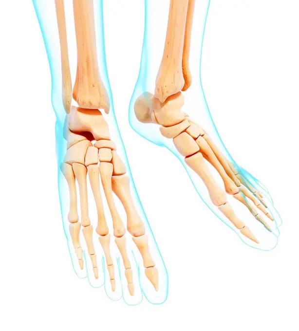
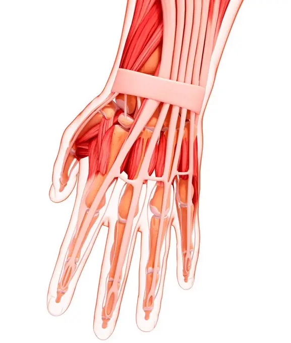
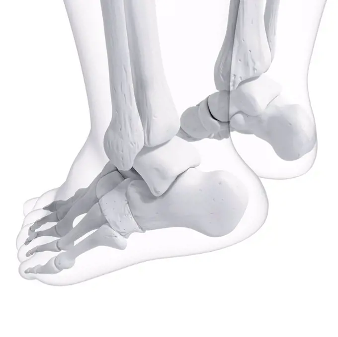
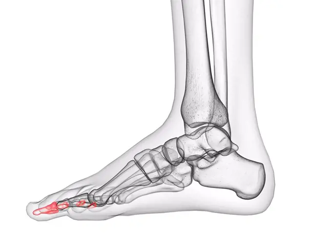
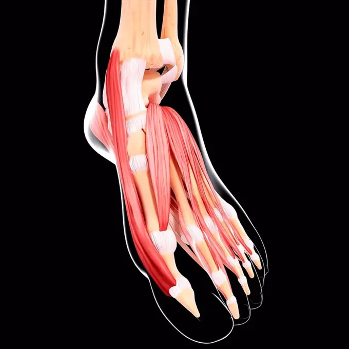
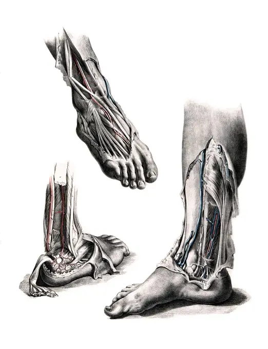
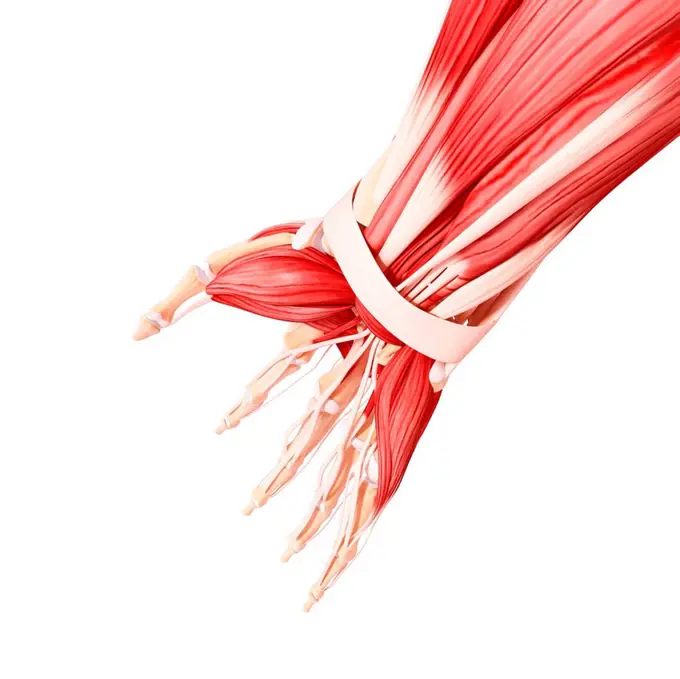
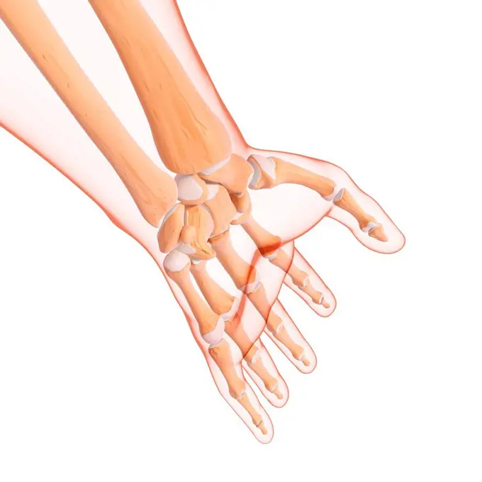
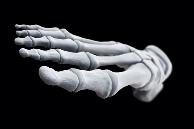
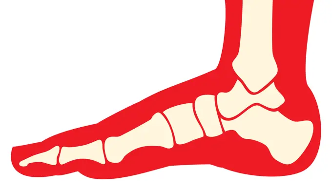
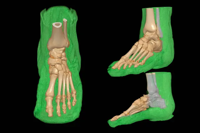
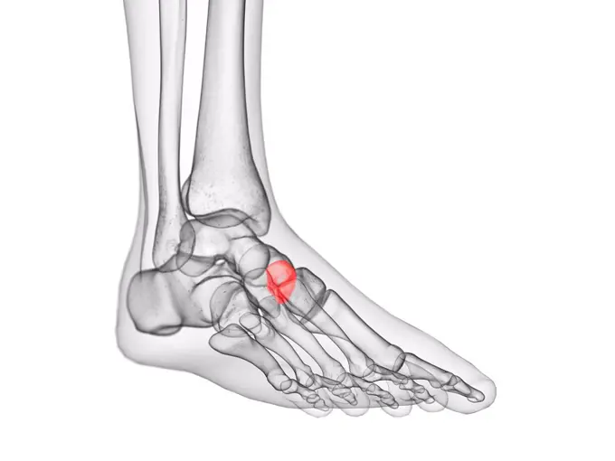
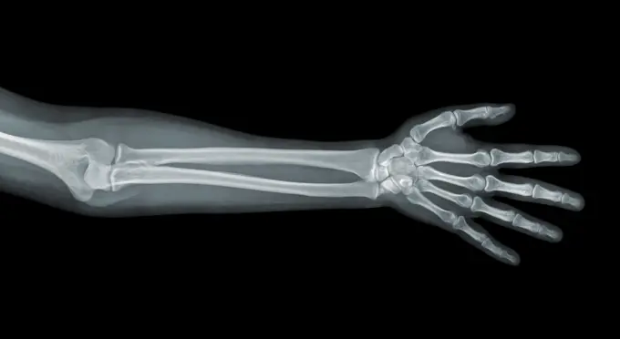
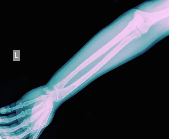
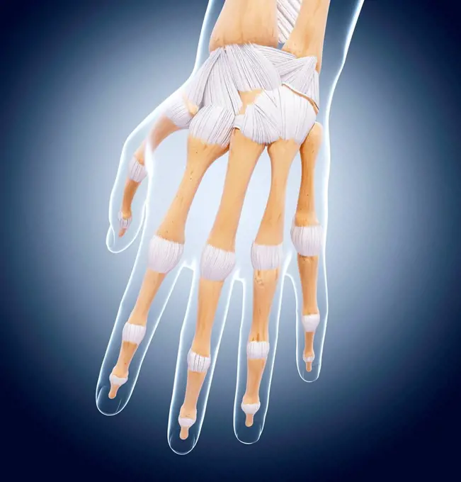
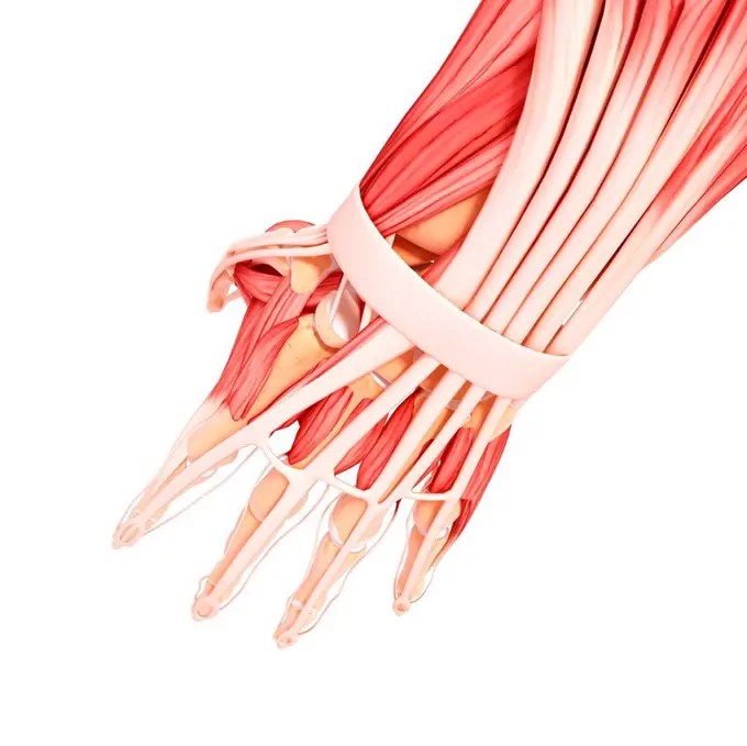
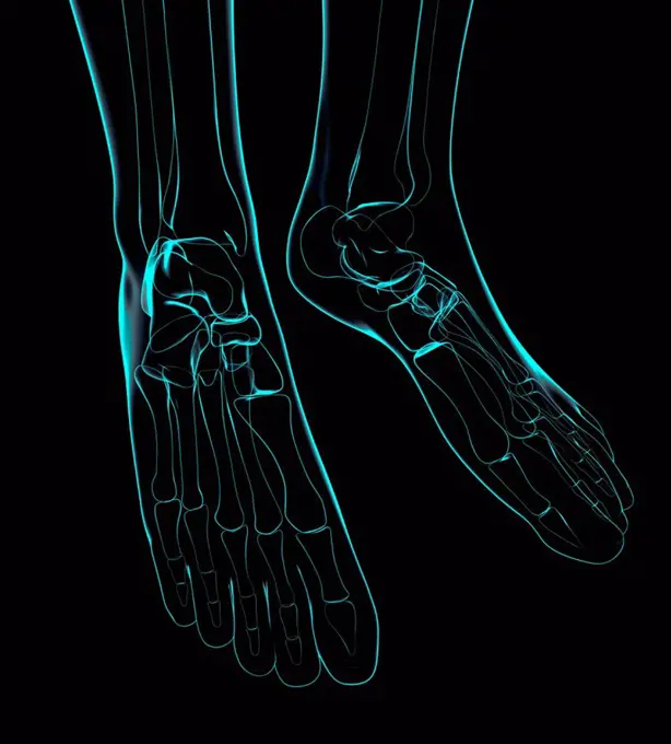
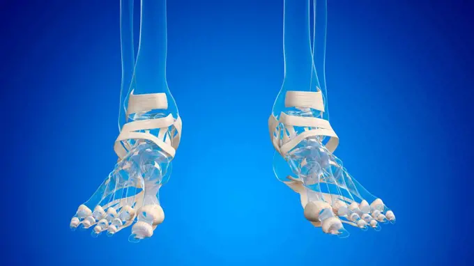
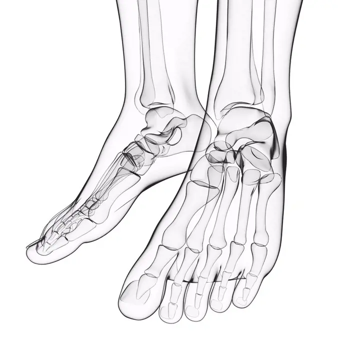
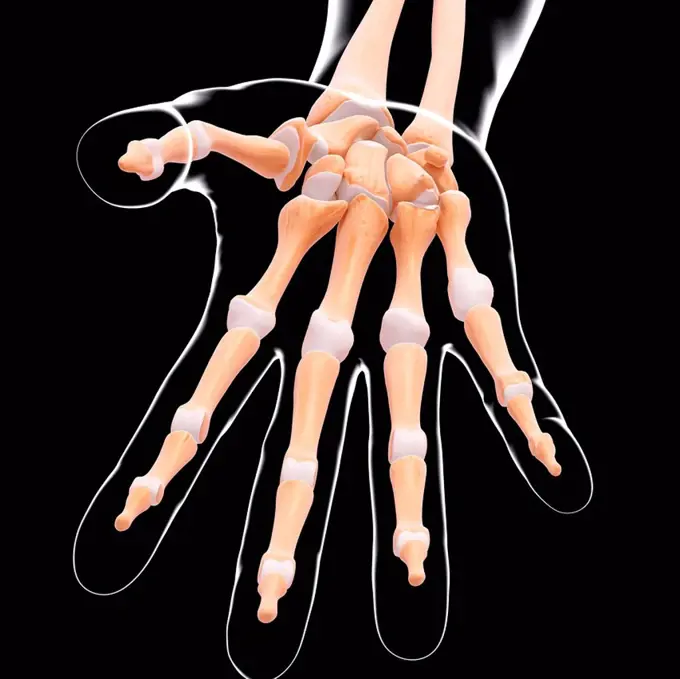
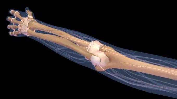
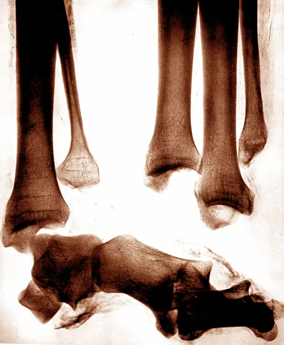
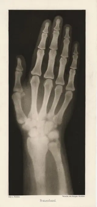
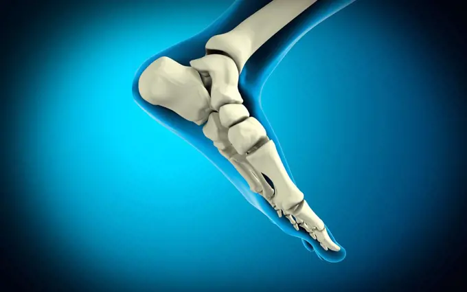
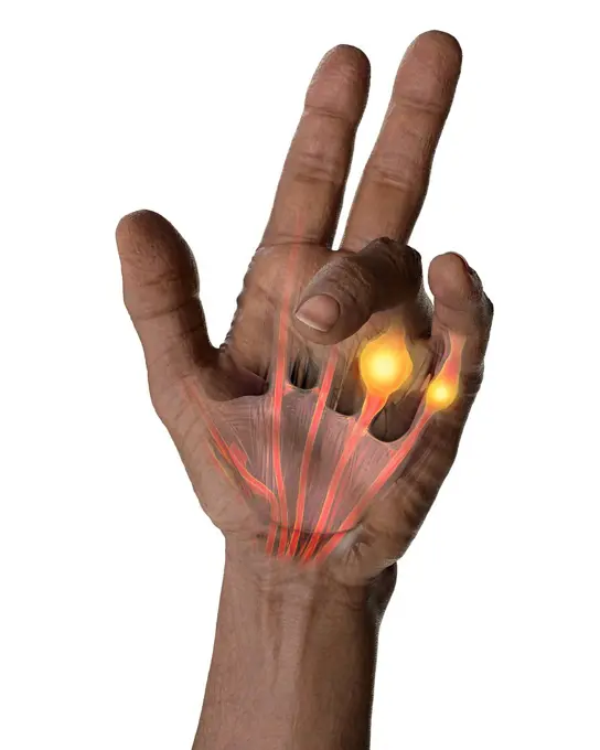
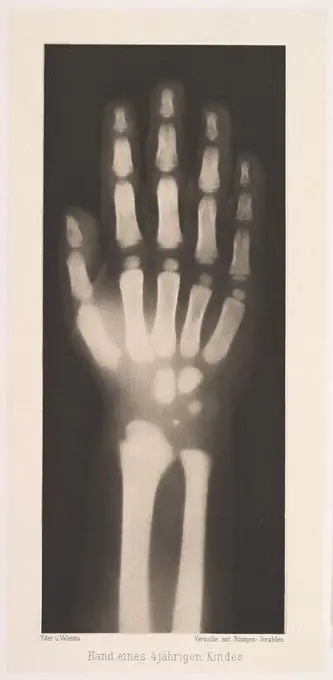
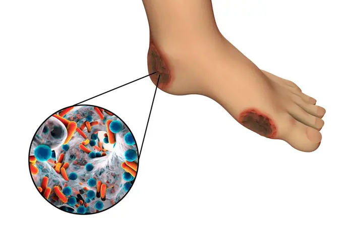
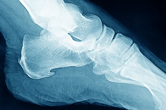
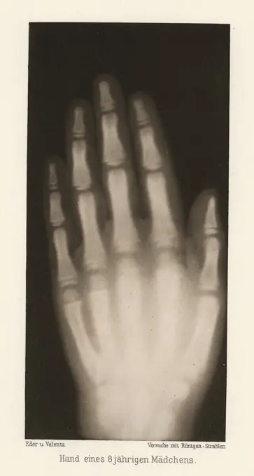
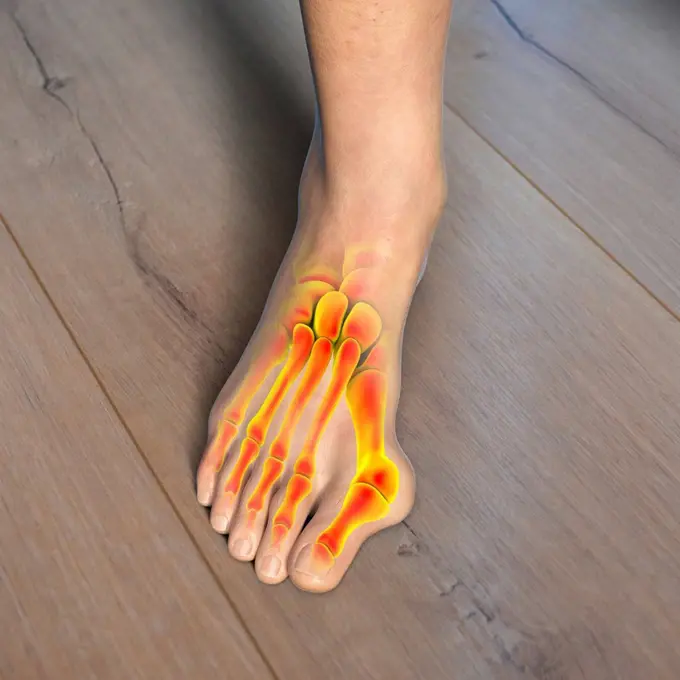
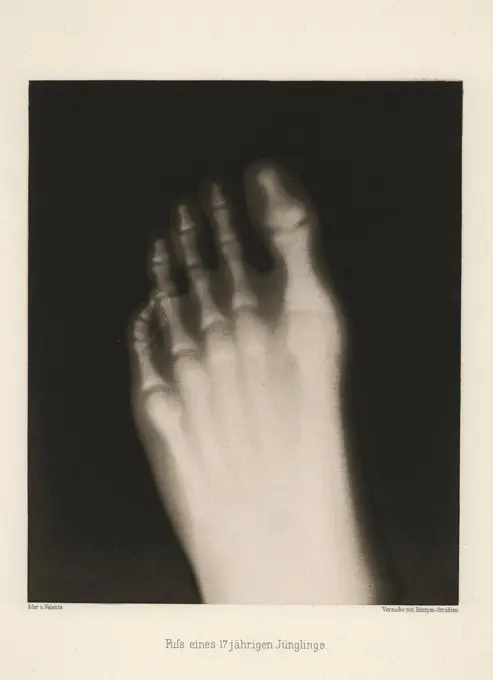
My Boards
View Pictures
  • My Boards

Human Anatomical Studies

Detailed illustrations of human hand and foot anatomy, highlighting muscles and nervous systems against dark backgrounds for clarity.

Human foot bones, computer artwork.
282 assets in this story
4239R-8214
Medical illustration of arteries, veins and lymphatic system in human hand and arm.
4128R-36635
Human hand musculature, computer artwork.
4128-30418410
Veins of the hand, illustration
4128-V58560724
4128-30415745
Feet bones, illustration
4128R-36648
Human leg bones, computer artwork.
4128R-12577309
Human hand bones, computer illustration.
6188-58114056
human radiography scan human radiography scan made in 3D Copyright: xZoonar.com/OxanaxRishnyakx 4219399
4128R-32482
Human foot bones, computer artwork.
4128-15666128
Distal phalanx bones, illustration
4128R-32405
Human foot musculature, computer artwork.
1899-61459179
Medicine, human anatomy, the circulatory system, blood circulation: sections of the foot, lower leg and ankle, with arteries and veins shown in red and blue. Colored lithograph by J. Maclise, 1841.
4128-20043139
Aneurysmal bone cysts of the hand, X-ray
4128R-13022219
Human hand bones, illustration.
4128R-11545074
Human hand bones, computer illustration.
4128R-33524
Human hand musculature, computer artwork.
4128-15666096
Talus bone, illustration
4128-30417342
Anatomy of the left hand, illustration
4128R-35721
Human hand bones, computer artwork.
4378-5714
Foot Bones
4128-30421494
Human hand, illustration
1525-24357336
Human foot anatomy (skeleton, bones)
4128-28514392
Foot, 3D CT scans
4239R-7982
X-ray view of human foot.
4128-30417346
Nerves of the left hand, illustration
4128-15666139
Intermediate cuneiform bone, illustration
4128R-13370686
Human hand anatomy, illustration.
6188-55508973
Hand x-ray view Hand x-ray view on a black background Copyright: xZoonar.com/NatalliaxYaumenenkax 3626563
1439-57940364
Radiant blue Xray of a human arm revealing bones and joints
4128-15950936
Metacarpal bone, illustration
4128-V58567651
4128R-33577
Human foot anatomy, computer artwork.
4128-30418406
Bones of the hand, illustration
4128-48285503
Healthy hand, X-ray
4128R-34608
Human hand bones, computer artwork.
4128R-32381
Human hand musculature, computer artwork.
4128R-35789
Human foot bones, computer artwork.
4128-19056256
Ligaments of the foot, illustration
1848-61029535
Human skeleton hand model for medical anatomy science Medical clinic concept
4128-19052609
Feet bones, illustration
4128-30421497
Lymphatic vessels of the hand, illustration
4128R-35252
Human foot bones, computer artwork.
4128-30421492
Anatomy of the hand, illustration
4128-30421493
Anatomy of the hand, illustration
1525-26034196
Hand drawn illustration of the foot tendons isolated on white, artistic anatomy graphic study
4128R-13371172
Human foot anatomy, illustration.
4128R-36871
Human hand bones, computer artwork.
4128-V58567644
4128R-12964437
Human hand muscles, Illustration
4378-1483
A human model showing the hand muscles.
4128R-32220
Human leg musculature, computer artwork.
1525R-81953
Low section view of human legs
4128-30418388
Bones of the hand, illustration
1428R-1397
Female elbow and forearm skeletal anatomy, back, posterior view. Full color 3D illustration on black background
4128R-32677
Human foot nervous system, computer artwork.
1848-49516152
Mid 20th century X-ray photograph, radiograph showing De Quervain syndrome, mucoid degeneration of two tendons that control movement of the thumb
1525R-193160
Close-up of xray of hand.
1570R-21137237
X-rays of the human hand and leg
6188-58104055
Transparent Human Body with Visible Bones medical science image of human skeleton bones Copyright: xZoonar.com/StanislavxRishnyakx 13861303
1746-28999145
X-ray images of the legs of an ancient Egyptian mummy created soon after the announcement of Wilhelm Röntgen's discovery of X-rays. Wilhelm Röntgen (1845-1923) a German/Dutch mechanical engineer and physicist. Dated 19th Century
824-63203253
Ensethopathy of the supeficial plantar fascia. Sagittal section CT scan of the foot.
4128R-13572652
Illustration of the proximal phalanges bones.
1848-49575371
Insulating rubber gloves on a metallic background
6145-52682702
Heliogravure depicting women's hands, showing anatomy and posture, created by Josef Maria Eder in 1896.
4128-30422909
Bones of the foot, illustration
4239R-7988
X-ray view of bones in human foot.
4128-30422451
Dupuytren's contracture, illustration
4128-30420416
Foot bones, illustration
6145-44588702
ProVision Technologies, a NASA research partnership center at Sternis Space Center in Mississippi, has developed a new hyperspectral imaging (HSI) system that is much smaller than the original large units used aboard remote sensing aircraft and satellites. The new apparatus is about the size of a breadbox. Health-related applications of HSI include non-invasive analysis of human skin to characterize wounds and wound healing rates (especially important for space travelers who heal more slowly), determining if burns are first-, second-, or third degree (rather than painful punch biopsies). The work is sponsored under NASA's Space Product Development (SPD) program.
1848-55997719
Radiography of a dog paw. Real x ray image of an injured dog paw
4128-30421523
Anatomy of the hand, illustration
4128-30422906
Bones of the foot, illustration
4409-17375820
Hand eines 4 jährigen Kindes. Artist: Josef Maria Eder (Austrian, Krems an der Donau, 1855-1944 Kitzbühel); Eduard Valenta (Austrian, 1857-1937). Dimensions: Image: 7 13/16 × 3 1/4 in. (19.9 × 8.2 cm)Plate: 8 13/16 × 4 3/16 in. (22.4 × 10.7 cm)Sheet: 19 9/16 × 13 7/8 in. (49.7 × 35.2 cm). Date: 1896.Eder was the director of an institute for graphic processes and the author of an early history of photography. With the photochemist Valenta, he produced a portfolio in January 1896, less than a month after Wilhelm Conrad Röntgen published his discovery of X-rays. Eder and Valenta's volume, from which this plate derives, demonstrated the X-ray's magical ability to reveal the hidden structure of living things. Human hands and feet, fish, frogs, a snake, a chameleon, a lizard, a rat, and a newborn rabbit are all presented in exquisitely printed photo-gravures, as are carved cameos and an assortment of natural materials. In an era when photography's ability to accurately depict the visible wor
4128R-13763279
Destruction of human cytomegalovirus (HCMV), computer illustration. Conceptual image for HCMV infection treatment and prevention. HCMV is a member of ...
824-63212842
Enthesopathy: calcifications of the plantar fascia. Visualisation on saggital plane x-ray.
6145-52682791
Heliogravure by Josef Maria Eder showing the hand of an eight-year-old girl, depicting bones, fingers, and anatomical structure, created in 1896.
4128-20045501
Bunion, illustration
6145-52890223
X-ray glass diapositive by Josef Maria Eder showing the foot of a 17-year-old student, captured in 1896, focusing on bone structure and anatomy.
6145-52682865
Heliogravure by Josef Maria Eder depicting the foot of a seventeen-year-old youth, showing bones, toes, and anatomical structure, created in 1896.
4239-69152014
Muscles of the Foot
6188-67431818
3D Isometrische flache konzeptionelle Illustration von Hallux Valgus, Ballenzeh im Fußbr3D-Isometrische flache konzeptionelle Illustration von Hallux Valgus, Ballenzeh im Fuß 3D Isometric Flat Conceptual Illustration of Hallux Valgus, Bunion in Foot 3D Isometric Flat Conceptual Illustration of Hallux Valgus, Bunion in Foot Copyright: xZoonar.com/AnastasiiaxTorianykx 22948837
4128-30420510
Athlete's foot, illustration
PREVIOUS
of 3
NEXT
2401 S. Ervay, Suite 206
Dallas, Texas 75215
United States
Get Started
Free ResearchMy BoardsMy Cart
For Creators
How To License Your ContentContributor PortalFrame of Mind
Resources
API accessPricing
Contact
+1 866 236 0087help@viewpictures.co.uk Contact form
©2026 View Pictures. All Rights Reserved. -A
This site is protected by reCAPTCHA and Google Privacy Policy and Terms of Service apply.