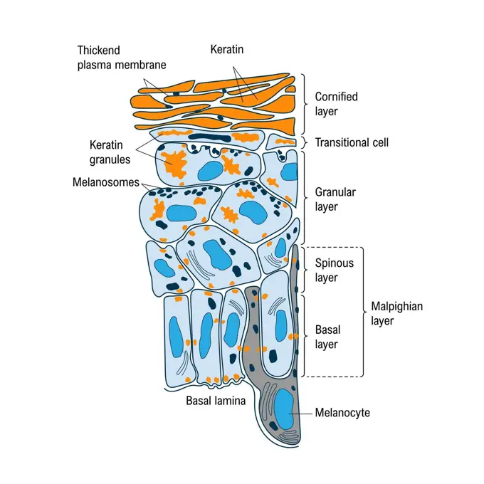
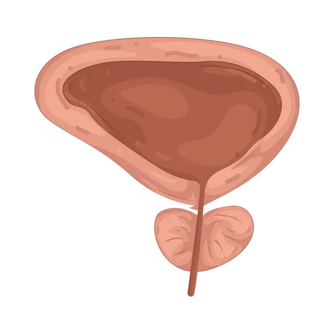
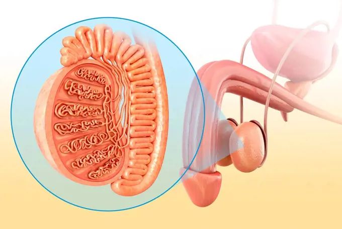
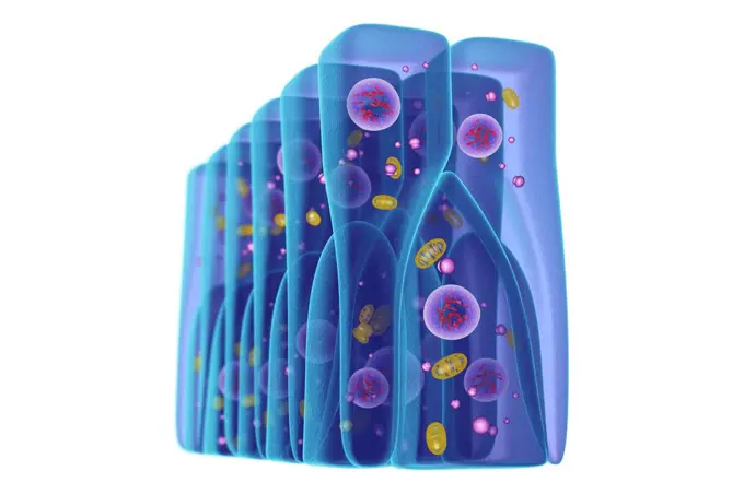
My Boards
View Pictures
  • My Boards

Human Anatomy Illustrations

Detailed illustrations showcasing human reproductive systems and anatomical structures, providing educational insights into human biology.

Female reproductive system, illustration.
181 assets in this story
824-63219930
Acne around the hair follicle with inflammation, sebaceous hypersecretion, hyperkeratinization. Skin section of the epidermis and dermis. Normal hair follicle and acne pimple in the center. The acne pimple is formed around the hair follicle with hyperkeratinization and inflammation of the epidermis caused by glycerides (in yellow) under the action of the bacteria Propionibacterium acnes (in red). The sebaceous gland is in hypersecretion. Polynuclear cells from blood vessels flock
824-63183449
Illustration of the ciliated cells of the sinus. These cells drain mucus that traps external particles. In the case of chronic sinusitis, the irrigation capacity of the ciliated nasal sinus system is altered, mucus stagnates in the mucous membrane and thickens.
1525-56853624
A blood clot in a vein, or venous thrombus, is the clumping of blood in vessels that bring blood back to the heart. 3D rendering. A blood clot in a vein, or venous thrombus, is the clumping of blood in vessels that bring blood back to the heart.
4128-20040400
Prostate cancer, illustration
1525-57151523
Virus and bacteria. Internal organ. Medical treatment. Health and illness. Cartoon flat illustration. Human stomach. Viral infection
824-63188670
Illustration of an artheroscletoric plaque which obstructs the light in the artery and slows blood flow.
824-63164021
From muscle to muscle fiber: tendon, muscle, muscle fiber.
824-63218274
Site of action of drugs in the treatment of acute otitis media. Frontal cut of the auditory apparatus passing through the outer ear and the middle ear with a purulent effusion in the middle ear and inflammation of the mucous membrane. Below, zoom on the mucosa with a Haemophilus influenzae bacterium seen in section allowing to see the different layers of the wall with the betalactamases of the periplasmic space and the PLP of the cytoplasmic membrane bacterial DNA with the ribosomes.
4128R-14638073
Heart muscle structure, illustration and micrograph
4128R-13723617
Illustration of the anatomy of a male testis (testicle).
824-63180573
A cross-section illustration of skin with a hair follicle (skin appendage), the sebaceous gland (next to the hair follicle), the arrector pili muscle and the blood vessels. In the dermis, seen enlarged, the fibroblasts (purple) and the collagen fibers (beige).
4128R-11295098
Human muscular structure, computer artwork.
824-63167629
Epidermis, dermis, and hypodermis (also known as the subcutaneous tissue or subcutis).
4128R-13372881
Fat cells, light micrograph and computer illustration. White adipose tissue composed of adipocytes (fat cells). Adipocytes form adipose tissue, which ...
1525-25848608
Coronary stent and angioplasty concept as a heart disease treatment symbol diagram with the stages of an implant procedure in an artery that has cholesterol plaque blockage being opened for increased blood flow as a 3D illustration.
4128-19358349
Inflamed appendix, conceptual illustration.
1899-53509460
Transmission of nerve impulses in the brain from one neuron to another.
4128-30418981
Stomach tumour, illustration
4239-20481107
Detail of a nerve synapse showing the release of neurotransmitters.
4128R-13763982
Tooth decay. Computer illustration of a tooth with a cavity and a close-up view of the bacteria that cause caries formation.
1899-30608848
Transmission of nerve impulses in the brain from one neuron to another.
1525-56722541
Atherothrombosis is a cardiovascular disease characterized by ruptured atherosclerotic plaque and clot formation. 3D rendering. Atherothrombosis is a cardiovascular disease characterized by ruptured atherosclerotic plaque and clot formation.
1525-56722597
Atherothrombosis is a cardiovascular disease characterized by ruptured atherosclerotic plaque and clot formation. 3D rendering. Atherothrombosis is a cardiovascular disease characterized by ruptured atherosclerotic plaque and clot formation.
824-63195451
Illustration of a sharks brain. At the front of the brain there are two olfactory bulbs and two ampullae of Lorenzini which allow the shark to detect its prey by perceiving its electrical field.
1848-77403229
3D Illustration Concept of Human Internal Digestive Organ Liver Anatomy
1899-54027820
Carbon monoxide is a toxic gas for the heart and arteries. Affinity between carbon monoxide and hemoglobin is 200 times stronger than between oxygen and hemoglobin. This lessens the blood's capacity to transport oxygen.
824-63217428
Carbon monoxide is a toxic gas for the heart and arteries. Affinity between carbon monoxide and hemoglobin is 200 times stronger than between oxygen and hemoglobin. This lessens the bloods capacity to transport oxygen.
1525-28110911
Bacteria in the intestine as gut probiotic bacterium inside small intestine and digestive microflora inside the colon or bowel as a health symbol for microbiome as a 3D render isolated on a white background.
5507-31002947
Anatomical structure and function of skin, medical illustration
4128R-13763366
Gram-positive bacterial cell wall, artwork. The horizontal layers include a plasma membrane (blue) containing transmembrane proteins (green, yellow an...
1525-56853467
Celiac Disease is a chronic condition characterized an inflammatory allergic response of the digestive tract to Gluten. 3D rendering. Celiac Disease is a chronic condition characterized an inflammatory allergic response of the digestive tract to Gluten.
1525-71581545
prostate
4128R-13725101
Illustration of male testis anatomy.
4128R-14181242
Lumbar vertebrae and spinal cord, illustration
1525-56565138
Cell of human organism. Layer of epidermis. Scientific material for education. Cartoon flat illustration. Skin system. Biology and Microbiology. Cell of human organism. Layer of epidermis.
824-63189030
Illustration of Couinaud liver segmentation. The liver is divided into sectors, which are in turn divided into segments. The hepatic veins divide the liver into sectors and the branches of the portal vein divide the liver in segments that are numbered from I to VIII.
4128R-13844668
Pseudostratified epithelium, illustration.
824-63164207
Urinary system from kidney to glomerulus with structures of kidney and ureter.
1525-75763404
Artificial insemination illustartion vector in flat style. Ovary cell in Petri dish. IVF procedure.. Artificial insemination illustartion vector in flat style. Ovary cell in Petri dish.
1525-27988746
Reduce Levels of cholesterol plaque in vessels vector illustration
824-63217374
Illustration of the signs of ageing in skin, dulling of the epidermis, brown spots, skin slackening and wrinkles. In the centre of the image: a cross-section illustration of young skin enabling a comparison with different types of ageing: -Top left illustrates the dulling of the epidermis, the skin is dry and cutaneous microrelief is irregular, causing the skin to become permeable to pollutants and dehydration. Superficial microcirculation is less good; skin cells become less oxygenated. -Top right illustrates spots. If they are brown it is due to melanin deposits, when red it is due to superficial microcirculation vessels, which are dilated. -Bottom left illustrates the loss of firmness due to diminished activity of the fibroblasts (blue). Fibroblasts are responsible for skins elasticity, producing elastin and collagen -Bottom right illustrates wrinkles due to the deterioration of collagen (white vertical) and elastin (pink horizontal) fibers.
4128-48286108
Lungs affected by miliary tuberculosis, illustration
1525-25848615
Coronary artery cholesterol therapy medical concept as a vein with gradual plaque removal from clogged arteries and atherosclerosis with with a 3D human anatomy diagram of the reduction of cholesterol buildup.
4128R-11292086
Female reproductive system, computer artwork.
1788-20762609
Circulatory system, drawing.
4378-4240
Dentine
4128R-11312728
Human lymph node, computer artwork.
1899-54027818
Carbon monoxide is a toxic gas for the heart and arteries. Affinity between carbon monoxide and hemoglobin is 200 times stronger than between oxygen and hemoglobin. This lessens the blood's capacity to transport oxygen.
4128-19249232
Mesenteric ischaemic, illustration.
5507-42694116
Stop Bacterial overgrowth in small  intestine  vector illustration
4128R-11291220
Breast anatomy, computer artwork.
5507-46944681
Cause of body odor vector flat illustration
1899-53509239
Lung, pulmonary alveolus with its capillary network allowing gas exchange.
824-63219959
Asthma, Normal bronchiole, asthmatic bronchiole with bronchoconstriction, hypersecretion, treatments. Representation of the lungs with the bronchial tree from which 3 zooms start. Top left, a healthy bronchiole, bottom, two asthmatic bronchioles. An allergen (green star) triggers an asthma attack by the activation of lymphocytes producing IgE (green Y) leading to the degranulation of mast cells (purple) which release leukotrienes (black beads) and cytokines (blue beads) mediators of inflammation of the mucosa which leads to bronchoconstriction of smooth muscle fibers, hypersecretion of mucus and obstruction of the airways.
4409-17349028
Blood brain barrier.
4128R-13723515
Illustration of a lung bronchus with excess mucus.
824-63180640
Illustration of the chain reaction involved in muscle cramp.
4128R-24460
Balloon angioplasty. Computer artwork of a stent being placed in a narrowed blood vessel.
4128R-13586583
Illustration of pancreas and gallbladder.
4128R-14181689
Aspergilloma of the brain, illustration
4128-19249245
Cholesterol in human blood vessels, illustration.
824-63220051
Infectious conjunctivitis and its treatments. View of an eye with inflammation of the conjunctiva. Zoom on the conjunctiva with from top to bottom 1 bacterial infection (green) and 1 viral infection (red-orange). In yellow, purulent discharge due to the presence of bacteria (green), the cells of the conjunctiva are destroyed. Goblet cells secreting mucus and edema of the conjunctiva in the area infected with the virus. Right from top to bottom, a bacteria seen in section, a window with an infected connective cell (Herpes Simplex virus) detailing the process of viral replication transcription of viral DNA, DNA polymerase, viral replication
824-63167618
Epithelia continually renews itself due to differentiating stem cells. Here, culture of bronchial stem cells destined for transplant and multiplication in the pulmonary alveoli. For the following reasons, adult stem cells may eventually prove to be better for cell therapy than embryonic stem cells: their greater availability in adult tissue, the absence of immune-response rejection, their decreased disposition for tumors. Removed from the patient, stem cells can be cultured and multiplied to attain a quantity that is sufficient to treat sickness or injuries.
4391-159
This illustration explains the process of metastasis. Once metastatic cells are attached to the basement membrane (a physical barrier that seperates tissue components), they break through with the help of an enzyme called type IV collagenase. Cancer cells then move through the blood stream enabling them to spread to other parts of the body. A secondary tumor may form at another site in the body. Illustration by Jane Hurd.
824-63164349
Cancerous tumor, stage 1: The tumor is still small.
4128R-11314938
Human digestive system, computer artwork.
4128R-12700049
Cross section through an model of an fracture in the neck of the femur (thigh bone).
4128-20040729
Prostatitis, illustration
4128R-13446632
Spinal cord, cross-section. Light micrograph and computer illustration of a spinal cord. At right it is seen inside the vertebrae (bones). The section...
5507-45611392
Colon cancer.
1525-22346082
Close-up of the human spine and a blister pack
5507-43624719
Type of obesity illustration (Japanese) . Abdominal sectional View (visceral fat )
5507-43624720
Type of obesity illustration (Japanese) . Abdominal sectional View (subcutaneous fat).
5507-46944683
Cause of body odor vector flat illustration
4128R-13386779
Illustration of child's spinal cord cross-section.
824-63211034
Cholera bacilli or cholera vibrio (Vibrio cholerae). Cholera is an infectious and contagious disease characterized by sudden and very abundant diarrhoea. In the absence of treatment, the classic major form is fatal in more than half of the cases. Optical microscopy X 1000.
4128-19358380
Bladder cancer stages, illustration.
4128-19358351
Inflamed appendix, conceptual illustration.
4128-19490537
Colonoscopy, illustration
4128-19249250
Angioplasty, illustration.
1525-27726679
Agate rock consisting primarily of cryptocrystalline silica chalcedony, alternating with microgranular quartz, vector stone isolated on white. Agate Rock Consisting Primarily Cryptocrystalline
PREVIOUS
of 2
NEXT
2401 S. Ervay, Suite 206
Dallas, Texas 75215
United States
Get Started
Free ResearchMy BoardsMy Cart
For Creators
How To License Your ContentContributor PortalFrame of Mind
Resources
API accessPricing
Contact
+1 866 236 0087help@viewpictures.co.uk Contact form
©2026 View Pictures. All Rights Reserved. -B
This site is protected by reCAPTCHA and Google Privacy Policy and Terms of Service apply.