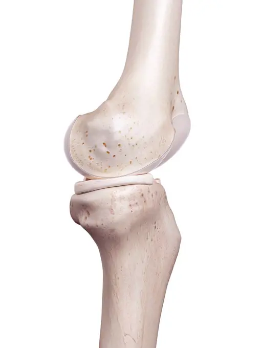
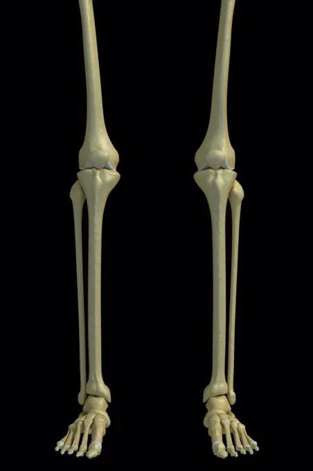
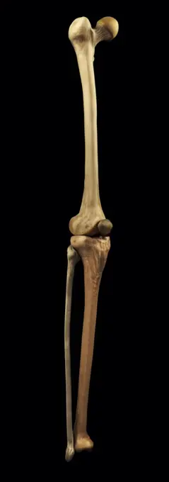
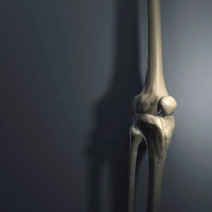
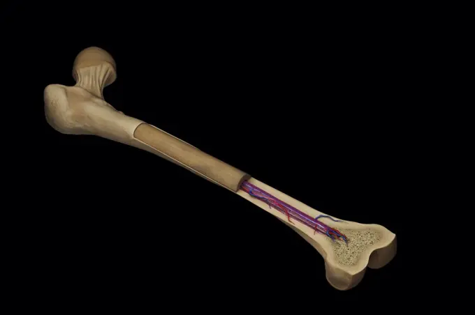
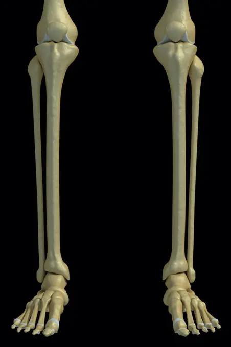
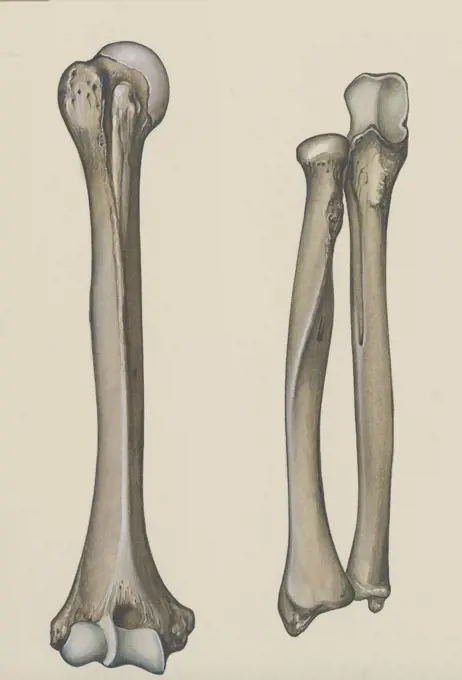
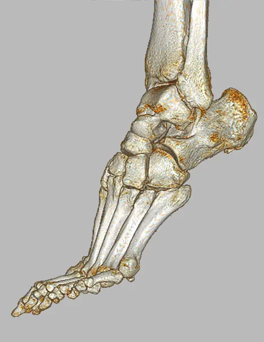
My Boards
View Pictures
  • My Boards

Human Anatomy Illustrations

Detailed illustrations of human arm, elbow, and knee anatomy, highlighting bones and muscles with anatomical accuracy in a clean, educational style.

Human elbow joint, computer illustration.
141 assets in this story
4128R-13385717
Human hand muscles, illustration.
4128R-13372502
Human hand anatomy, illustration.
4128R-13412317
Leg bones
4128R-13372464
Human hand bones, illustration.
4128R-12576931
Human foot anatomy, computer illustration.
4128R-12964199
Human leg bones, Illustration
4128R-32006
Human leg bones, computer artwork.
4128-19247929
Elbow joint, illustration.
4128R-12577393
Human hip muscles, computer illustration.
4128R-12580481
Hand muscle, computer illustration.
4128R-11322989
Human femur head, computer artwork.
4128R-33616
Human arm bones, computer artwork.
1848-54708927
Human foot, anatomical model
4128-15982214
Opponens pollicis muscle, computer illustration.
4128R-12580545
Human hand anatomy, computer illustration.
4378-5157
Foot Bones
4128R-15515032
Illustration of the human knee
4128R-12580525
Human hand anatomy, computer illustration.
4409-17348486
Bones of the hand.
4128R-12580538
Human hand anatomy, computer illustration.
4128R-13370977
Muscles of the human arm, illustration.
4128-19053087
Femur, illustration
4128-28968782
Femur bone, illustration
4128-30421522
Bones of the hand, illustration
4378-315
Front view of the bones of the legs, knee joints, ankle joints and feet.
4378-2893
Bones and joint that form the skeletal structure of the human leg
4128-111577872
Abductor digiti minimi muscle, computer illustration.
4378-3664
The bones of the knee viewed from a three-quarter perspective against a gray background.
4128R-13385647
Human abdominal muscles, illustration.
4378-2823
Bones and joint that form the skeletal structure of the human leg
4128R-26164
Thigh bone. Computer artwork showing the femur bone.
4128-19247290
Human foetus anatomy at week 15, illustration.
4378-2566
Cross section of a femur bone showing the anatomical structure including cancellous bone and marrow.
4378-122
Bones of the lower legs, ankle joints and feet.
4128-30418082
Frog's skeletal system, illustration
1788-21803409
Humerus, radius and ulna, human body, drawing.
4128R-11322791
Human femur, computer artwork.
824-63198902
Human foot seen on a 3D CT scan.
4239R-20483515
Renal Corpuscle and the filtration membrane. Glomerular capillary surrounded by podocytes.
4378-2637
Model showing the human knee joint and its connecting bones.
1899-53513448
Bone development on a humerus at birth, in children and adults. This illustration makes it possible to compare the development of the bone during these three ages. From birth to childhood, the bone structure is cartilaginous and flexible. We see it thanks to the aspect of the bone at the level of the epiphyses and the diaphysis. But we can also see it thanks to the area shown in pink. This area constitutes the epiphyseal plate. It is this epiphyseal plate that produces cartilage. This allows the bone to gradually grow and ossify. Bone growth ends between the ages of 18 and 25. In adults, the epiphyseal cartilage has ossified. We can no longer distinguish an epiphyseal line. This line is visible with a lighter color between the epiphysis (head of the humerus) and the metaphysis. The metaphysis is the intermediate zone between the epiphysis and the diaphysis. The diaphysis represents the body of the bone. On each section of the humerus is shown a section of horizontal section in blue. At
PREVIOUS
of 2
NEXT
2401 S. Ervay, Suite 206
Dallas, Texas 75215
United States
Get Started
Free ResearchMy BoardsMy Cart
For Creators
How To License Your ContentContributor PortalFrame of Mind
Resources
API accessPricing
Contact
+1 866 236 0087help@viewpictures.co.uk Contact form
©2026 View Pictures. All Rights Reserved. -A
This site is protected by reCAPTCHA and Google Privacy Policy and Terms of Service apply.