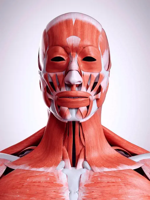
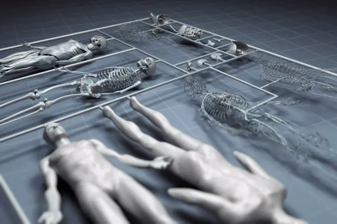
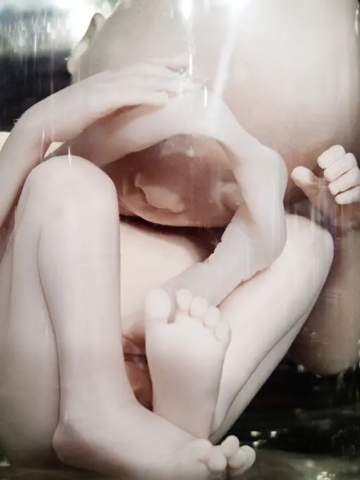
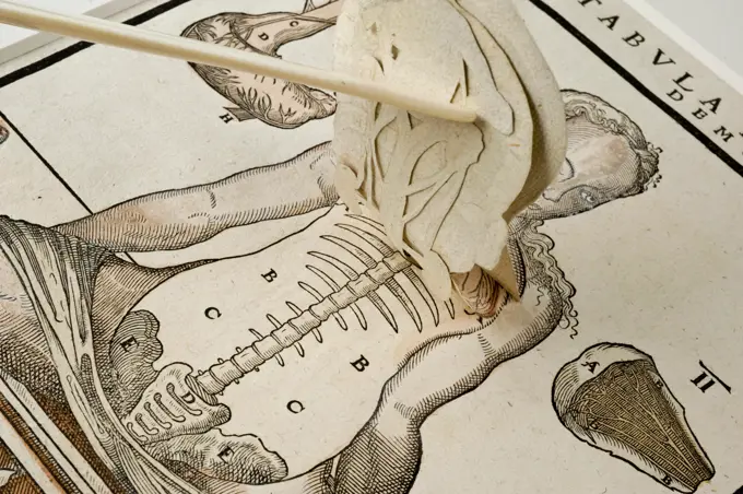
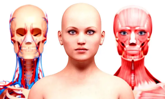
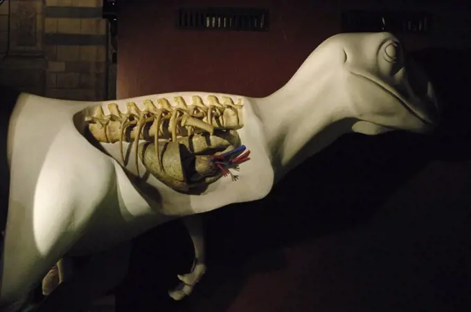
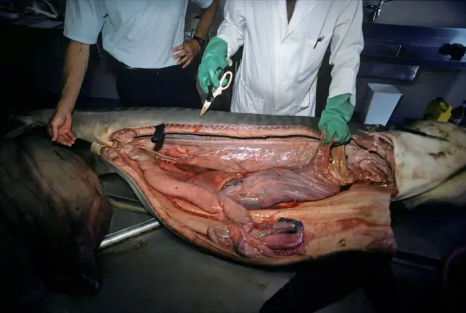
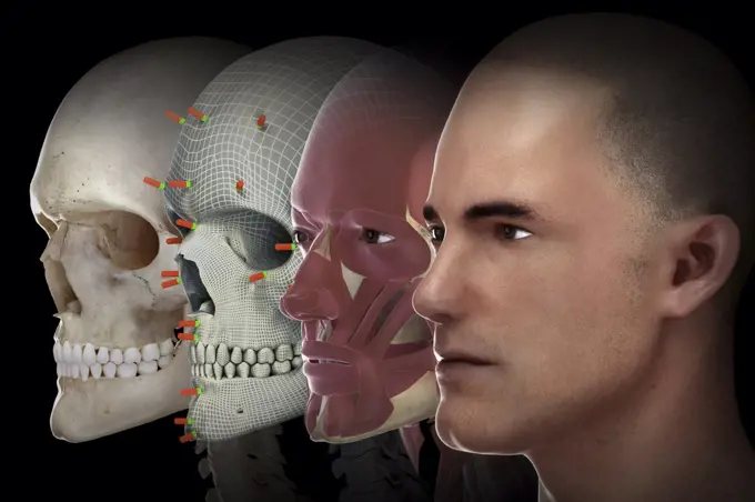
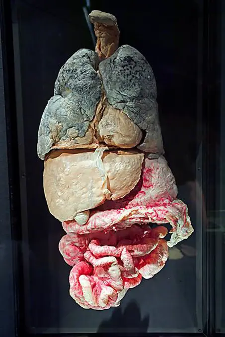
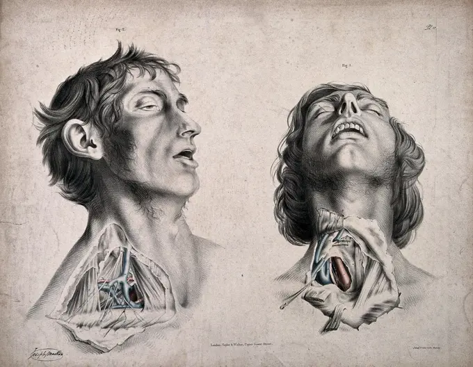
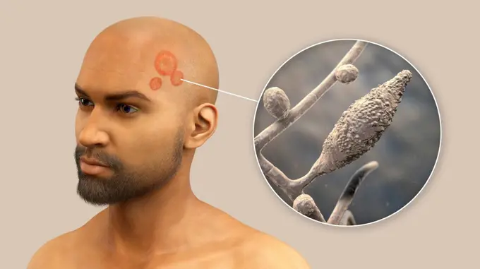
My Boards
View Pictures
  • My Boards
← Science

Human Anatomy Illustrations

Detailed digital illustrations of human skeletal and anatomical systems, showcasing structures like ribs, lungs, and neural pathways.

Human skeletal system, computer illustration.
168 assets in this story
4297-1693
Colorized x-ray of the foot of a person with diabetes who has a calcified artery
4128R-15061506
Baby's brain as it grows inside the womb, illustration
1848-65798244
Human skeleton with cigarette. World No Tobacco Day quit or stop smoking!
4128R-13726084
Human vertebrae, illustration.
6188-68124845
Nahaufnahme von Austernpilzen mit ihren einzigartigen Kiemen und Texturen Nahaufnahme von Austernpilzen mit ihren einzigartigen Kiemen und Texturen LicenseRF Copyright: xZoonar.com/NerijusxLiobex 23202221
4128R-13372403
Human facial muscles, illustration.
4128R-13574404
Illustration of human spinal cord cross-section.
1428-137
Rear view of a human skeleton
6188-58105736
bones of the Human skeleton bones of the Human skeleton Copyright: xZoonar.com/StanislavxRishnyakx 14776955
1525-26275292
Bronchopulmonary segmental anatomy. 3D illustration. Bronchopulmonary segmental anatomy
1848-49408374
Skeleton preparation for biology lessons, human skull, model by the Hedinger company, Didacta education fair, Stuttgart, Baden-Wuerttemberg, Germany, Europe
4128R-15515294
Illustration of the head and neck muscles
824-28706037
Anatomical mannequin for teaching human organs
824-63225284
Anatomical model in front of an x-ray of the lungs.
4378-4657
Human Cloning
824-28706013
Anatomical mannequin for teaching human organs
824-63225286
Anatomical model in front of an x-ray of the lungs.
1990-29048673
close up of a human fetus 20 weeks old
6145-29256117
Anatomical fugitive sheet,female
824-28705980
Anatomical mannequin for teaching human organs
1525-56184342
When light hits the retina , special cells called photoreceptors turn the light into electrical signals. These electrical signals travel from the retina through the optic nerve to the brain. Then the brain turns the signals into the images you see. 3d illustration. The mechanism of vision in the human eye
824-28706033
Anatomical mannequin for teaching human organs
824-28705985
Anatomical mannequin for teaching human organs
4128R-11294915
Female anatomy, computer artwork.
824-28706016
Anatomical mannequin for teaching human organs
824-28706014
Anatomical mannequin for teaching human organs
1525-27019825
digital visualization of the dna
4128R-11544885
Female neck bones, computer illustration.
824-28706010
Anatomical mannequin for teaching human organs
1525-24925389
alien man portrait futuristic silver skin extraterrestrial space metaphor
4128-V58571005
824-28706030
Anatomical mannequin for teaching human organs
4378-2414
Anatomical model showing the head and internal structure of the nervous system.
4128R-13726088
Human digestive system, illustration.
4128R-13244814
Human brain, illustration.
6145-29246916
Muscles and tendons of the upper arm: two écorchés. Watercolour, 18--().
4128-30416866
Lymphatic vessels of the head, illustration
1525-20460833
human bones radiography scan. x-ray  image
5514-71372565
Dissection Scenes
4128R-15535916
Illustration of an obese man's throat anatomy
1899-53508332
Anatomical mannequin for teaching human organs
6145-29291919
A diseased part of the body. Watercolour by Juan Wandesforde, 1844.
6145-46715153
Skiopticon image from the Department of Photography at the Royal Institute of Technology. Use by Professor Helmer Bäckström as lecture material. Bäckström was Sweden's first professor in photography at the Royal Institute of Technology in Stockholm 1948-1958.KvinnoBröst Photographed on infrared tank. The vein (veins) appear clearly. Compare SKC7324.For more info See: Bäckström, Helmer. Photographic manual. Other revised edition. Nature and Culture. Stockholm. 1948. p. 1070.
6188-58516805
Stuckskulpturen im großen Saal des Theaterhauses Walhalla Stuckskulpturen im großen Saal des Theaterhauses Walhalla ,property released Copyright: xZoonar.com/wolfgangxcezannex 21608845 ,model released, Symbolfoto ,property released
1899-53508568
3D reconstruction from patient images, 3D printed pelvis.
824-28706005
Anatomical mannequin for teaching human organs
1439R-1001729
Skeleton and whiteboard
6145-29251705
Female genitalia showing severely diseased tissue all over the pelvic region. Watercolour by Christopher D' Alton, 1860.
6188-67137188
ARCHÄOLOGIE ANTHROPOLOGIE FORSCHUNG Anthropology - hands of an anthropologist examining human remains Copyright: xZoonar.com/AntonxChalakovx 22558970
824-63186244
Reportage in Descartes University, Paris, France where Vizua 3D is based. Vizua provides real-time 3D visualisations for doctors using imagery from patients. Doctors can then print these visualisations in 3D. A scan using the Vizua system that is due to be printed in 3D.
4128R-3711
Electroencephalography.
4409-61168
Replica model of a dinosaur heart. Natural History Museum. London. United Kingdom.
4413-93117
Dissection of 3 meter Tiger Shark Natal Sharks Board
442-12382
Ginseng roots in bottles, Namdaemun Market, Seoul, South Korea
442-21984422
England, London, The Wellcome Collection, Transparant Model of the Human Body
4378-20365100
Facial Reconstruction
1848-51501676
Plastinate, human abdomen, internal organs with metastases, Body Worlds, Menschen Museum, Berlin, Germany, Europe
6188-58114158
x-ray examination x-ray examination made in 3D graphics Copyright: xZoonar.com/OxanaxRishnyakx 4281015
6145-29270590
The circulatory system: two dissections of the neck of a man, with arteries, blood vessels and veins indicated in red and blue. Coloured lithograph by J. Maclise, 1841/1844.
4413-108407
Three meter Tiger Shark being prepared for dissection RSA
4409-28580179
Infographic on the functioning of the mammary glands of mammals. QuarkXPress (.qxp); 6259x4015.
1815R-72349
Alligator skull with stethoscope and syringe in background
4409-17250067
EARLY MAN (2018), directed by NICK PARK. Copyright: Editorial use only. No merchandising or book covers. This is a publicly distributed handout. Access rights only, no license of copyright provided. Only to be reproduced in conjunction with promotion of this film.
4128-20042038
Ringworm, illustration
1525-24004194
Underground fossil. Cartoon Earth ground layers with prehistoric dinosaur skeleton and skull. Ancient extinct reptile bones science exploration. Vector geology and paleontology excavation illustration. Underground fossil. Cartoon Earth ground layers with dinosaur skeleton and skull. Extinct reptile bones science exploration. Vector geology and paleontology excavation illustration
7208-70657808
Egypt, X-rays, Iufaa, 26th dynasty priest to pharaohs Amasis and Psamtek III, Abu Sir
1848-49408371
Torso for biology lessons, human body with muscles and organs, model by Hedinger, Didacta education fair, Stuttgart, Baden-Wuerttemberg, Germany, Europe
1525R-73914
Girl looking at skeleton of pelvis
PREVIOUS
of 2
NEXT
2401 S. Ervay, Suite 206
Dallas, Texas 75215
United States
Get Started
Free ResearchMy BoardsMy Cart
For Creators
How To License Your ContentContributor PortalFrame of Mind
Resources
API accessPricing
Contact
+1 866 236 0087help@viewpictures.co.uk Contact form
©2026 View Pictures. All Rights Reserved. -A
This site is protected by reCAPTCHA and Google Privacy Policy and Terms of Service apply.