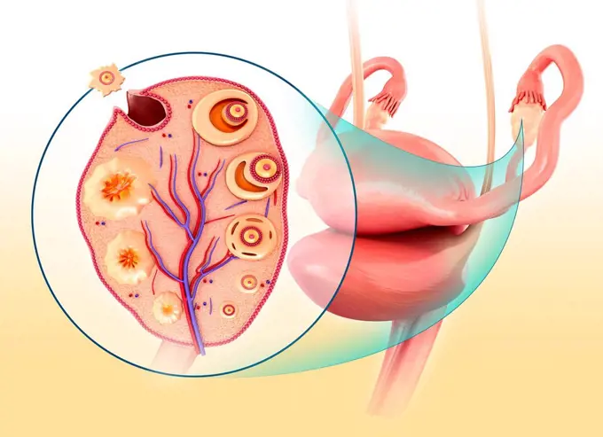
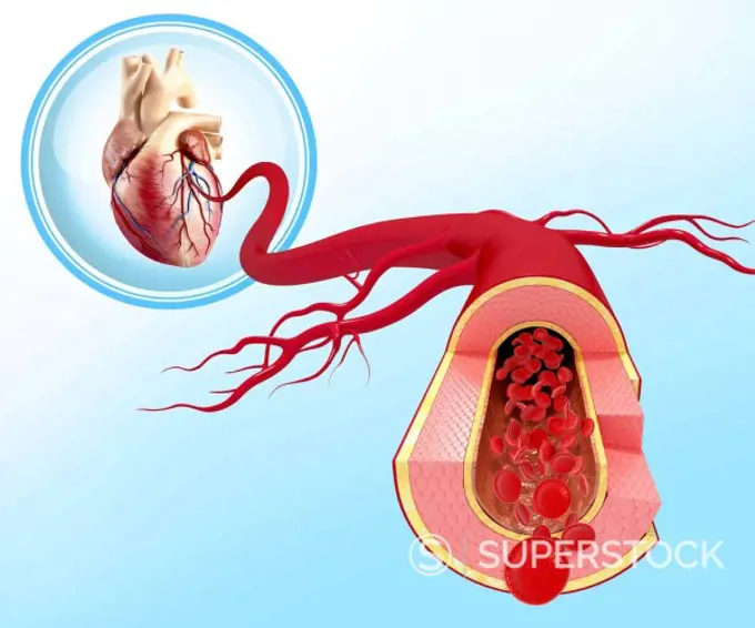
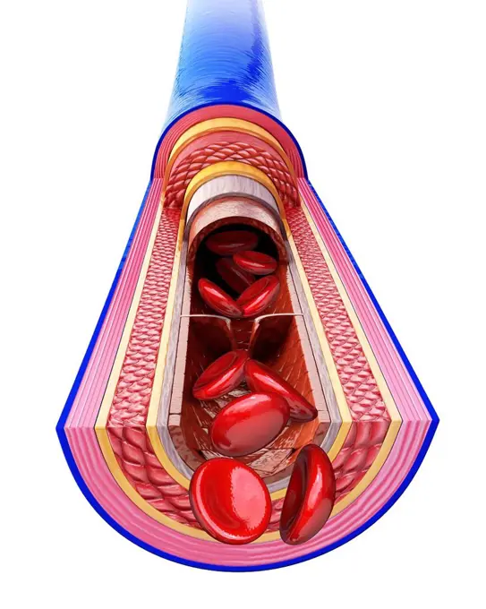
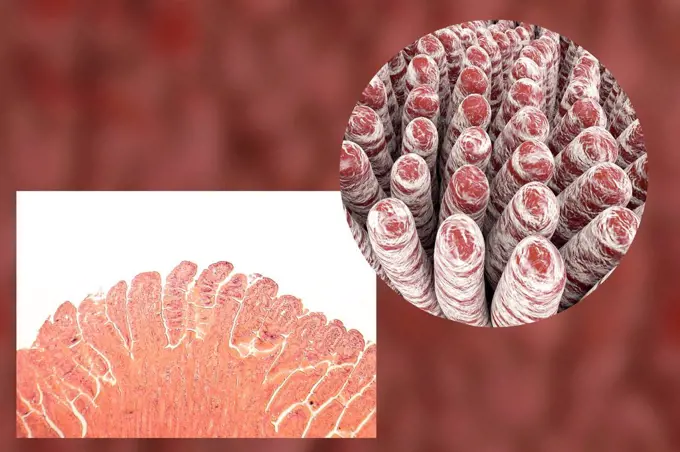
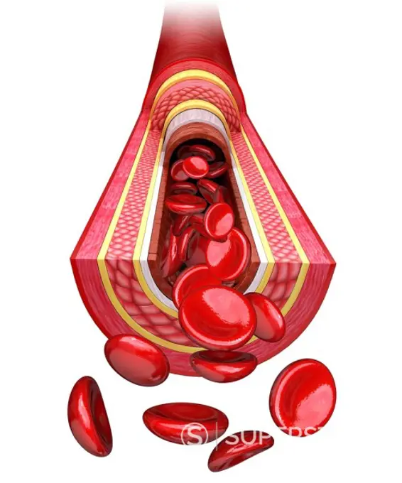
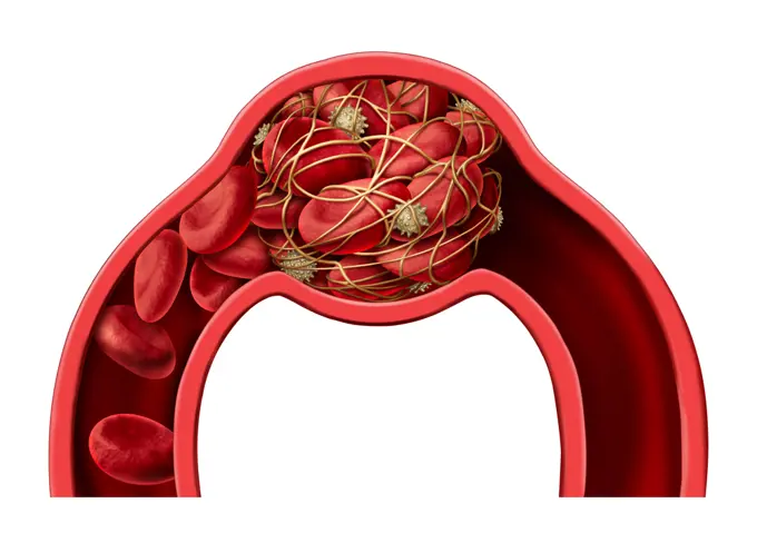
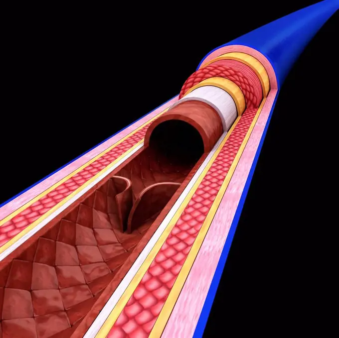
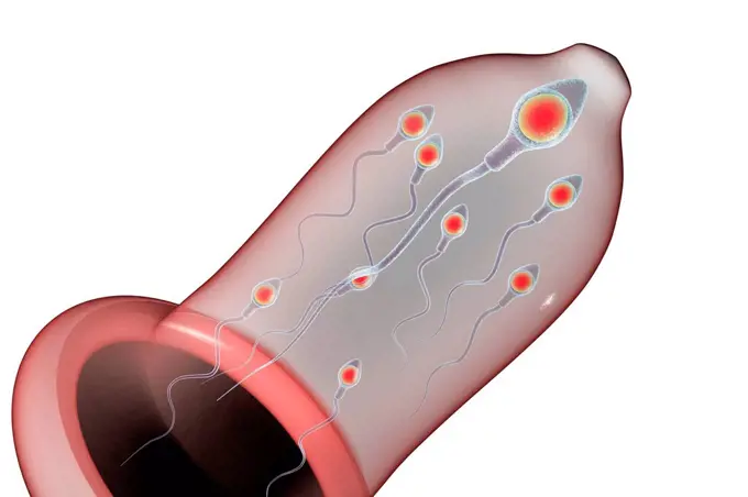
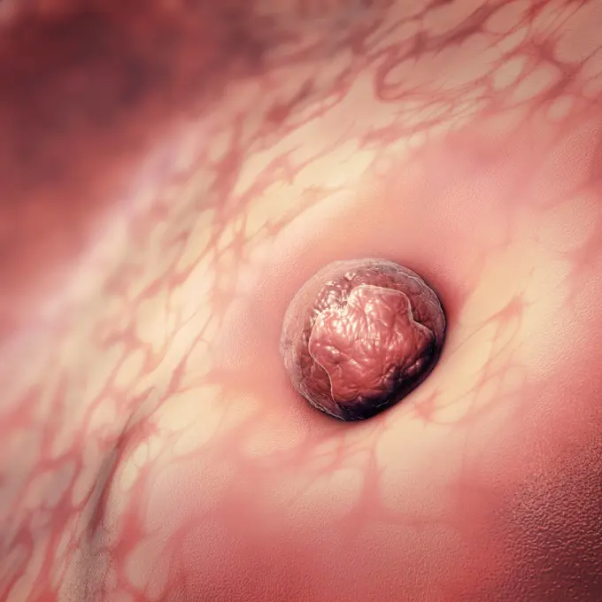
My Boards
View Pictures
  • My Boards

Human Anatomy Illustrations

Detailed illustrations of human anatomy, showcasing systems like the blood-brain barrier and reproductive structures, highlighted in vibrant and soft color gradients.

Female reproductive system, illustration.
159 assets in this story
4409-17348997
Pulmonary ventilation. Gas exchange.
4128R-28165
Healthy artery, artwork
1525-56996908
3D illustration. Fresh slices or meat. Marbling of pork or beef tenderloin for steak.
1428-160
Close-up of a human digestive system
824-63214050
Illustration of the formation of metastases : The primary tumour is in the breast. The primary cancer tumour develops : Step 1, the tumour is still small in size Step 2, the tumour has grown, the cancer cells have multiplied and transformed. Step 3, the tumour has grown even more. The cells with irregular nucleus are mature cancer cells (metastases) which will spread to other parts of the body through the blood vessel network. In the case of breast cancer, the metastases can remain dormant for several years in the brain, liver and bones, or can proliferate immediately and form a secondary cancer tumour.
4128R-11292317
Human femur, computer artwork.
824-63198624
Illustration of amyloid plaques forming. From the APP membrane protein (amyloid precursor protein), two enzymes, alpha secretase and beta secretase, isolate the Aß peptide (yellow), which sticks to it to form oligomers, before migrating towards dendrites on the surrounding neurons to form amyloid plaques.
1525-56722545
Glucose is the body primary source of fuel and is either converted into energy within cells 3D rendering. Glucose is the body primary source of fuel and is either converted into energy within cells
824-63189044
Illustration of the second phase of malaria transmission from one organism to another. The parasites (merozoites) move through blood vessels to the lungs where immune surveillance is lower and blood flow slower. They break out of their envelope (made by the liver). They penetrate red blood cells, ultra rapid transporters that allow them to avoid the filtering spleen. They proliferate in their shelter until, after 48h, they blow up the cell membrane, spread through the blood and quickly parasite other red blood cells to proliferate once more. An immune reaction starts up and the infected person has a high temperature. After a few cycles, the person is tired as the red blood cells no longer carry enough oxygen. Cell residue stuck to blood vessel lining risks blocking the vessels and producing a coma, or even killing the infected person. With each cycle, some merozoites change into asexual male or female gametes who cling to the lining of blood vessels close to the skin and wait for the r
824-63219043
Myocardial infarction, coronary artery with thrombus, atheromatous plaques, treatments. The atheromatous plaques (yellow) in the wall of the coronary artery cause coronary disease. In the zoom of the coronary vessel, a thrombus forms at the bottom right, in contact with the blood by platelet aggregation. Treatments act on platelets to prevent platelet aggregation, on the secretion of aldosterone by the kidneys and adrenals, but also upstream, on the liver by limiting the synthesis of cholesterol at the origin of atheromatous plaques.
824-63220101
Coronavirus replication, CoVid-19, SARS-Cov2. This scientific illustration explains how the 2019 coronavirus replicates thanks to a host cell. FUSION: When the virus reaches the respiratory system by inhalation, it fuses with a host cell thanks to its protein S (spicule), viral surface protein and a membrane receptor of the host cell. TRANSLATION: Viral RNA (positive strand) binds to ribosomes. It is transcribed to form an RNA replicase which will make RNA (negative strand) from RNA (positive strand). TRANSLATION: Viral RNA (positive strand) binds to ribosomes. It is transcribed to form an RNA replicase which will make RNA (negative strand) from RNA (positive strand). TRANSCRIPTION.ET RÉPLICATION: This negative strand is used to make RNA under genomics It allows to replicate viral RNA (positive strand). TRANSLATION:. Thanks to the ribosomes of the host cell, the viral RNA (positive strand) produces the viral proteins (N, M, E and S) which fuse with the endoplasmic membrane. The latter
1525-56195161
Heart cholesterol vector image
4128R-11289380
Human vein, cut-away computer artwork.
1525-26243989
High-density lipoprotein (HDL) cholesterol is known as good cholesterol because it helps remove other forms of cholesterol from your bloodstream. 3D illustration. High-density lipoprotein (HDL) cholesterol
1525-27083940
Group of Spermatozoons Flowing Toward Female Ovum Egg Isolated on White Background. Natural Fertilization Process Microscopic View. Reproductive Medicine Scientific 3d Vector Realistic Illustration.. Spermatozoons Group Flowing Toward Female Ovum Egg
4128R-13372973
Intestinal villi, light micrograph and 3D computer illustration. Villi are finger-like projections that greatly increase the intestinal surface area f...
1525-56722704
The mammary glands as modified sweat glands designed to produce and secrete milk. 3D rendering. The mammary glands as modified sweat glands designed to produce and secrete milk.
4128R-11479019
Female breast, cut away computer artwork.
4128R-11289322
Human artery, cut-away computer artwork.
1525-25421077
Blood clot disease symbol and thrombosis medical 3D illustration concept as a group of human blood cells clumped together by sticky platelets and fibrin creating a blockage in an artery or vein as a health disorder.
824-63175500
Exchange at the level of the pulmonary alveoli. Representation of a pulmonary alveolus and schematic zoom of the alveolus to show the exchanges between air and blood.
4239R-8148
Relation of thyroid and pituitary gland.
4128R-11289769
Human vein, cut-away computer artwork.
4128R-13575515
Contraception. Conceptual illustration showing a condom protecting against spermatozoans.
4239R-8126
Blood vessel anatomy.
6188-66486323
3D-Illustration einer Eizelle oder Eizelle, die an der Gebärmutter klebt 3D Illustration of an egg cell or ovum sticking to the uterus Copyright: xZoonar.com/ChristophxBurgstedtx 22602903
4128R-11313677
Human artery, cut-away computer artwork.
6188-65540500
Eine mit Malaria infizierte Stechmücke überträgt die Malariaerreger bei einem Mückenstich in die menschliche Blutbahn - 3D-Illustration A malaria-infected mosquito transmits malaria pathogens to the human bloodstream during mosquito bites - 3D illustration Copyright: xZoonar.com/ChristophxBurgstedtx 22579426
824-63188502
Representation of the transmission of nerve impulses at the level of the synaptic space, with dopamine (green) which activates postsynaptic dopaminergic receptors (beige).
5507-32893954
Human digestive system anatomy, infographics banner. Liver, stomach, pancreas, gallbladder, intestines.
6188-65540337
Eine mit Malaria infizierte Stechmücke überträgt die Malariaerreger bei einem Mückenstich in die menschliche Blutbahn - 3D-Illustration A malaria-infected mosquito transmits malaria pathogens to the human bloodstream during mosquito bites - 3D illustration Copyright: xZoonar.com/ChristophxBurgstedtx 22579341
824-63218990
Hematopoiesis, physiological process allowing the creation and renewal of blood cells or hematocytes. Stem cells come from the red bone marrow of the flat and short bones represented here by the pelvic bone. These stem cells give two types of cells: myeloid stem cells which give the differentiated cells following red blood cells or erythrocytes, platelets, white blood cells or leukocytes (polynuclear and monocytes) and lymphoid stem cells which give white blood cells (lymphocytes, which give NK cells (natural killers), T lymphocytes, B lymphocytes, plasma cells).
4128R-28814
Atherosclerosis. Computer artwork of a narrowed artery, due to a cholesterol plaque.
4128R-13376731
Tongue surface, illustration
824-63220048
Infectious conjunctivitis and its treatments. View of an eye with inflammation of the conjunctiva. Zoom on the conjunctiva with from top to bottom 1 bacterial infection (green) and 1 viral infection (red-orange). In yellow, purulent discharge due to the presence of bacteria (green), the cells of the conjunctiva are destroyed. Goblet cells secreting mucus and edema of the conjunctiva in the area infected with the virus. Right from top to bottom, a bacteria seen in section, a window with an infected connective cell (Herpes Simplex virus) detailing the process of viral replication transcription of viral DNA, DNA polymerase, viral replication
4128-38524194
Escherichia coli anatomy, illustration
4128R-11297058
Female reproductive system, computer artwork.
1525-56857786
Young Blood Transfusion and reversing aging process concept by transfusing younger plasma with healthy Chromosomes with long telomeres with old shorter telomere chromosome as a 3D illustration.
4128R-11315129
Human digestive system, computer artwork.
4128R-11289342
Human artery, cut-away computer artwork.
4128R-15290730
Haemorrhoids, illustration
4409-17349053
Capillary network.
1788-21803443
Gas exchange of oxygen and carbon dioxide in the pulmonary alveoli, human body, drawing.
4239-20481134
Detail of deep back muscles with a close-up of sprain, strain and spasm.
1525-25977424
Bone health and osteoperosis prevention concept as a healthy anatomy in a heart shape as a strong normal spongy tissue from a human skeleton structure as an icon for orthopedic and an orthopedist with 3D illustration elements.
824-63219981
Breast cancer and its treatments. A silhouette of a woman with a brain section with the pituitary gland. The pituitary gland stimulates the secretion of estrogens by the ovary (uterus in frontal section), which will bind to the estrogen receptors of cancer cells (bottom center). On the silhouette a mammary gland with the axillary ganglia which relate to it. A first zoom starts from this gland to show the anatomical structure of the mammary gland with a main galactophore channel receiving the lobules, themselves made up of acinus. An enlarged acinus is seen in section. A cancerous tumor is located on the main milk duct. In the foreground, zoom of a blood vessel surrounding cancer cells seen in section the tumor with its numerous vessels in the background. The HER2 receptor (green), EGFR (pink) and the estrogen receptor (orange) are present on the cancer cell whose nucleus is cut to show the mitotic spindle. A strand of DNA in the replication phase is extracted from a chromosome.
4128R-13244776
Infected human bronchus of the lungs, illustration.
824-63208818
Illustration of hemochromatosis, a hereditary genetic disorder affecting the HFE gene of chromosome 6 and causes an imbalance in transferrin carrying iron in the blood. This imbalance leads to excess iron absorption and an overload in certain organs capacity to store iron.
824-63175504
Representation of the intestine with cutout highlighting the villi and the external and internal blood circulation indicating the direction of circulation and the exchanges during the absorption of nutrients.
4128R-13387090
Illustration of teenager's infected bronchi.
4128R-11291112
Human muscular structure, computer artwork.
4239R-8149
Relation of thyroid and pituitary gland.
5507-47101827
Human veins and arteries illustration design
4128-18800943
Fibroadenoma in female breast, illustration
4128-20040455
Prostate cancer, illustration
4128-19358359
Bladder cancer stages, illustration.
4128R-14060517
Stages of liver disease in hepatitis C, illustration
5507-31222747
Vestor  illustration of normal aorta
5507-46321278
Atherosclerotic plaque in blood vessel, good and bad lipids
PREVIOUS
of 2
NEXT
2401 S. Ervay, Suite 206
Dallas, Texas 75215
United States
Get Started
Free ResearchMy BoardsMy Cart
For Creators
How To License Your ContentContributor PortalFrame of Mind
Resources
API accessPricing
Contact
+1 866 236 0087help@viewpictures.co.uk Contact form
©2026 View Pictures. All Rights Reserved. -B
This site is protected by reCAPTCHA and Google Privacy Policy and Terms of Service apply.