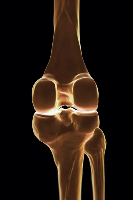
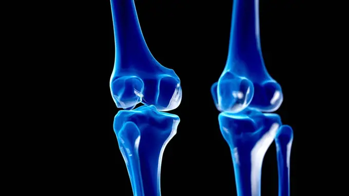
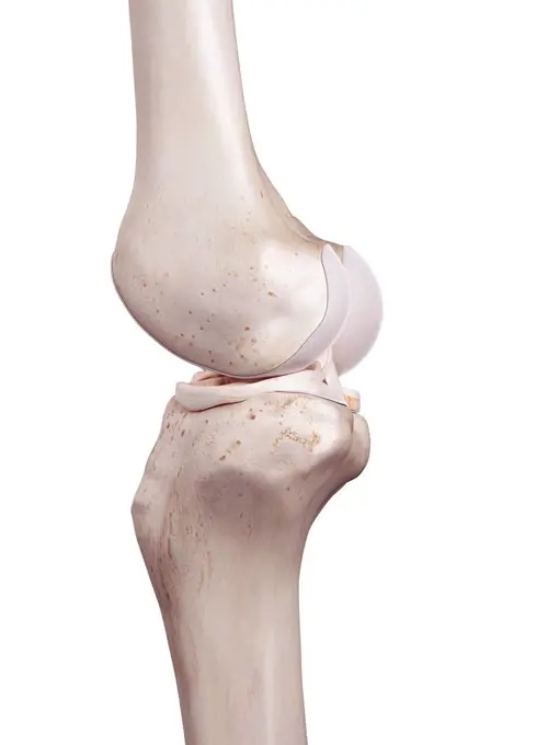
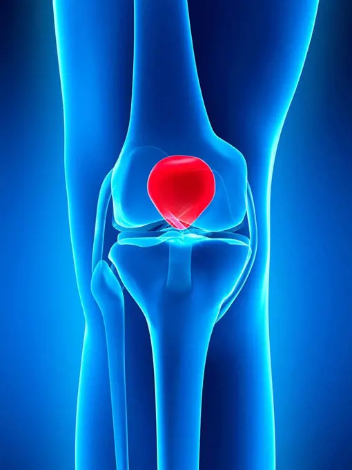
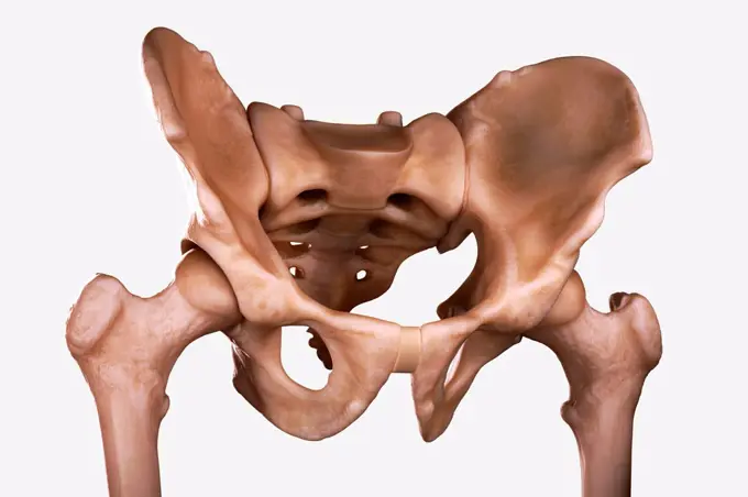
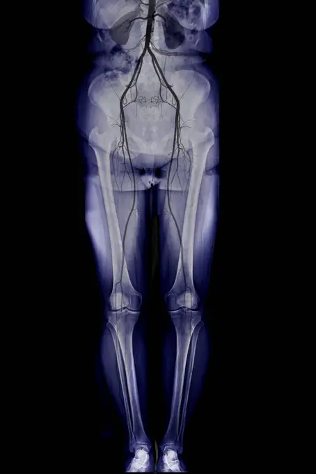
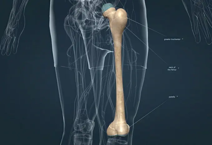
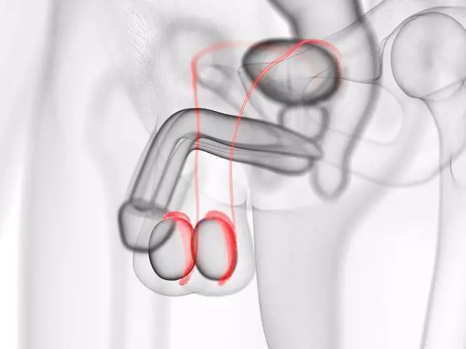
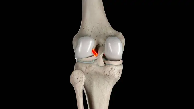
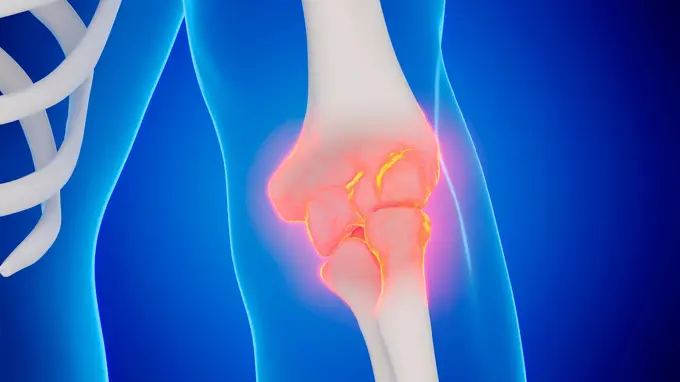
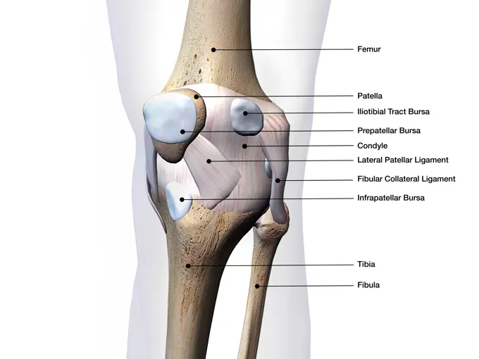
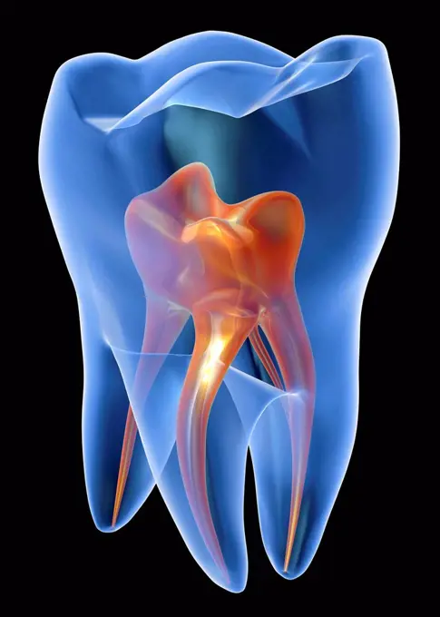
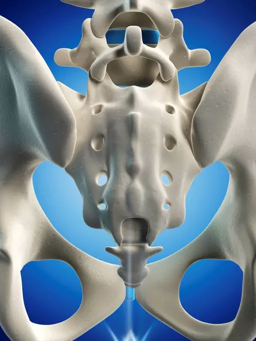
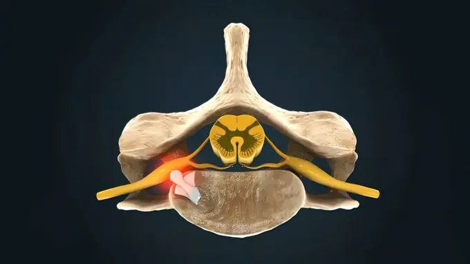
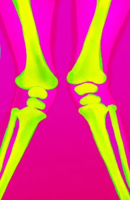
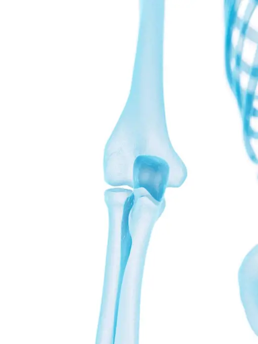
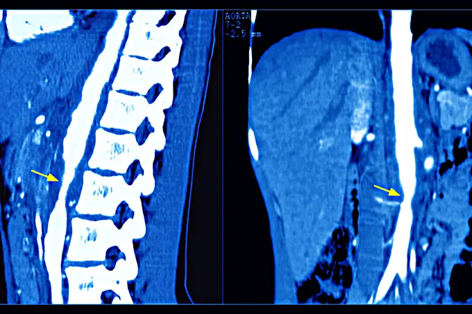
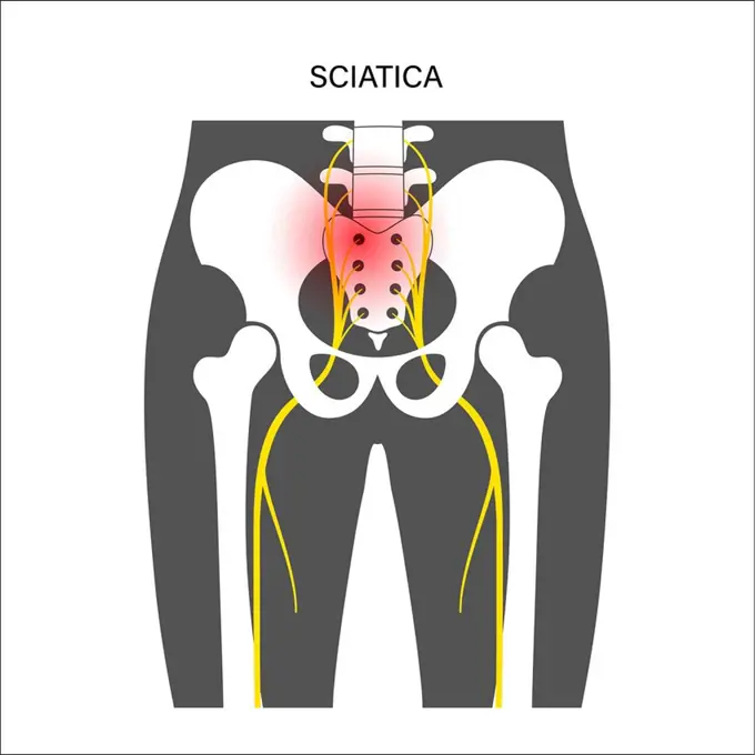
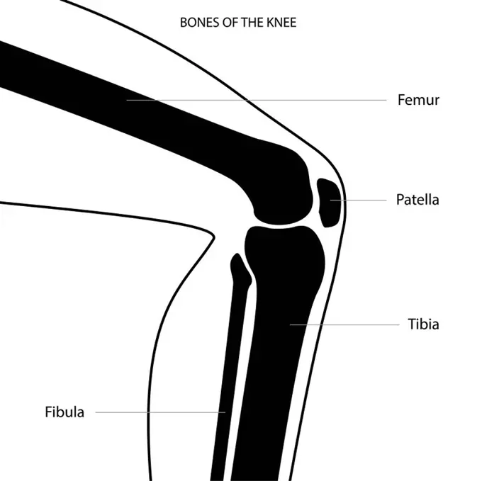
My Boards
View Pictures
  • My Boards

Human Anatomy Illustrations

Illustrations of various muscle groups and bones, including knee joints and pelvic structures, showcasing anatomical features with detailed graphics.

Model showing the human knee joint and its connecting bones.
161 assets in this story
4128-19056432
Knee joints, illustration
824-63170777
This sagital X-ray image of the knee shows a exostosis of the thighbone.
4128R-15515031
Illustration of the human knee
4128-19246806
Inflamed hip joint, illustration.
4128R-11472523
Human knee cap, computer artwork.
4128R-11541576
Human skeletal system, computer illustration.
4128R-13374637
Human knee joint
4128-15665707
Lower leg bones, illustration
4128-28968655
Fracture of the femur neck, illustration
4128R-13574806
Illustration of the vastus lateralis muscles.
4378-5176
Pelvis Joints
4128-19248131
Skeletal knee, illustration.
4128-28766923
Healthy torso and lower body, X-ray
4128R-11309504
Human knee tendons, computer artwork.
4128-111578576
Elbow joint, computer illustration.
4128R-30120
Knee joint, computer artwork.
4128-28968807
Anatomy of the pelvis bones, illustration
1899-85841
Pelvis X Ray
1525-56184120
The patella, also known as the kneecap, is a flat, rounded triangular bone which articulates with the femur and covers and protects the anterior articular surface of the knee joint. 3d illustration. Human Anatomy, Bony Pelvis and Lower Limb, Knee Patella
4128-15666150
Epididymis, illustration
4128R-13577537
Illustration of the iliofemoral ligament.
4128-30422358
Anterior cruciate ligament, illustration
1439-57940799
Monochrome Xray image of a human knee joint
4128R-11309488
Human knee tendons, computer artwork.
824-63225295
Osteosynthesis of the hip by a nail-plate prosthesis, following a fracture of the neck of the femoral head. x-ray of the left hip.
4128-24795315
Cyamella, X-ray
824-63178441
Coloured Colour Magnetic Resonance Imaging (MRI) scan of a side view of the knee joint of an adult man.
824-63197038
X-ray of the pelvis showing an intrauterine device (IUD).
4128R-13022259
Male penis anatomy, illustration.
4128-30415823
Skeletal knee, illustration
4297-1895
Colorized x-ray of a woman wearing high-heeled shoes
4128-28514569
Legs, CT scan
4128-30420432
Knee bones, illustration
4128-30422362
Posterior cruciate ligament, illustration
4128-30416469
Male elbow joint, illustration
4128R-12964599
Human knee joint, Illustration
1428-67304574
Knee Joint Bone and Connective Tissue Labeled on white background, 3D Rendering
4128-111577862
Adductor brevis muscle, computer illustration.
1525-20460812
human bones radiography scan. x-ray  image
4128R-13572785
Illustration of the gluteus maximus muscle.
1848-73120245
Image converted using ifftoany
4128-30415820
Knee joint, illustration
4128R-11827
Molar tooth
4128R-26457
Male skeleton, computer artwork.
1525-23843047
Human Spine In Details: Vertebra, Bone Marrow, Disc And Nerves 3d illustration. Human spine in details: Vertebra, bone marrow, disc and nerves
4252-4215
Legs deviation
4128R-11324432
Human elbow joint, computer artwork.
4128R-13572666
Illustration of the medial cuneiform bone.
4269-27541
Fractured pelvis. 3D Computed Tomographic (CT) reconstruction scan of the pelvis showing a fracture of the left ischium (at right).
4269-25433
Epicondylitis. 3D computed tomographic (CT) scan reconstruction of the elbow showing the head of the humerus (top), and the tips of the radius and ulna (below), the bones of the forearm. The inflammation of the epicondyle and surrounding tendons further to muscle overwork is called epicondylitis, or tennis elbow.
4252-4227
The pelvis
824-63206987
Takayasus arthritis, seen by 2 adominal scanners (sagittal section on the left and frontal on the right), at the descending aorta.
824-63178611
Recolorized radiograph of the left hip showing a leg fracture with displacement in a 66-year-old patient.
1525-26274970
Cross section through a typical synovial joint showing the bone synovial membrane synovial fluid cartilage and ligament 3d illustration. Cross section through a typical synovial joint showing the bone synovial membran
4269-25434
Epicondylitis. 3D computed tomographic (CT) scan reconstruction of the elbow showing the head of the humerus (top), and the tips of the radius and ulna (below), the bones of the forearm. The inflammation of the epicondyle and surrounding tendons further to muscle overwork is called epicondylitis, or tennis elbow.
4269-27530
Fractured pelvis. 3D Computed Tomographic (CT) reconstruction scan of the pelvis showing a fractured left iliac wing (at right) and a fractured ischium.
1525R-175136
4128-20042520
Sciatica, illustration
4128-19490286
Knee anatomy, illustration
6188-58103614
Transparent Human Body with Visible Bones medical science image of human skeleton bones Copyright: xZoonar.com/StanislavxRishnyakx 13526667
4413-17244
Large plan of the draft of a Cow of race Holstein France
PREVIOUS
of 2
NEXT
2401 S. Ervay, Suite 206
Dallas, Texas 75215
United States
Get Started
Free ResearchMy BoardsMy Cart
For Creators
How To License Your ContentContributor PortalFrame of Mind
Resources
API accessPricing
Contact
+1 866 236 0087help@viewpictures.co.uk Contact form
©2026 View Pictures. All Rights Reserved. -A
This site is protected by reCAPTCHA and Google Privacy Policy and Terms of Service apply.