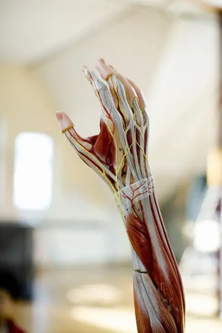
My Boards
View Pictures
  • My Boards

Human Anatomy Muscular Illustrations

Detailed illustrations of human muscles, including biceps and triceps, showcasing muscle structure and movement in a clear, educational style.

Human Extensor hallucis longus muscle computer artwork.
114 assets in this story
4128R-13375141
Neck muscle
4128R-13374505
Human neck muscles, illustration.
4128R-11477782
Human sacrum, computer artwork.
4128-28766989
Femoral artery, CT scan
4128R-13374890
Human skull
4128R-12700041
Cross section through an model of an normal upper right femur or leg bone.
4378-1455
A human model showing the leg and foot muscles.
1899-53513449
Newborn bone tissue, bone development, cartilage structure, pediatrics. This illustration shows the anatomy of a humerus in frontal section. From birth to childhood, the bone structure is cartilaginous and flexible. The center of the bone presents a vascularized medullary cavity. Compact bone is formed around this cavity (beige area in the center). At each end of this area is the epiphyseal cartilage (in pink). This area is more important in infants than in children. It has disappeared in the adult. Then the ends of the bone (epiphyses) are very vascularized. It is at this level that the secondary ossification center is located. The bone can thus grow gradually with the child. A cut section is indicated at the level of the shaft of the bone. It is shown top view at the bottom of the illustration. This makes it possible to see the structure of the bone in horizontal section. The bone is covered with a membrane, the periosteum. Then a part of compact bone, and in the center the medullary
4128R-11292328
Human femur, computer artwork.
6145-45250557
BALLISTIC IMPACT OF MONOLYTHIC AND COMPOSITE PLATES
824-63224373
Bone development on a humerus at birth, in children and adults. This illustration makes it possible to compare the development of the bone during these three ages. From birth to childhood, the bone structure is cartilaginous and flexible. We see it thanks to the aspect of the bone at the level of the epiphyses and the diaphysis. But we can also see it thanks to the area shown in pink. This area constitutes the epiphyseal plate. It is this epiphyseal plate that produces cartilage. This allows the bone to gradually grow and ossify. Bone growth ends between the ages of 18 and 25. In adults, the epiphyseal cartilage has ossified. We can no longer distinguish an epiphyseal line. This line is visible with a lighter color between the epiphysis (head of the humerus) and the metaphysis. The metaphysis is the intermediate zone between the epiphysis and the diaphysis. The diaphysis represents the body of the bone. On each section of the humerus is shown a section of horizontal section in blue. At
4306R-9778
Anatomical model of an arm.
1525-56198101
Thoracic vertebrae compose the middle segment of the vertebral column, between the cervical vertebrae and the lumbar vertebrae. 3D illustration. Thoracic vertebrae compose the middle segment of the vertebral column, between the cervical vertebrae and the lumbar vertebrae.
4378-2210
Skeleton layered over a female body in a variation of the dolphin plank pose showing the skeletal alignment in this particular yoga posture.
PREVIOUS
of 2
NEXT
2401 S. Ervay, Suite 206
Dallas, Texas 75215
United States
Get Started
Free ResearchMy BoardsMy Cart
For Creators
How To License Your ContentContributor PortalFrame of Mind
Resources
API accessPricing
Contact
+1 866 236 0087help@viewpictures.co.uk Contact form
©2026 View Pictures. All Rights Reserved. -A
This site is protected by reCAPTCHA and Google Privacy Policy and Terms of Service apply.