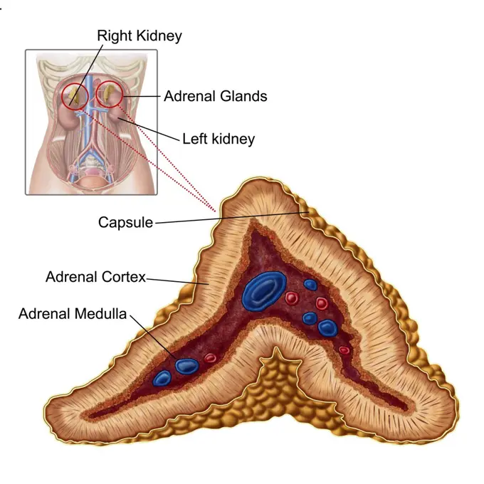
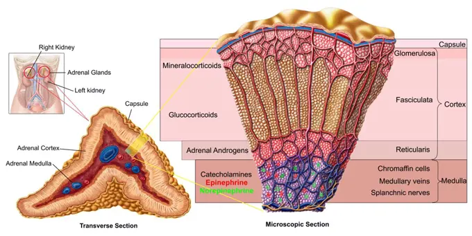
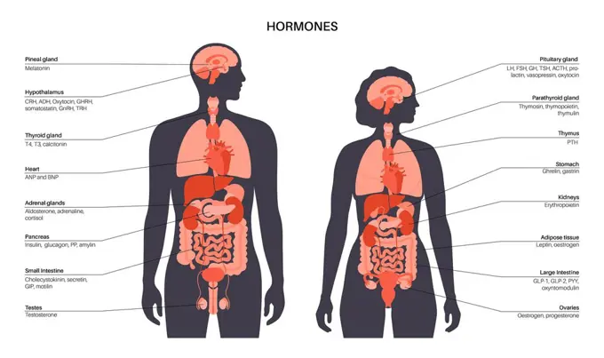
My Boards
View Pictures
  • My Boards

Human Anatomy Overview

Detailed illustrations of human organ systems, including the colon and kidneys, with labeled anatomical structures and functions.

Anatomy of adrenal gland, transverse section.
209 assets in this story
4239R-8139
Anatomy of human kidney function.
4128-28682162
Micturition neural control, illustration
824-63190803
Illustration of 2 nephrons and the urine collection system in the kidney.
4239-18641153
General colon anatomy, with labels.
824-57656239
Male genitals. Anatomy of the male genital organs: - in blue, corpus spongiosum and corpora cavernosa of the penis - behind the penis, in grey, testicle surmounted by the epididymis in pink, then by the vas deferens passing behind the seminal vesicle and opening into the prostate. - above the prostate, it is the bladder - in front of the bladder, the pubic symphysis, behind, the colon.
4239R-8147
Anatomy of prostate gland.
824-63175510
Representation of the circulatory system with the direction of circulation and the exchanges with the different organs, brain, lungs, kidney, intestine, muscles: oxygen supply, carbon dioxide rejection and waste evacuation.
824-63224374
Infant bone growth, bone development, cartilage structure, pediatrics. This illustration shows the anatomy of a humerus in frontal section. From birth to childhood, the bone structure is cartilaginous and flexible. The center of the bone presents a vascularized medullary cavity. Compact bone is formed around this cavity (beige area in the center). At each end of this area is the epiphyseal cartilage (in pink). This area is more important in infants than in children. It has disappeared in the adult. Then the ends of the bone (epiphyses) are very vascularized. It is at this level that the secondary ossification center is located. The bone can thus grow gradually with the child. A cut section is indicated at the level of the shaft of the bone. It is shown top view at the bottom of the illustration. This makes it possible to see the structure of the bone in horizontal section. The bone is covered with a membrane, the periosteum. Then a part of compact bone, and in the center the medullary
4239-18641937
Common locations of anal abscesses.
4239-18641139
General colon anatomy, with labels.
4239R-20483631
Anatomy of the organ of Corti, part of the cochlea of the inner ear.
4239R-8129
Anatomy of adrenal gland, transverse section.
4128-19358567
Breast cancer stages, illustration.
824-63219957
Asthma and its treatments. Representation of the lungs with the bronchial tree from which 3 zooms depart. Above left, a healthy bronchiole, below, two asthmatic bronchioles. An allergen (green star) triggers the asthma attack by activating lymphocytes producing IgE (green Y) leading to the degranulation of mast cells (purple) which release leukotrienes (black beads) and cytokines (blue beads) mediating inflammation of the mucous membrane which leads to bronchoconstriction of smooth muscle fibers, hypersecretion of mucus and obstruction of the respiratory tract.
824-63219040
Myocardial infarction, coronary artery with thrombus, atheromatous plaques, treatments. The atheromatous plaques (yellow) in the wall of the coronary artery cause coronary disease. In the zoom of the coronary vessel, a thrombus forms at the bottom right, in contact with the blood by platelet aggregation. Treatments act on platelets to prevent platelet aggregation, on the secretion of aldosterone by the kidneys and adrenals, but also upstream, on the liver by limiting the synthesis of cholesterol at the origin of atheromatous plaques.
1899-61461906
The periodontal pocket. Representation of the anatomy of the tooth and of a dental pathology, the periodontal pocket
824-63189084
Illustration of hormone control over the menstrual cycle. - The hypothalamic cycle : gonadotropin-releasing hormone (GnRH) released in the hypothalamus causes the hypophyseal secretion of two gonadotropin hormones (LH and FSH). - The hypophyseal cycle : The FSH (freen losange) intervenes in the development and maturing of one follicle per cycle. The LH (blue triangle) allows the formation of the corpus luteum. LH and FSH trigger ovulation. - The ovarian cycle : Oestradiol (purple square) causes positive retrocontrol on the secretion of LH that is responsible for ovulation. The corpus luteum secretes progesterone (pink circle) which causes negative retrocontrol on the hypothalamo-hypothyseal complex during the luteal phase.
4239-18642014
Structures of the inner ear.
4128-20042384
Arm anatomy, illustration
4128-19358509
Female breast anatomy, illustration.
1899-61461905
Path of sound from the ear to the brain. Anatomy of the ear, from left to right: - Outer ear and external auditory canal - Middle ear, eardrum (light purple), ossicles (beige), hammer, incus, stirrup, Eustachian tube (burgundy) - Inner ear, cochlea, vestibules and semicircular canals (purple) - Auditory nerve (yellow) With Zoom of the organ of hearing: - Cochlear canal (pink) containing the organ of Corti, including the hair cells (orange) - The cochlear nerve (yellow) transmits the electrical signal to the auditory area of the brain
4128-20042293
Arm anatomy, illustration
4128-19358575
Female radical mastectomy, illustration.
824-63220055
Brain and ovary, uterine endometrium, cervical mucus, targets of hormonal contraceptives. The hypothalamus regulates the secretion of FSH and LH from the pituitary gland. FSH and LH occur at the time of follicular phase and ovulation. In case of fertilization, the egg fertilized by the spermatozoa migrates to the uterus for its nesting in the endometrium. Hormonal contraception acts by blocking the hypothalamic-pituitary axis, blocking ovulation, rendering the endometrium unsuitable for nesting and / or modifying cervical mucus, making it unfit for sperm migration.
4239-18641946
Biomedical illustration of mammary duct ectasia in the female breast.
824-57656263
The digestive system. Representation of the digestive system from the mouth to the anus. We see the pancreas in transparency at the back of the stomach.
1899-61461914
Representation of the anatomy of the ear, from left to right: - external: pavilion, auditory canal, eardrum - middle: ossicles, Eustachian tube - internal: cochlea, auditory nerve
1525-56854468
The female breast anatomy includes internal milk ducts and glands and external nipples. 3D rendering. The female breast anatomy includes internal milk ducts and glands and external nipples.
4391-217
A diagram of the human female breast, including a cutaway, and subcutaneous details.
4128-19358576
Female radical mastectomy, illustration.
4239R-8122
Anatomy of adrenal gland, cross section.
824-63164266
Tooth: formation and development of dental caries.
4239-18641968
Full gastrointestinal system on female figure with detail of hiatal hernia.
4128R-28155
Head anatomy, computer artwork.
1848-53930910
Diphtheria of the tonsils and uvula, illustration from Meyers Konversationslexikon, a German encyclopedia, 1897
4128-19358519
Female breast cancer, illustration.
824-63175511
Representation of the circulatory system with the direction of circulation and the exchanges with the different organs (supply of oxygen and evacuation of waste in the form of carbon dioxide and urine.
824-63181807
Illustration of a network of presynaptic and postsynaptic neurons attached to each other by the intersynaptic space which allows nervous impulses to flow, from the upper neuron (top) to the lower neuron (bottom).
4128-19358511
Female breast anatomy, illustration.
1525-26034201
Hand drawn illustration of the torso muscles isolated on white, artistic anatomy graphic study
1525-27129021
GASTROINTESTINAL TRACT Medicine Education Diagram Vector Set
1848-77405786
3D Illustration Concept of Female Urinary System Kidneys with Bladder Described with Labels Anatomy
4128-19490877
Nasal epithelium, illustration
5507-33678349
Cycle of human hair growth, infographic process with layers of skin, hair root, epidermis
4239-18641122
Colored base of human skull, inferior view, with labels.
4391-321
A cutaway drawing of the male urogential system, from the kidneys to the testicles.
824-63190801
Illustration of the various cerebral herniations (complications due to intracranial pressure). From top to bottom: -cingulate herniation -uncal herniation -central herniation -tonsillar herniation
4128-19358505
Female breast anatomy, illustration.
4128-19358540
Mastitis, illustration.
4128-20040724
Prostatitis, illustration
4239-18641608
Facial muscles of the human face (with labels).
824-63219973
Colorectal cancer and its treatments. Above right, the colon in radiological type from where a zoom of a colon seen in section with 2 cancerous tumors at 2 different stages highlighting the vascularization of the tumor. In the window at the bottom left, zoom in on one of the blood capillaries of the tumor with details of the transcription replication process thanks to VEGF (blue beads), a vascular growth factor produced by the tumor, which will bind to the VEGF receptor ( blue) present in the vessel wall. The tumor cell also produces the growth factors EGF (yellow beads) which, by binding to the EGF receptor (yellow) of the tumor cell, causes a cascade of signals responsible for cell proliferation and resistance to apoptosis.
4128-28575188
Lymphatic system, illustration
1525-27084008
Woman Breasts Anatomy. Cross Section Close Up View of Female Breast with all Important Components. Highly Detailed Vector Realistic Illustration. Medical Aid Banner, Visual Educational Material.. Woman Breasts Anatomy. Cross Section Close Up View
4128-19358521
Female breast cancer, illustration.
1525-27160149
Hand drawn vector illustration of Wuhan corona virus, covid-19. The entry of the virus into the lungs through breathing. Protect with medical mask. White background and black outlines.
4128-20040739
Prostatitis, illustration
824-63219034
Asthma, Normal bronchiole, asthmatic bronchiole with bronchoconstriction, hypersecretion, treatments. Representation of the lungs with the bronchial tree from which 3 zooms depart. Above left, a healthy bronchiole, below, two asthmatic bronchioles. An allergen (green star) triggers the asthma attack by activating lymphocytes producing IgE (green Y) causing the degranulation of mast cells (purple) which release leukotrienes (black beads) and cytokines (blue beads) mediating inflammation of the mucous membrane which leads to bronchoconstriction of smooth muscle fibers, hypersecretion of mucus and obstruction of the respiratory tract.
1525-26529698
Skin anatomy. Human normal skin dermis epidermis adipose layers recent vector biological infographic. Illustration anatomy skin human epidermis structure. Skin anatomy. Human normal skin dermis epidermis adipose layers recent vector biological infographic
6188-67437915
3D Isometrische flache konzeptionelle Illustration des Oropharynx, Bildungs-Schemabr3D Isometrische flache konzeptionelle Illustration des Oropharynx, Bildungsschema 3D Isometric Flat Conceptual Illustration of Oropharynx, Educational Schema 3D Isometric Flat Conceptual Illustration of Oropharynx, Educational Schema Copyright: xZoonar.com/AnastasiiaxTorianykx 22973335
824-63219979
Breast cancer and its treatments. A silhouette of a woman with a brain section with the pituitary gland. The pituitary gland stimulates the secretion of estrogens by the ovary (uterus in frontal section), which will bind to the estrogen receptors of cancer cells (bottom center). On the silhouette a mammary gland with the axillary ganglia which relate to it. A first zoom starts from this gland to show the anatomical structure of the mammary gland with a main galactophore channel receiving the lobules, themselves made up of acinus. An enlarged acinus is seen in section. A cancerous tumor is located on the main milk duct. In the foreground, zoom of a blood vessel surrounding cancer cells seen in section the tumor with its numerous vessels in the background. The HER2 receptor (green), EGFR (pink) and the estrogen receptor (orange) are present on the cancer cell whose nucleus is cut to show the mitotic spindle. A strand of DNA in the replication phase is extracted from a chromosome.
4239R-20483612
Anatomy of the cochlear duct in the human ear.
824-63175498
Representation of the respiratory system during inspiration (left silhouette) and expiration (right silhouette), left side, lung lodged in the rib cage, right side lung in section with bronchioles and some alveoli. Zoom in on the level of the pulmonary alveoli.
4409-17349067
Salivary glands.
4128-19358536
Fibrocystic breast disease, illustration.
4239-18641605
Facial muscles of the human head (with labels).
824-63219960
Asthma, Normal bronchiole, asthmatic bronchiole with bronchoconstriction, hypersecretion, treatments. Representation of the lungs with the bronchial tree from which 3 zooms depart. Above left, a healthy bronchiole, below, two asthmatic bronchioles. An allergen (green star) triggers the asthma attack by activating lymphocytes producing IgE (green Y) causing the degranulation of mast cells (purple) which release leukotrienes (black beads) and cytokines (blue beads) mediating inflammation of the mucous membrane which leads to bronchoconstriction of smooth muscle fibers, hypersecretion of mucus and obstruction of the respiratory tract.
1848-78927468
Anatomy in the animal kingdom, detailed anatomical overview of an earthworm, including its internal structure and a cocoon, Lumbricus terrestris, reproduction of a historical school picture, mural or teaching board, authentic, exact date unknown
824-63190784
Illustration of the various sensitivities of the lemniscus system. They are made of axons that leave special skin receptors (fine touch) and receptors located in the joint capsules and ligaments (conscious proprioreceptive sensitivity). They ascend the spinal cord, penetrating the medulla oblongata and relay to the nucleus gracilis and cuneiform. After crossing on the median line (sensitive decussation), these pathways form the medial lemniscus (Reils ribbon) which goes all the way up the brainstem behind the pyramidal tracts to reach the thalamus, the last relay before the parietal lobe in the cortex.
5507-43151542
Irritable bowel syndrome (IBS) in a large intestine
824-63220059
Brain and ovary, uterine endometrium, cervical mucus, targets of hormonal contraceptives. The hypothalamus regulates the secretion of FSH and LH from the pituitary gland. FSH and LH occur at the time of follicular phase and ovulation. In case of fertilization, the egg fertilized by the spermatozoa migrates to the uterus for its nesting in the endometrium. Hormonal contraception acts by blocking the hypothalamic-pituitary axis, blocking ovulation, rendering the endometrium unsuitable for nesting and / or modifying cervical mucus, making it unfit for sperm migration.
4128-28575155
Lymphatic system, illustration
5507-33664153
Human head hair growth cycle. Biological catagen, telogen phases. Hair infographics.
4128-19490367
Human hormones, illustration
4128-20040826
Prostatitis, illustration
1538R-57922
4430-10695
zoology/animals monkeys chimpanzees left: musculature of head & neck right: median cut through head drawing after Professor G. Ruge to Hans Kraemer ""Weltall und Menschheit"" Berlin 1900,
824-57656258
Vascularization of the retina. Highlighting the retina, the cells of the retina and its vascularization.
5507-31177560
Sectional view illustration of stomach cancer (gastric cancer)
5507-31071928
Human skin anatomy. Layered epidermis with hair bulb, sweat and sebaceous glands, artery, nerve and veins. Epidermis, dermis, hypodermis. Vector illustration
4128-20042326
Arm anatomy, illustration
4128-20040817
Male reproductive system, illustration
4128-19497191
Arterial line catheter, illustration
1525-25872591
Human hair anatomy infographics with cross section of skin layers hair follicle bulb and shaft and sebaceous gland realistic vector illustration. Realistic Human Hair Anatomy Infographics
4128-19358556
Invasive ductal carcinoma, illustration.
4128-20040539
Pituitary gland anatomy, illustration
5507-33665527
3D structure of the hair skin scalp, anatomical education infographic information poster vector illustration.
4409-17349022
Digestive process-1.
824-63190810
Lateral illustration of the larynx, from top to bottom: - hyoid bone - thyrohyoid membrane - epiglottis - the aryepiglottic muscle - the oblique and transverse arythenoid muscles - the thyroarytenoid muscle - thyroid cartilage - the lateral cricoarytenoid muscle - the posterior cricoarytenoid muscle - cricoid cartilage - the cricothyroid muscles
4297-1678
Anatomical illustration of the underside of the human brain
5507-42307650
Diagram of human bone anatomy
4128-19358436
Serotonin pathway in the human brain, illustration.
4128-19358546
Types of breast cancer, illustration.
4128-20042511
Pelvis anatomy, illustration
4409-17349019
Swallow-1.
4128-19490906
Pineal gland, illustration
4128-18498290
Hair strand structure, illustration
5507-45577502
Anatomy of the ear.
4128-19358416
Serotonin pathway in the human brain, illustration.
824-63175538
Representation of the organization of the visual cortex. In the vision mechanism, the hypothalamus (small purple circle) regulates circadian rhythms (cycles of active and non-active periods), the pretectum (small orange circle) controls the pupil and lens reflex, the colliculus upper (green oval) directs eye and head movements.
PREVIOUS
of 3
NEXT
2401 S. Ervay, Suite 206
Dallas, Texas 75215
United States
Get Started
Free ResearchMy BoardsMy Cart
For Creators
How To License Your ContentContributor PortalFrame of Mind
Resources
API accessPricing
Contact
+1 866 236 0087help@viewpictures.co.uk Contact form
©2026 View Pictures. All Rights Reserved. -A
This site is protected by reCAPTCHA and Google Privacy Policy and Terms of Service apply.