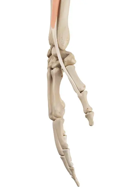
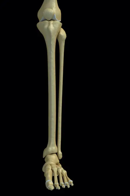
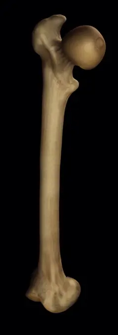
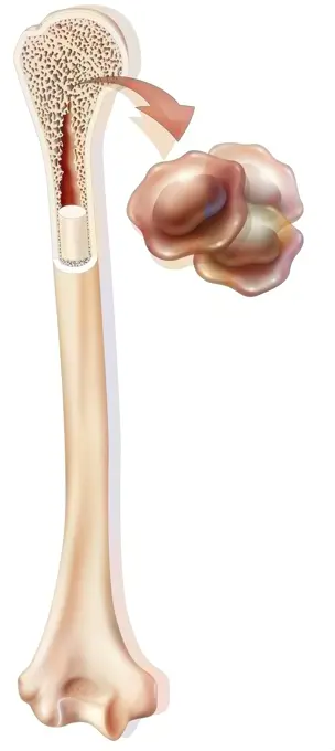
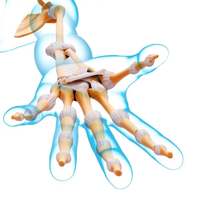
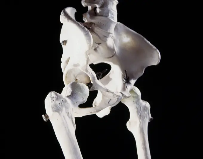
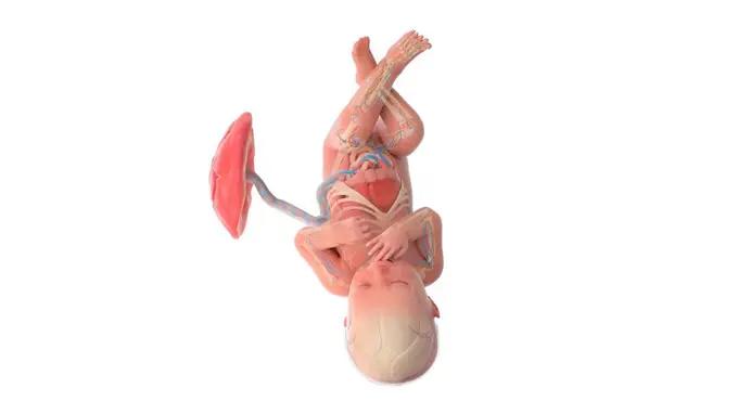
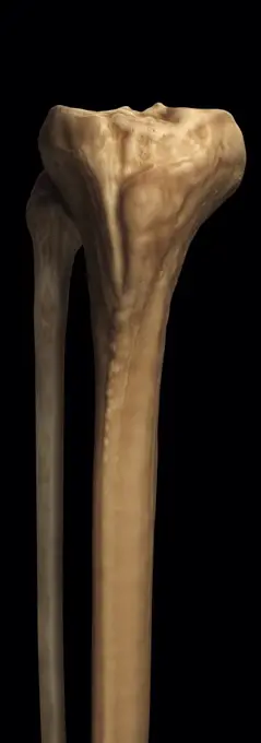
My Boards
View Pictures
  • My Boards

Human Arm Anatomy Illustrations

Detailed illustrations of human arm anatomy, highlighting muscles, bones, and the elbow joint. The images showcase a scientific and educational style.

Human hand anatomy, computer illustration.
162 assets in this story
4128R-12577457
Human spine, computer illustration.
4378-570
Front view of the bones of the left lower leg.
4128R-12964595
Human foot bones, Illustration
4128R-13370736
Human hand anatomy, illustration.
4128R-12580703
Human leg artery, computer illustration.
824-63167656
Spinal column. The spinal column is constituted of 7 cervical vertebrae, 12 thoracic vertebrae, 5 lumbar vertebrae, sacrum and coccyx. The sacrum results from the junction of the five vertebrae, the sacral vertebrae. The sacrum ends by the coccyx, also formed by the junction of various vertebrae.
4378-2996
Model of the right femur bone.
4462-21958589
Human hand bones
4378-1436
A human model showing the hand muscles.
1899-53509147
Hematopoiesis, bone marrow in the humerus producing blood cells.
4128R-32392
Human hand bones, computer artwork.
4128R-11287446
Thigh (femur) bone, computer artwork.
6188-60019174
3d rendering of a isolated human arm in on white background 3d rendering of a isolated arm in on white background Copyright: xZoonar.com/HugoxKurkx 14605285
4128-19053254
Lumbar spine, illustration
4128R-12576954
Human foot anatomy, computer illustration.
4128-30418081
Frog's skeletal system, illustration
1899-13156
Muscles of the calf
4128R-31581
Human hand bones, computer artwork.
4128R-12581033
Human hand anatomy, computer illustration.
4378-20013633
Pelvis Joints
4220-21334636
Human Skeleton.     Date:
4128-19247486
Human foetus anatomy at week 36, illustration.
4128-19053186
Scapula, illustration
824-63224379
Adult bone tissue, bone structure, bone development, cartilage structure, humerus. This illustration represents the bony structures of the humerus in an adult. At the top, the proximal epiphysis is covered with cartilage. An epiphyseal line (whiter) separates the epiphysis from the metaphysis. Epiphysis and metaphysis are made up of cancellous bone. Then the body of the bone is the diaphysis made up of compact bone. In the center, the medullary canal is largely vascularized. The bony areas of spongy and compact bone are covered by the periosteum (in dark beige). The lower part of the humerus is full, with the disc epiphysis covered with articular cartilage (trochlea). It constitutes the upper part of the elbow joint with the ulna and radius. A horizontal section is shown at the level of the shaft of the humerus. This cut at the bottom of the illustration shows the bone structure with the periosteum on the periphery (dark beige). Then the interior is made up of compact bone around the m
4128R-12580702
Human arm arteries, computer illustration.
4378-2804
Model of the tibia and fibula bones that form part of the human leg.
4378-2688
Model showing the human knee joint and its connecting bones.
1746-19665179
Ancestor figure from Latmul region of Papua New Guinea, 20th Century. This was a person of high rank indicated by the scars on his thighs
1525-27722609
Arm bones and its parts with wrist fingers, human body bone of different types humerus, radius ulna internal framework isolated on vector illustration. Arm Bones and Parts with Wrist Vector Illustration
4378-5259
Pelvis Joints
4378-289
Rear view of the bones of the lower legs.
4378-2462
Model showing the human knee joint and its connecting bones.
4128R-31896
Human foot musculature, computer artwork.
4128R-12581031
Human arm muscles, computer illustration.
4128-19053208
Atlas vertebrae, illustration
4128R-11287434
Thigh (femur) bone, computer artwork.
4128R-11323714
Human serratus anterior muscles, computer artwork.
4413-124134
Longitudinal cut of a Corn seed during germination
4128-19247394
Human foetus at week 24, illustration.
4128R-11292033
Human femur, computer artwork.
4128R-11289196
Leg bones, computer artwork.
4128-30418125
Male skeletal system, illustration
4128R-11322814
Human sacrum bone, computer artwork.
4128R-13723599
Illustration of male urinary system.
4128-30418075
Frog's nervous system, illustration
4128-30418549
Shark's skeletal system, illustration
1746-30004511
Fossil of a Pteranodon (flying reptile) of Late Cretaceous period.
4128R-32808
Human hand bones, computer artwork.
4443-73253681
Gautier, Jean-Baptiste, Essai d'anatomy en tableaux imprimés: Figure 15, verso - stamped, Anatomical muscle model of a torso, print, prints, height 606 mm, width 460 mm, French, 1775 - 1825, print maker, 1745 - 1745, paper, color mezzotint
4128-19247505
Human foetus anatomy at week 38, illustration.
1899-53513455
Bone structure at birth, bone development, cartilage structure, pediatrics. Infant skull in top view, fontanel, sutures, pediatrics. This anatomical illustration shows a newborn baby's skull viewed from above. The front of the skull is visible at the top of the drawing thanks to the nose represented. The skull is made up of bony plates, but also of fontanelles and sutures made of connective tissue. These structures give it great elasticity, allowing the baby to pass through the pelvic strait during birth. From top to bottom, we can distinguish the metopic suture, then the anterior fontanel. Coronal sutures are drawn on each side of the anterior fontanel. Then behind the anterior fontanel, it is the sagittal suture. This sagittal suture runs from the anterior fontanel to the posterior fontanel. Two lambdoid sutures, not shown here, start on each side of the posterior fontanel. In the baby, the bones of the skull are not united. This allows the skull to grow according to the rapid growth
1525-56198104
Thoracic vertebrae compose the middle segment of the vertebral column, between the cervical vertebrae and the lumbar vertebrae. 3D illustration. Thoracic vertebrae compose the middle segment of the vertebral column, between the cervical vertebrae and the lumbar vertebrae.
4128R-36563
Human leg bones, computer artwork.
4128-30415617
Bladder and urethra, illustration
1525-21250958
Human thorax
4128-19053206
Atlas vertebrae, illustration
4413-124132
Longitudinal cut of a Corn seed during germination
824-63148692
Heart, illustration
4239-69146188
Cordaites prehistoric plant from the Carboniferous period.
4409-61880666
Triceratops foot, Paleontological Center , Enciso, La Rioja , Spain, Europe.
1525-21045137
man dressed as a pilot on a white background
1848-77403949
3D Illustration Concept of Human Skeleton System Appendicular Skeleton Anatomy
PREVIOUS
of 2
NEXT
2401 S. Ervay, Suite 206
Dallas, Texas 75215
United States
Get Started
Free ResearchMy BoardsMy Cart
For Creators
How To License Your ContentContributor PortalFrame of Mind
Resources
API accessPricing
Contact
+1 866 236 0087help@viewpictures.co.uk Contact form
©2026 View Pictures. All Rights Reserved. -A
This site is protected by reCAPTCHA and Google Privacy Policy and Terms of Service apply.