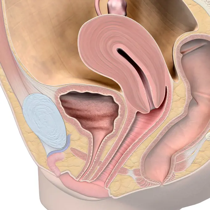
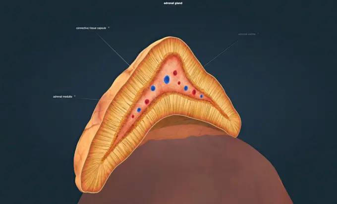
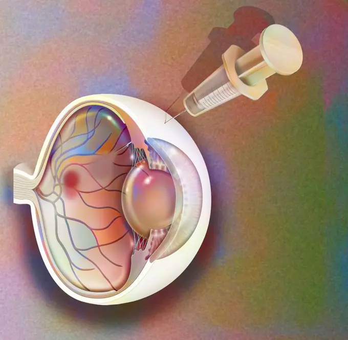
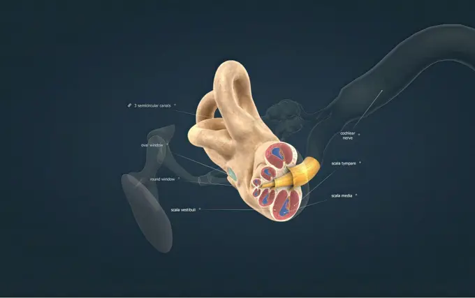
My Boards
View Pictures
  • My Boards
← Science

Human Body Anatomy Illustrations

Medical illustrations depicting the anatomy of the nose, male and female reproductive systems, and ear structures, showcasing internal body features.

Female reproductive system, medical illustration
215 assets in this story
1525-56211211
The adrenal glands produce corticosteroid hormon and epinephrine, which maintain blood pressure and regulate metabolism 3D illustration. The adrenal glands produce corticosteroid hormon and epinephrine, which maintain blood pressure and regulate metabolism
4128R-13723556
Illustration of male urinary system.
824-57656230
Urinary incontinence. Representation of the urinary system seen from the front in a woman's body with two zooms showing: - at the top the anatomy of a non-incontinent woman with a well-toned pelvic floor. - below, the pelvic floor is distended and allows urinary leakage: incontinence during effort (sports activity, cough, etc.)
824-63164593
Treatment of macular degeneration by injection of RNA interference.
1525-56211171
The semicircular canals of each ear contain three main parts: anterior, posterior, and horizontal canals. 3D illustration. The semicircular canals of each ear contain three main parts: anterior, posterior, and horizontal canals.
4239R-20483608
Medical ilustration of an appendix with appendicitis.
1848-54945823
Display of the upper jaw model on the monitor
1848-54919801
Depiction of an upper jaw model
1525-20579545
Coronary artery disease medical concept as a circular vein with gradual plaque formation resulting in clogged arteries and atherosclerosis with with a three dimensional human anatomy diagram showing the risks of cholesterol buildup.
4128R-11295049
Male reproductive system, computer artwork.
824-57656256
The path of sound in the ear. Representation of the path of sound in the ear. The sound wave in the outer ear (in orange) captured by the pinna of the ear enters the auditory canal, vibrates the eardrum which isolates the outer ear from the inner ear, and vibrates in the middle ear (in blue) the ossicles (hammer, anvil, stirrup). The vibrations of the stapes vibrate in the inner ear (in pink) the membrane of the oval window which generates hydraulic waves in the perilymph of the cochlea, which exerts pressures of the endolymph inside the cochlear canal , displacing the basilar membrane, causing the hair cells to move against the tectorial membrane producing nerve impulses in the cochlear nerve fibers. This nerve impulse travels through the auditory nerve, then from neuron to neuron via the spinal cord to the auditory area of the brain which analyzes the data.
1525-23789206
The vagina is a muscular, hollow tube that extends from the vaginal opening to the uterus.3d illustration. A female’s internal reproductive organs are the vagina, uterus, fallopian tubes, and ovaries.
824-63164036
Anatomy of the inner ear showing the eardrum, the cochlea.
824-63190782
Illustration of a paravertebral block (blue area), locoregional anaesthesia in the thoracic and lumbar regions.
1574R-018962
Earwax
PREVIOUS
of 3
NEXT
2401 S. Ervay, Suite 206
Dallas, Texas 75215
United States
Get Started
Free ResearchMy BoardsMy Cart
For Creators
How To License Your ContentContributor PortalFrame of Mind
Resources
API accessPricing
Contact
+1 866 236 0087help@viewpictures.co.uk Contact form
©2026 View Pictures. All Rights Reserved. -A
This site is protected by reCAPTCHA and Google Privacy Policy and Terms of Service apply.