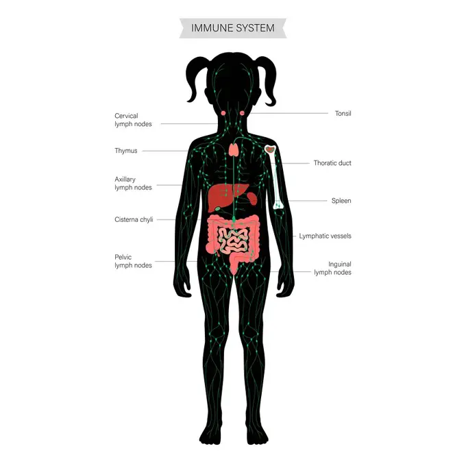
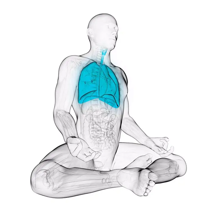
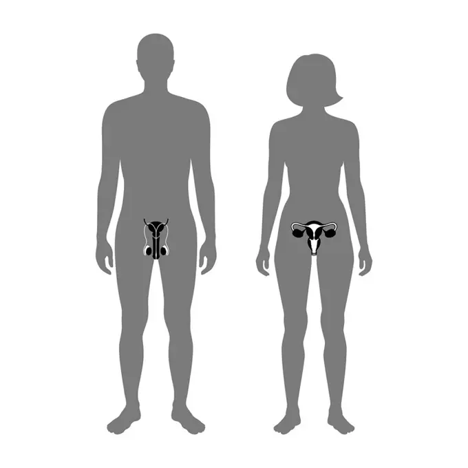
My Boards
View Pictures
  • My Boards
← Science

Human Body Systems Illustration

Detailed illustrations of the human body focusing on cardiovascular and respiratory systems, highlighting organs like the heart and lungs in various styles.

Human respiratory system, illustration.
280 assets in this story
824-63147239
Depiction of a cancerous tumour in the left lung.
824-63148716
Respiratory tract, drawing
4128-28970658
Cardiovascular system, illustration
4128R-11544320
Human chest muscles, computer illustration.
4128R-14058310
Male gallbladder, illustration
5507-31546821
Digestive system illustration. Female body anatomy map
4128-16171521
Computer illustration showing a female body with the internal organs.
4128R-11296033
Male anatomy, computer artwork.
4128R-12577512
Human arteries, computer illustration.
4128R-11296695
Male urinary system, computer artwork.
4128R-12577439
Human arteries, computer illustration.
4128R-11313326
Human respiratory system, computer artwork.
4239-18642099
Female anatomy of cardiovascular system with skeleton and internal organs. Ghost effect on white background.
4378-261
The upper body showing the skeleton and respiratory system.
4128R-13269322
Human spleen, illustration.
4128-30416706
Alimentary tract, illustration
4128-19055707
Illustration of a boy's lungs
4128-20040590
Pancreas, illustration
1525-56853869
Stage III testicular cancer is characterized by spread to distant lymph nodes or any solid organ. 3D rendering. Stage III testicular cancer is characterized by spread to distant lymph nodes or any solid organ.
824-63148697
Thorax, anatomy
1899-53510748
Lymphatic system in children, lymphatic network, lymph nodes, lymphoid organs. Medical illustration depicting the lymphatic system in a silhouette of a 3 year old child. This system is made up of a network of lymphatic vessels and lymphoid organs. There are two types of lymphoid organs. The primary lymphoid organs are the thymus and bone marrow. Lymphocytes are produced, develop and are selected from primary lymphoid organs. But there are also the secondary lymphoid organs. The secondary lymphoid organs are the spleen, lymph nodes, lymphoid tissues associated with mucous membranes (Peyer's plaque). These secondary lymphoid organs are the site of activation of naive lymphocytes, and therefore the starting point of the adaptive immune response. (Photo by: JACOPIN/BSIP/UIG)
6188-62298893
Male naked body standing, with full digestive system superimposed. Male naked body standing, with full digestive system superimposed. On white background with clipping path included. Copyright: xZoonar.com/Pixelchaosx 9887693
4128R-11542398
Human liver, computer illustration.
4128-28575196
Lymphatic system, illustration
4128-28575178
Lymphatic system and organs, illustration
4128R-10520
Digestive system, artwork
4128R-13723768
Illustration of male gallbladder.
824-63187037
Illustration of an aortic aneurysm.
4128-111582674
Lung anatomy, computer illustration.
4128R-14060229
Child's artery anatomy, illustration
4128R-13387270
Illustration of human lungs cross-section.
4128R-12580613
Human artery, computer illustration.
4128-28575207
Lymphatic system and organs, illustration
4128-28575220
Lymphatic system and organs, illustration
4128R-14060042
Baby's digestive system, illustration
4128R-13244879
Human kidneys, illustration.
4128R-11296279
Male anatomy, computer artwork.
4128-28682295
Gut brain connection, illustration
4128R-13268558
Human heart anatomy, illustration.
5507-43771070
child digestive anatomy
6188-66529005
Blood circulation, illustration Blood circulation, illustration 016889_244
1525R-176985
4128R-26631
Healthy stomach, computer artwork.
4128R-11295545
Male anatomy, computer artwork.
1899-53510763
Position of the heart of a 7-year-old child, pediatrics. The heart is located in the rib cage. It is shifted slightly to the left relative to the central axis represented by the sternum. Its apex is oriented downwards to the left. In the 7-year-old child, the apex is located at the level of the 5th intercostal space. While this cardiac apex is located at the level of the fourth intercostal space in the infant. In the 7-year-old child, the heart is in the same axis as that of the adult. (Photo by: JACOPIN/BSIP/UIG)
4128R-11475092
Baby's anatomy, computer artwork.
4128R-14060251
Child's heart and diaphragm, illustration
6188-66528846
Depiction of a healthy bronchial tube left and a bronchial tube during an asthma attack right. ASTHMA, DRAWING 016889_098
5507-31911099
Digestive system and intestines concept.
4128R-13244886
Human skeletal system, illustration.
4239R-20482704
Artist depcition of abdominal aortic aneuryism.
4128R-14060209
Baby's head and chest anatomy, illustration
4128R-13446738
Illustration of male lung anatomy.
1848-49200630
Medicine, human anatomy, dissection of the torso of a seated white man, showing the large blood vessels, organs, artery. Coloured lithograph by J. Maclise, 1841, 1844, Historic, digitally restored reproduction from a 19th century original
824-63147180
Depiction of a healthy bronchial tube (left) and a bronchial tube during an asthma attack (right).
4128-28575152
Lymphatic system, illustration
1501R-783
Acid Reflux 2 Linda Braucht (b.20th C. American) Computer graphics
4128R-26643
Healthy large intestine, computer artwork.
4128-28575195
Lymphatic system, illustration
4128-20045228
Human endocrine system, illustration
6145-29245344
Anatomical Illustration
6188-55458695
Anatomy Human Body Model Isolated Medicine human body model isolated on white background Copyright: xZoonar.com/DanielxFerreira-LeitesxCiccarinox 12726038
4128-30415620
Bronchi, illustration
4128-28575229
Lymphatic system and organs, illustration
824-63147139
Depiction of a healthy bronchial tube (left) and a bronchial tube during an asthma attack (right).
4128-16171507
Computer illustration showing a female torso with the internal organs, front view.
824-63187579
Illustration of the sites that metastases will migrate to in the case of breast cancer. Metastases from a primary tumour in the breast will migrate to the brain, bones and lioves. Nevertheless, the metastases are mammary in origin.
4128R-13267118
Respiratory system of a child, illustration.
4239R-9473
Pulmonary embolism, pathway of embolus to the heart and lungs.
824-63175533
The disturbing elements on the brain and the motor response. Representation of the mechanism of interfering elements on the motor response. Alcohol is involved in the transmission of nerve impulses (purple flash on the right). It modifies the perception of the environment and modifies motor commands. Fatigue intervenes directly at the level of the brain (violet flash above), modifies the perception of the environment and modifies motor commands. The noise via the receiving organ (ear - violet flash on the left), modifies the perception of the environment and modifies the motor commands.
4128-28575218
Lymphatic system and organs, illustration
4128R-22149
Healthy lungs, computer artwork.
4128-20040767
Male and female reproductive organs, illustration
4128-19053063
Lung, illustration
4128R-26637
Healthy liver, computer artwork.
4128R-13586499
Illustration of male pancreas.
4128R-12572251
Lungs of a boy, computer illustration.
1788-39549
Medicine - Pathology - Atherosclerosis, drawing
4391-327
A line drawing of the central area of the human digestive tract, including the intestines, liver, stomach, and pancreas.
4128-19497217
Venous access port, illustration
PREVIOUS
of 3
NEXT
2401 S. Ervay, Suite 206
Dallas, Texas 75215
United States
Get Started
Free ResearchMy BoardsMy Cart
For Creators
How To License Your ContentContributor PortalFrame of Mind
Resources
API accessPricing
Contact
+1 866 236 0087help@viewpictures.co.uk Contact form
©2026 View Pictures. All Rights Reserved. -A
This site is protected by reCAPTCHA and Google Privacy Policy and Terms of Service apply.