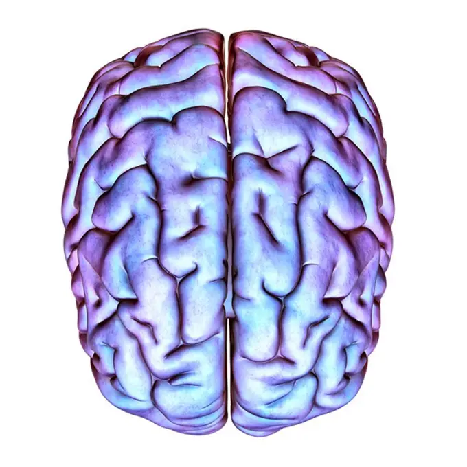
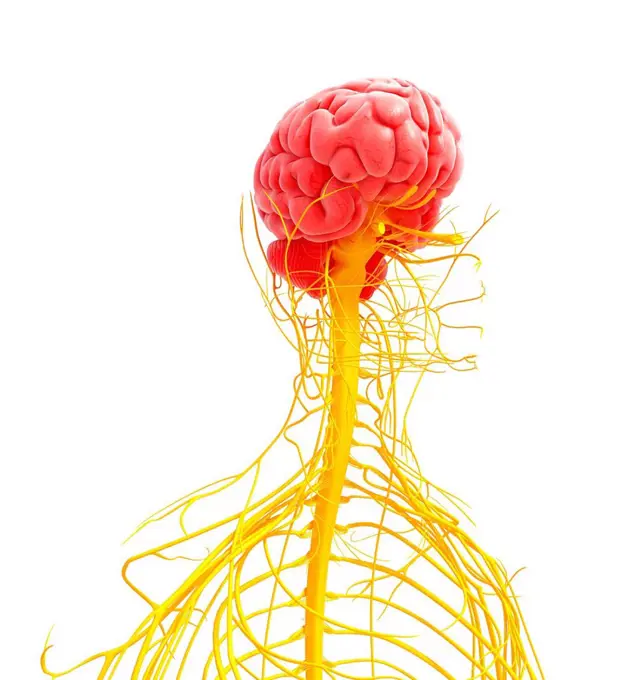
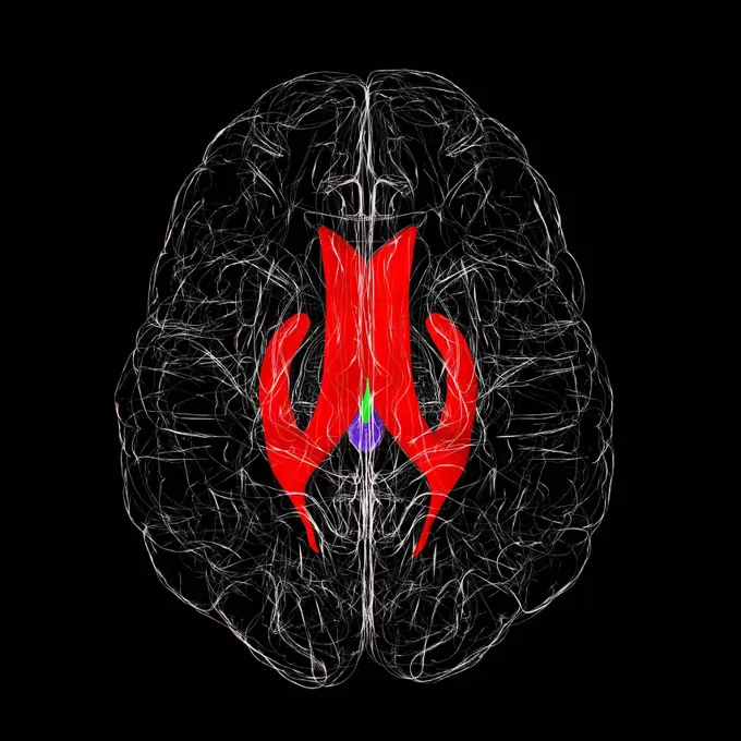
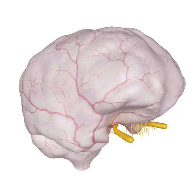
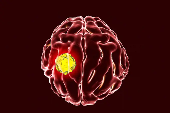
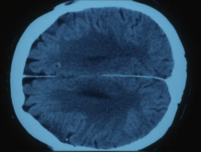
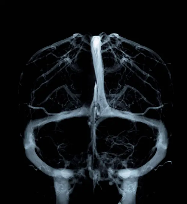
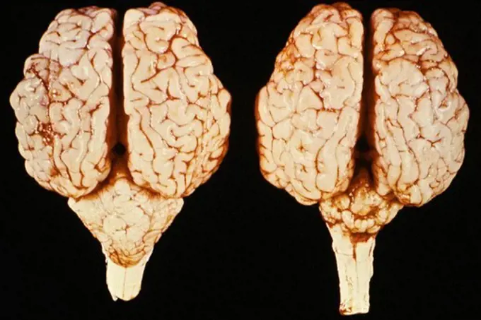
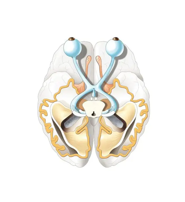
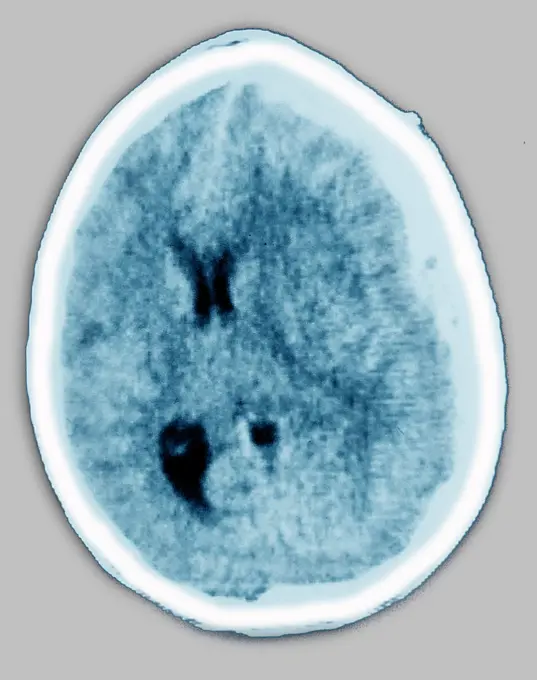
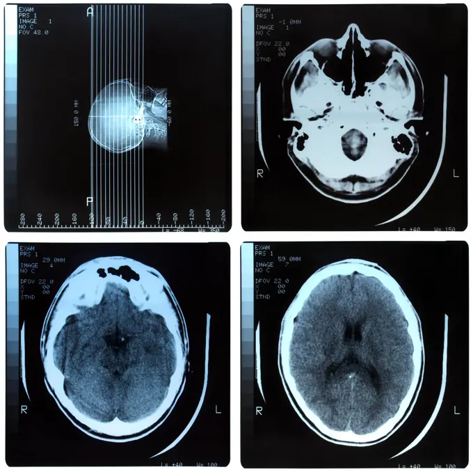
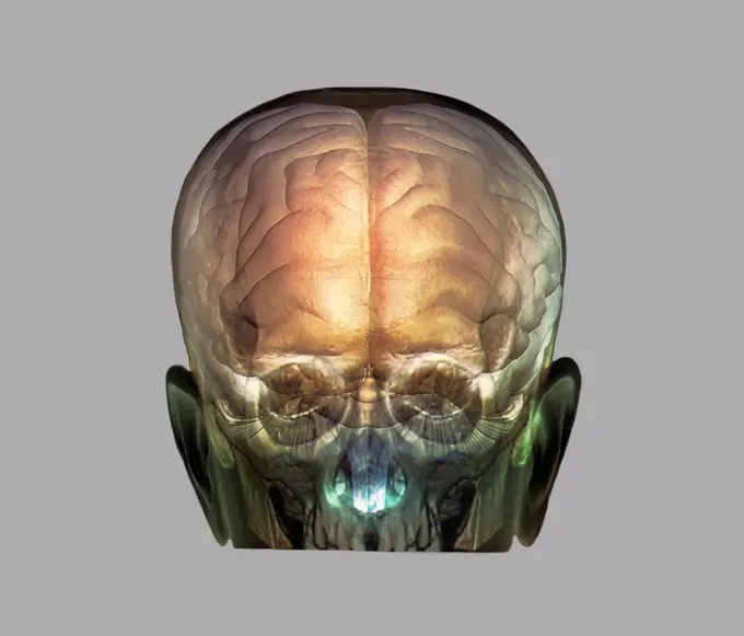
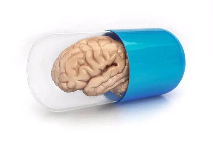
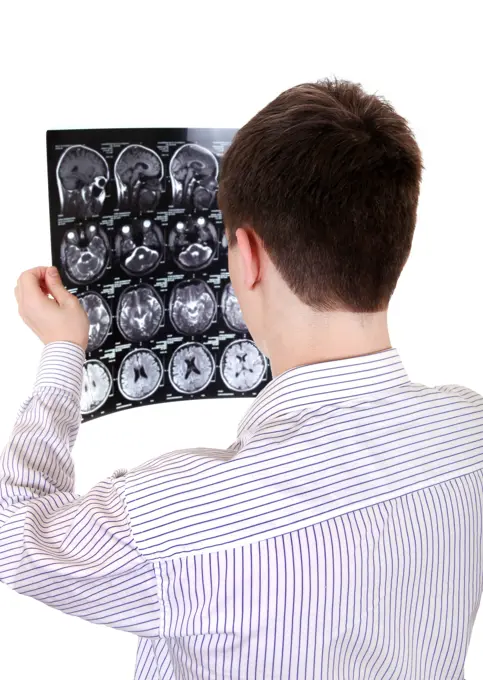
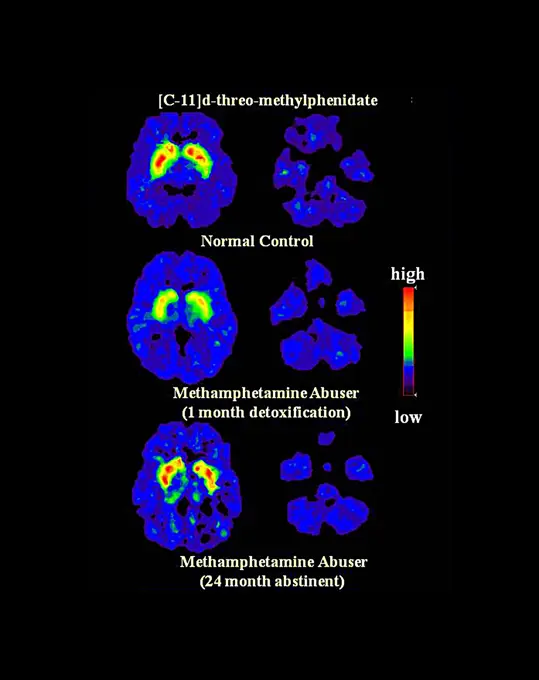
My Boards
View Pictures
  • My Boards

Human Brain Artwork

Vibrant computer-generated images of the human brain, illustrating various perspectives and highlighting intricate structures and electrical activity.

Human brain, artwork
248 assets in this story
4128R-14637810
Human brain arteries, illustration
6188-68116055
Zentrales Organ des menschlichen Nervensystems Anatomie des Gehirns 3D Illustration Concept of Central Organ of Human Nervous System Brain Anatomy LicenseRF Copyright: xZoonar.com/Magicxminex 23022887
4128R-11316447
Human nervous system, computer artwork.
4128-30422566
Ventricular system of the brain, illustration
4128R-15514929
Illustration of the dura mater
4128R-14057413
Brain cancer, illustration
1525-26034570
Brain with eyes. 3d render of brain with eyes on white background
824-63224714
CT scan of brain edema (abnormal accumulation of fluid in brain tissue in the left hemisphere). Radial CT scan of the skull.
4128-V58614919
6188-55645336
Computed tomography of the human head Computer tomography of the human head
4128R-30931
Human skull. Coloured 3D computer tomography (CT) and magnetic resonance imaging (MRI) scans of the skull and brain of a 35 year old patient.
4128R-13682547
Human brain, illustration.
824-63203262
Cerebral atrophy. Cortical-sub-cortical parietal atrophy as a result of an ischemic stroke, seen in a radial section MRI.
4128-30419901
Cerebral veins, MRV scan
4128-30415839
Skull and brain, illustration
1899-53511980
Thrombosis of the cerebral venous sinuses of the dura mater, visualized by cranial MRI angiography.
824-29057205
Thrombosis of the cerebral venous sinuses of the dura mater, visualized by cranial MRI angiography.
824-63188600
Localised malignant astrocytoma in the left hemisphere of the brain. Transversal MRI brain scan.
4384-289
One Healthy and one Infected Calf Brain
4128R-15516205
Illustration of a glass brain
4409-17348455
Optic nerve.
4128R-28946
Brain anatomy, computer artwork.
1899-53511479
CT scan of brain edema (abnormal accumulation of fluid in brain tissue in the left hemisphere). Radial CT scan of the skull.
824-63175226
Axial CT scan of the brain showing a subdural hematoma, a collection of blood between the dura and the arachnoid.
824-63128411
Scanner of a man pelvis and perineum. 1.Sartorius muscle (Great sartorius). 2.Right muscle of the thigh. 3.Tensor of the fascia lata. 4.Lateral great muscles of the lower limb. 5.Vastus intermedius muscle of the lower limb. 6.Obturator externus musc
4128R-12577880
Human brain with DNA (deoxyribonucleic acid), computer artwork.
824-63126591
Brain, drawing
6188-63344931
Intestine and digestive system protection and healthy bowel concept. Colon with arrow. Intestine and digestive system protection and healthy bowel concept. Colon with arrow. 3d illustration Copyright: xZoonar.com/MaksymxYemelyanovx 19630279
4128R-14057412
Brain cancer, illustration
4128-16506476
Brain neural network, computer illustration.
824-63188623
Damaged meniscus seen on a saggital plane MRI scan.
1899-20318750
A human brain left frontal view, emphasizing Broca's area, the first language area in the left hemisphere,
6188-55568422
Medical Xray Medical X ray imaging of human brain skull bones Copyright: xZoonar.com/ClaudioxDiviziax 2671693
4128R-30932
Human skull. Coloured 3D computer tomography (CT) and magnetic resonance imaging (MRI) scans of the skull and brain of a 35 year old patient.
4128R-14059028
Illustration of brain in transparent capsule
824-63193295
Using computer software programs, scientists combined brain MRIs from 20 healthy people into this composite image, in which ellipsoids represent normal anatomical variations. Pink purple ellipsoids, signifying the greatest variation, occur in brain regions that are uniquely human for example, regions that control language and logical reasoning. Blue ellipsoids, representing slight variations, occur in brain regions that control sensation and movements. Ultimately, this baseline data on interpersonal variability will allow scientists to distinguish normal anatomical variation from abnormal brain loss, such as that seen in Alzheimer's disease. Source: Paul Thompson, University of California, Los Angeles
4128-19055618
Illustration of the brain of a baby
6188-56081387
Human brain with measure tape around. 3D rendering Human brain with measure tape around. 3D rendering isolated on white background Copyright: xZoonar.com/alexlmxx 21257718
6188-67651558
Human brain with Ukrainian flag. Scientific research and education in Ukraine concept, 3D rendering Human brain with Ukrainian flag. Scientific research and education in Ukraine concept, 3D rendering Copyright: xZoonar.com/alexlmxx 23056773
6188-54894338
Teenager with Tomography Rear view of the Young Man with Tomography Isolated on the white background ,model released, Symbolfoto Copyright: xZoonar.com/AndreyxShadrinx 5754932 ,model released, Symbolfoto ,property released
824-66065387
Comparison of a healthy brain and a brain with severe Alzheimer's Disease.
1525R-58312
4128R-24440
Human brain, computer artwork.
824-66065389
NIH-funded scientists found that our brains may be uniquely sensitive to pitch, the harmonic sounds we hear when listening to speech or music.
824-63191274
Methamphetamine abuse decreases dopamine transporter activity and compromises mental function.
824-63194390
Images of dopamine transporter (DAT) binding in three brains: (1) a healthy control (top), (2) a methamphetamine abuser one month after discontinuing drug abuse (middle), and (3) a methamphetamine abuser after 24 months of abstinence (bottom).
824-63191276
Dopamine transporter loss after heavy methamphetamine use. Chronic methamphetamine abusers have significant reductions in the level of dopamine transporters.
824-57660094
Gross specimen of brain tissue from a patient who died of granulomatous amebic encephalitis caused by Balamuthia mandrillaris. The autopsy specimen revealed extensive necrotizing (mixed inflammatory, occasional giant cells, vasculitic) granulomatous encephalitis with a subependymal necroinflammatory process. Image courtesy of Cook Childrens Hospital, Fort Worth, Texas.
PREVIOUS
of 3
NEXT
2401 S. Ervay, Suite 206
Dallas, Texas 75215
United States
Get Started
Free ResearchMy BoardsMy Cart
For Creators
How To License Your ContentContributor PortalFrame of Mind
Resources
API accessPricing
Contact
+1 866 236 0087help@viewpictures.co.uk Contact form
©2026 View Pictures. All Rights Reserved. -B
This site is protected by reCAPTCHA and Google Privacy Policy and Terms of Service apply.