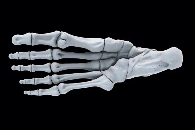
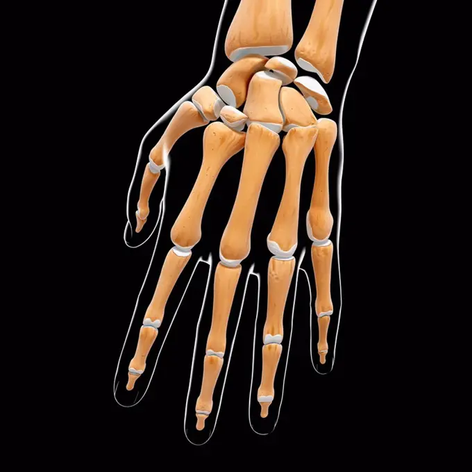
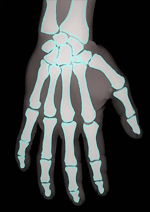
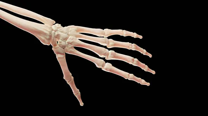
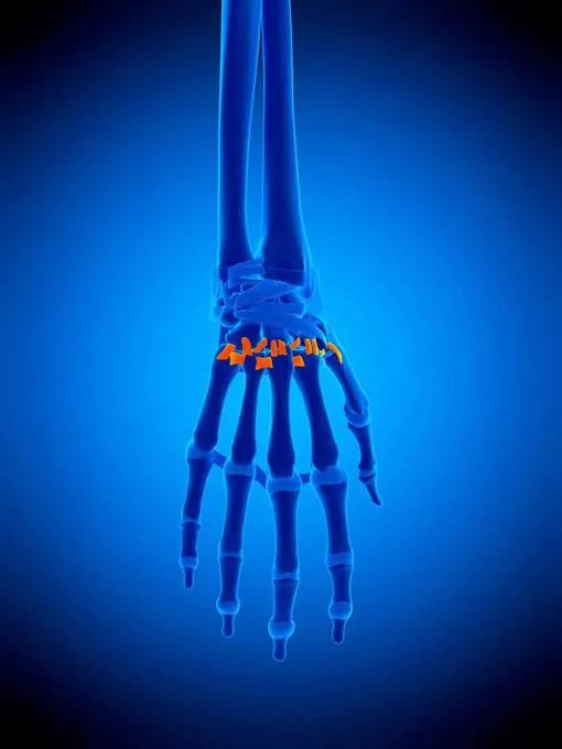
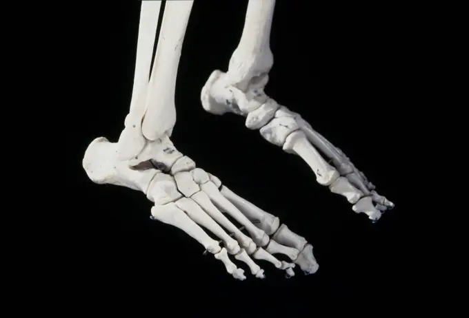
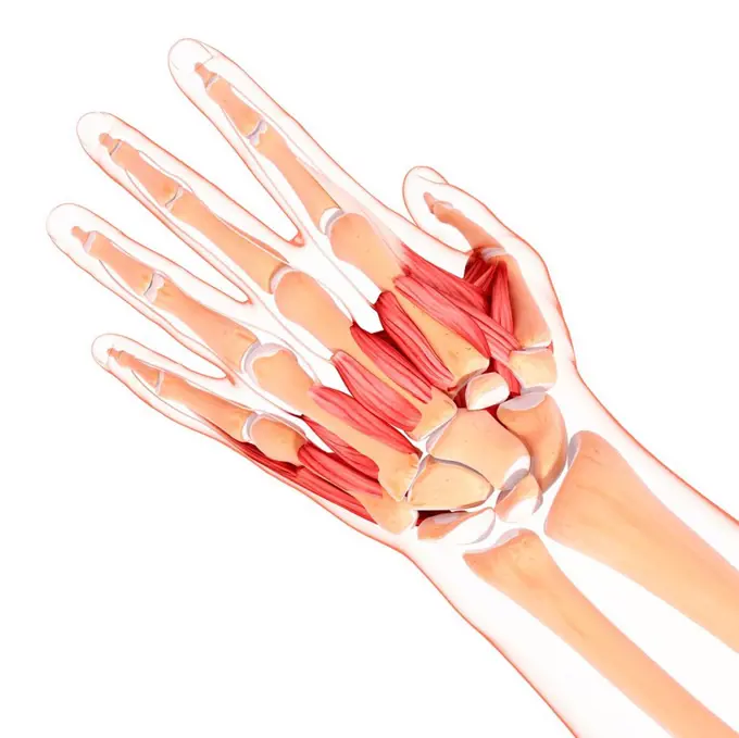
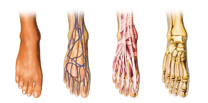
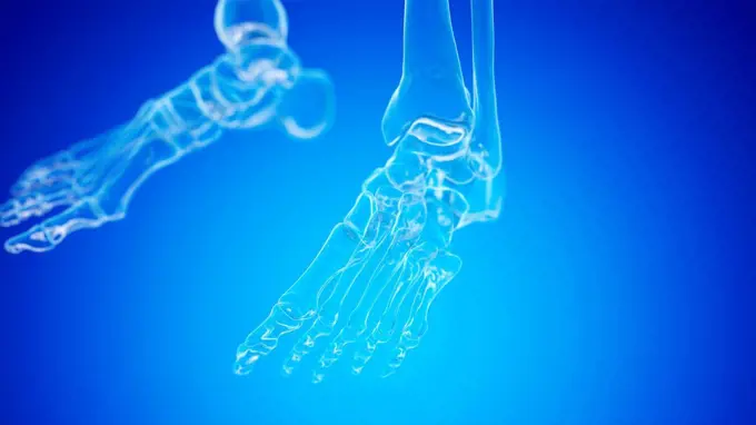
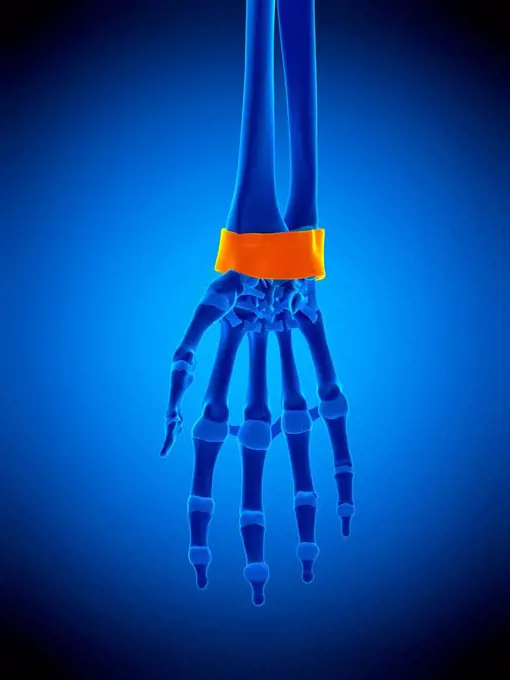
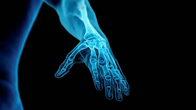
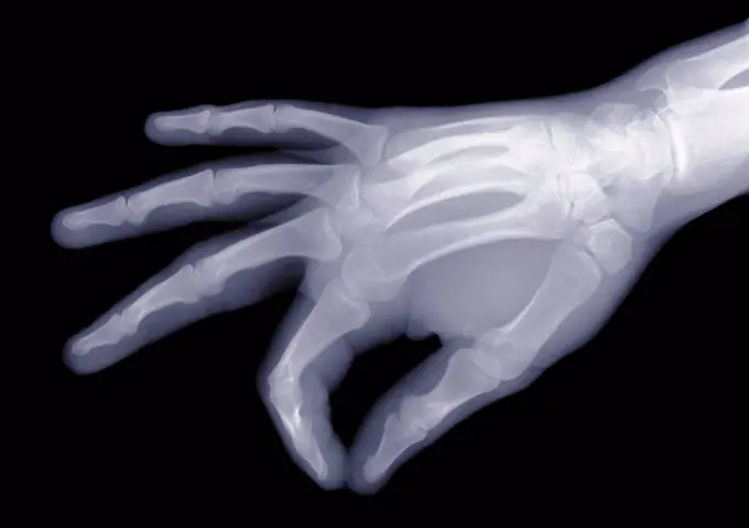
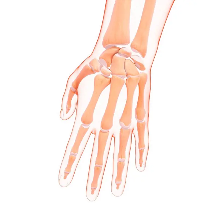
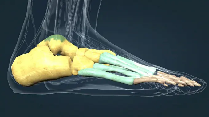
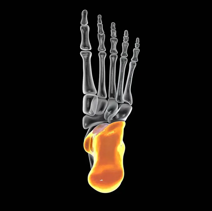
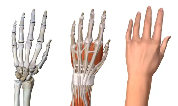
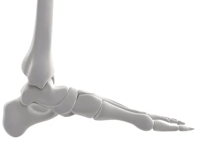
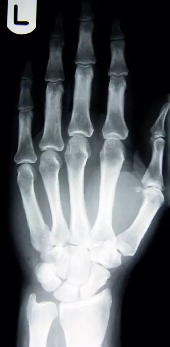
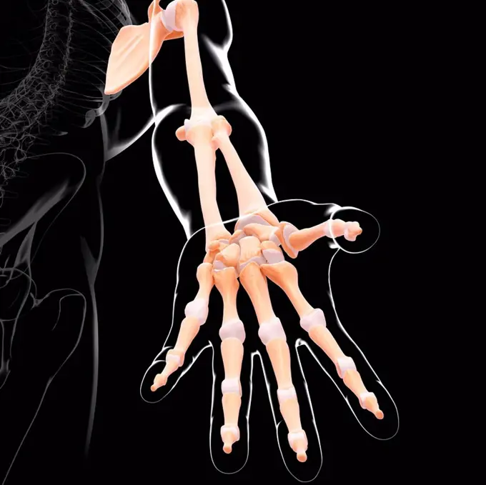
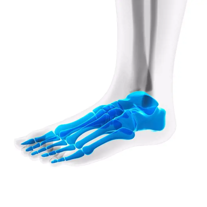
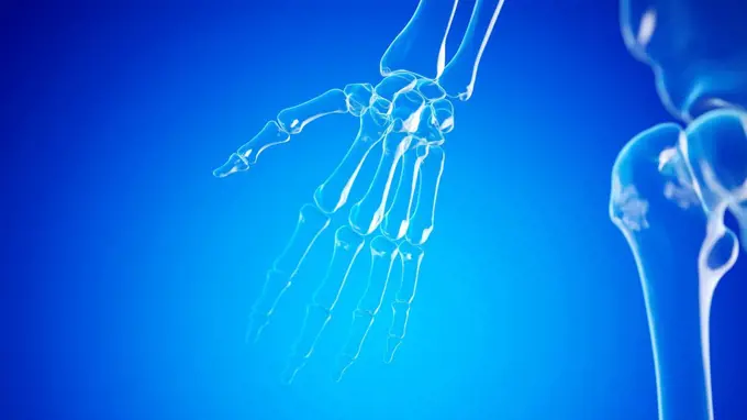
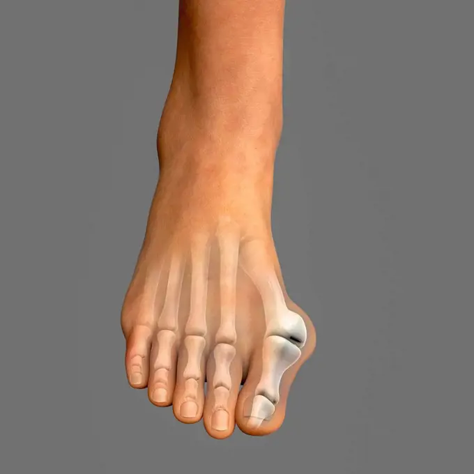
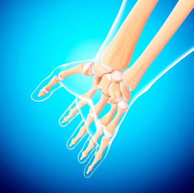
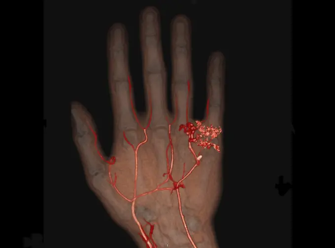
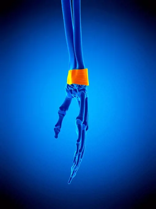
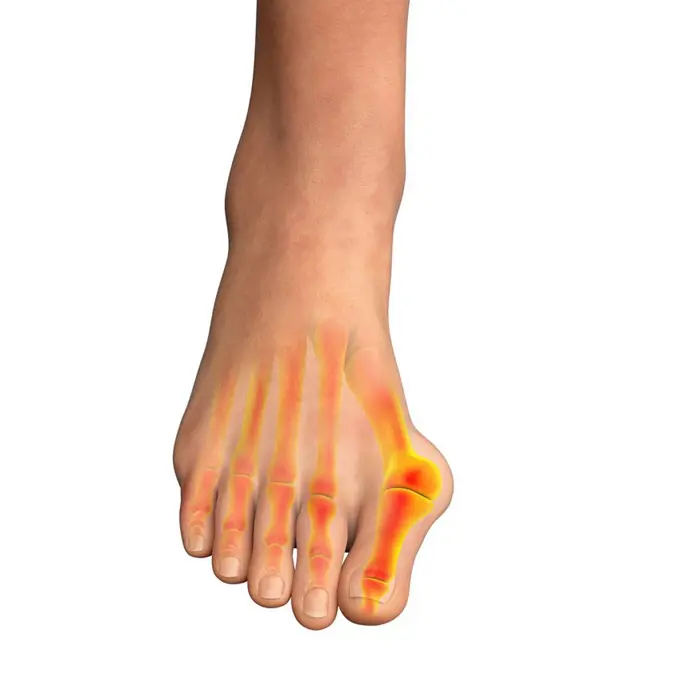
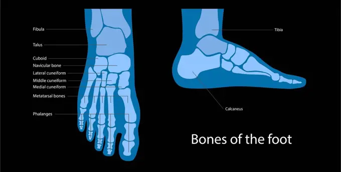
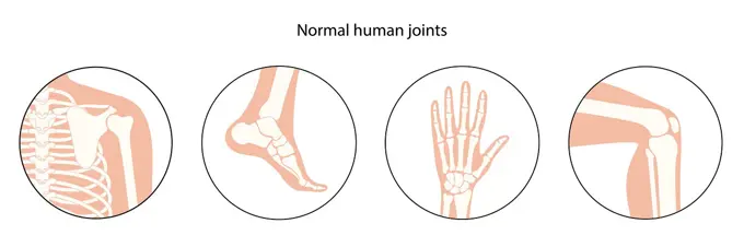
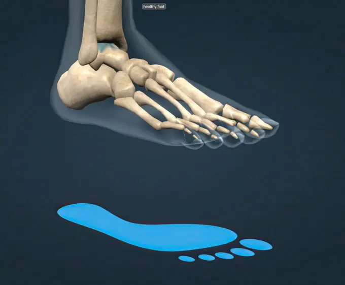
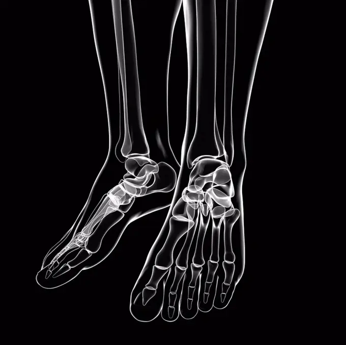
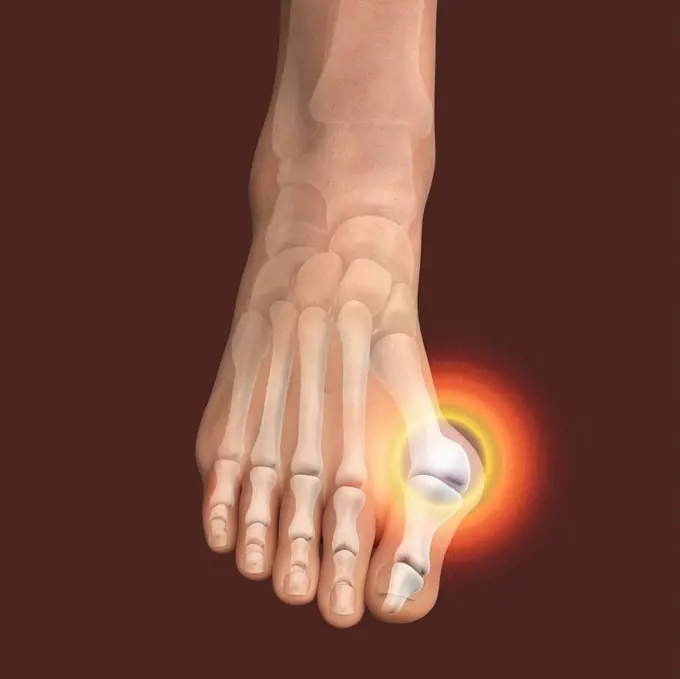
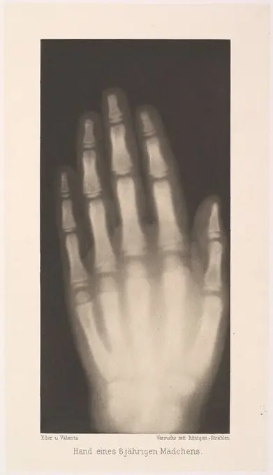
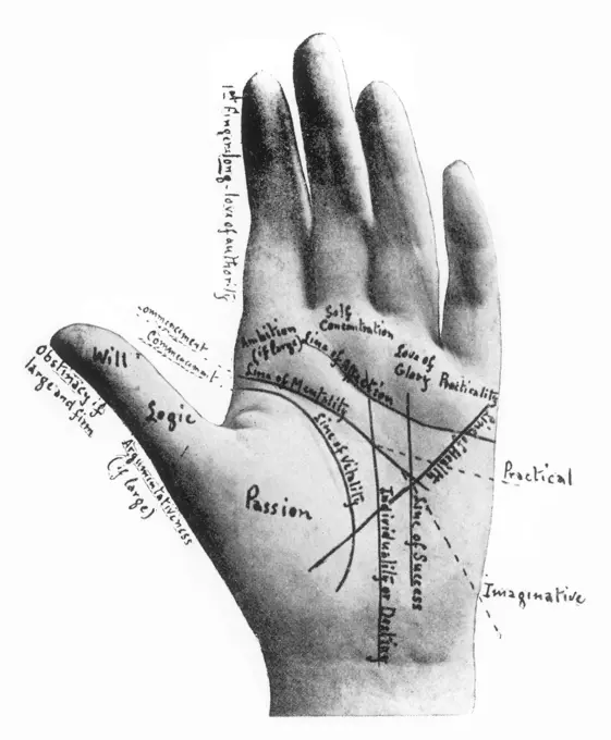
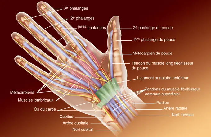
My Boards
View Pictures
  • My Boards
← Science

Human Hand and Foot Anatomy

Detailed illustrations of human hand and foot anatomy, showcasing bones, muscles, and ligaments, rendered in a clear and educational style.

Foot Bones
Foot Bones
173 assets in this story
4128R-35203
Human hand bones, computer artwork.
4128-V58567632
4128-30418420
Muscles of the hand, illustration
4128-48285505
Healthy hand, X-ray
4128-30417341
Bones of the left hand, illustration
4128-30418390
Male hand, illustration
4128R-13587643
Illustration of the palmar metacarpal ligaments.
4378-20013568
Foot Bones
4128R-11545067
Human foot bones, computer illustration.
4128R-12580850
Human foot anatomy, computer illustration.
4220-21334624
Human Skeleton body structure.     Date:
4128R-33550
Human hand musculature, computer artwork.
4128R-19693
Human lower leg anatomy, computer artwork.
4128-30418359
Structure of the foot, illustration
4128R-29291
Hand anatomy, computer artwork.
6188-62298913
Human foot anatomy cutaway representation. Human foot anatomy cutaway representation, showing skin, veins and arterias, muscles, bones. With clipping path included. Copyright: xZoonar.com/Pixelchaosx 9887821
4128-19056302
Ankle joint, illustration
4128R-13572678
Illustration of the navicular bone.
4128R-13587627
Illustration of the retinaculum ligament.
1848-65702858
Human skeleton hand model for medical anatomy science Medical clinic concept
4128-30421526
Bones of the hand, illustration
4128R-35929
Human arm bones, computer artwork.
4269-24872
Hand x-ray. X-ray of the hand of a 10 years old child revealing its bone age. The bone age shows the process of ossification (tranformation of cartilage into bone) of specific parts of the skeleton. Compared with chronoligical age, it reveals pathlogies like growth delay.
4128-16071982
Hand, X-ray
4128R-11286305
Male vascular system, computer artwork.
4128R-35965
Human hand bones, computer artwork.
1525-56183632
Anatomy of the Human Foot, Metatarsal bones 3d illustration. Anatomy of the Human Foot, Metatarsal bones
4128-20045437
Calcaneus bone, illustration
4128-V58567666
1428R-1395
Three views of the female hand anatomy: skeletal, muscular, and skin. Close up, detailed anatomy, full color 3D illustration on white background
4128-30418419
Bones of the hand, illustration
1899-65662229
X-ray of a normal hand.
4128R-26468
Foot bones, computer artwork.
1899-66339
X-ray image of female hand
4128R-36874
Human arm bones, computer artwork.
4128R-26189
Foot bones, computer artwork.
4128-19056312
Finger bones, illustration
4128-30421479
Skeletal anatomy of the foot, illustration
1848-77397860
3D Illustration Concept of Human Skeleton System Palm Hand Bone Joints Anatomy
4128-20042011
Bunion, illustration
1848-49200681
Medicine, human anatomy, the circulatory system, blood circulation: sections of the foot, lower leg and ankle, with arteries and veins shown in red and blue. Coloured lithograph by J. Maclise, 1841, 1844, Historic, digitally restored reproduction from a 19th century original
4128R-2092
Hand blood supply, artwork
4128R-36969
Human arm bones, computer artwork.
1439-57940621
Three phases of a hand xray image
4128-30418346
Veins of the foot, illustration
1788-39602
Medicine: Human anatomy, hand and foot bones
4128-30421496
Human hand, illustration
4269-27151
Capillaro-veinous malformation. 3D Computed Tomography (CT) scan of of the right hand showing a malformation of the capillaro-veinous vessels seen as red spots under the little finger.
4128R-12964684
Human hand bones, Illustration
4128-V58567571
4128R-13587635
Illustration of the retinaculum ligaments.
1848-55997722
Radiography of a dog paw. Real x ray image of an injured dog paw
5507-47244848
Human skeleton. Vector
4128R-14324140
Penicillium fungus, illustration
4128-20042004
Bunion, illustration
4128-15825268
Robotic arm, illustration.
4128-30415864
Feet bones, illustration
1788-39582
Medicine - Anatomy - Musculoskeletal (locomotor) system - Skeleton - Hand and foot. Drawing.
4128-19490334
Foot anatomy, illustration
4128-30420344
Healthy joints, illustration
1525-56184093
Healthy foot skeleton and blue footprint 3D illustration. Healthy foot skeleton and blue footprint
4128R-35030
Human foot bones, computer artwork.
1848-54708905
Anatomy of the hand, historical woodcut
6145-52682836
Heliogravure by Josef Maria Eder showing the hand of a four-year-old child using X-ray photography, depicting bones, fingers, and joint structure, created in 1896.
4413-8324
Worm parasitizes Cat Toxocara mystax in an intestine France
4128R-33619
Human musculature, computer artwork.
4128-20042007
Bunion, illustration
6145-29253007
Sir Gervas Powell Glyn, 6th Bart.: radiograph of his hand. Photograph by Sir G.P. Glyn, 1896.
4409-17377030
Hand eines 8 jährigen Mädchens. Artist: Josef Maria Eder (Austrian, Krems an der Donau, 1855-1944 Kitzbühel); Eduard Valenta (Austrian, 1857-1937). Dimensions: Image: 6 7/8 × 3 5/16 in. (17.4 × 8.4 cm)Plate: 8 1/16 × 4 9/16 in. (20.5 × 11.6 cm)Sheet: 19 1/2 × 13 3/4 in. (49.5 × 34.9 cm). Date: 1896.Eder was the director of an institute for graphic processes and the author of an early history of photography. With the photochemist Valenta, he produced a portfolio in January 1896, less than a month after Wilhelm Conrad Röntgen published his discovery of X-rays. Eder and Valenta's volume, from which this plate derives, demonstrated the X-ray's magical ability to reveal the hidden structure of living things. Human hands and feet, fish, frogs, a snake, a chameleon, a lizard, a rat, and a newborn rabbit are all presented in exquisitely printed photo-gravures, as are carved cameos and an assortment of natural materials. In an era when photography's ability to accurately depict the visible w
4220-21826390
A palmistry chart of the hand by Cheiro (Count Louis Hamon,  1866-1936), with lines and annotations.      Date: 1900
1899-61461939
Hand anatomy. Representation of the anatomy of the palmar face of the hand highlighting the carpal tunnel closed in front by the anterior annular ligament and its contents: the median nerve, the tendons of the flexor muscle of the hand, the radial artery
824-63186175
Palm view of the hand flexor sheaths.
824-63214030
Medical illustration showing the measuring of blood pressure in the brachial artery in the arm and the tibial arteries in the leg. Measuring blood pressure consists of measuring the systolic pressure relating to the hearts contraction in the arm or leg artery. The ultrasound probe enables the sound of the arterial blood flow to be amplified.
PREVIOUS
of 2
NEXT
2401 S. Ervay, Suite 206
Dallas, Texas 75215
United States
Get Started
Free ResearchMy BoardsMy Cart
For Creators
How To License Your ContentContributor PortalFrame of Mind
Resources
API accessPricing
Contact
+1 866 236 0087help@viewpictures.co.uk Contact form
©2026 View Pictures. All Rights Reserved. -A
This site is protected by reCAPTCHA and Google Privacy Policy and Terms of Service apply.