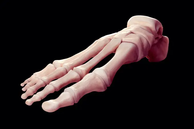
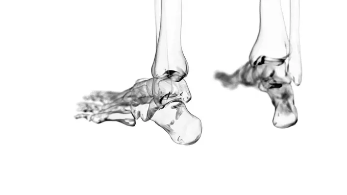
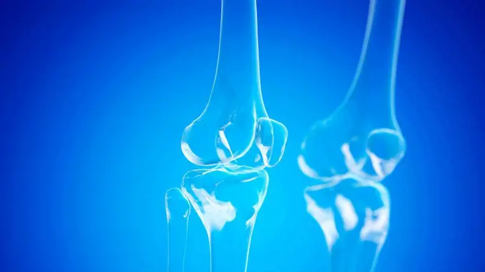
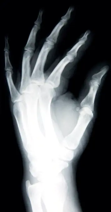
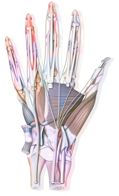
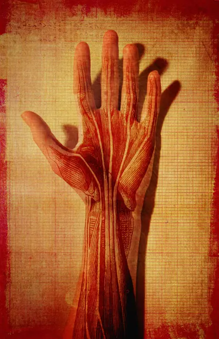
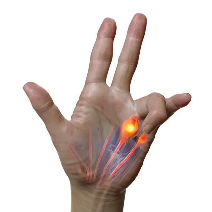
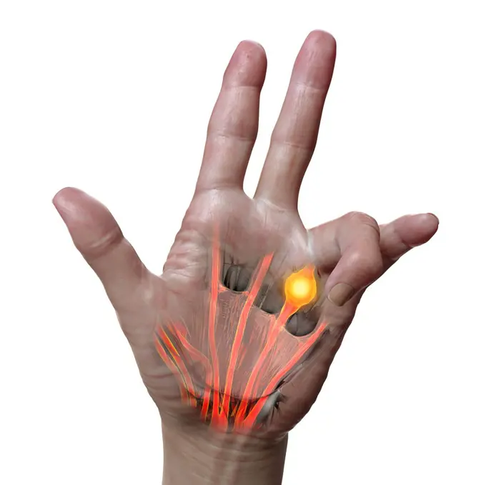
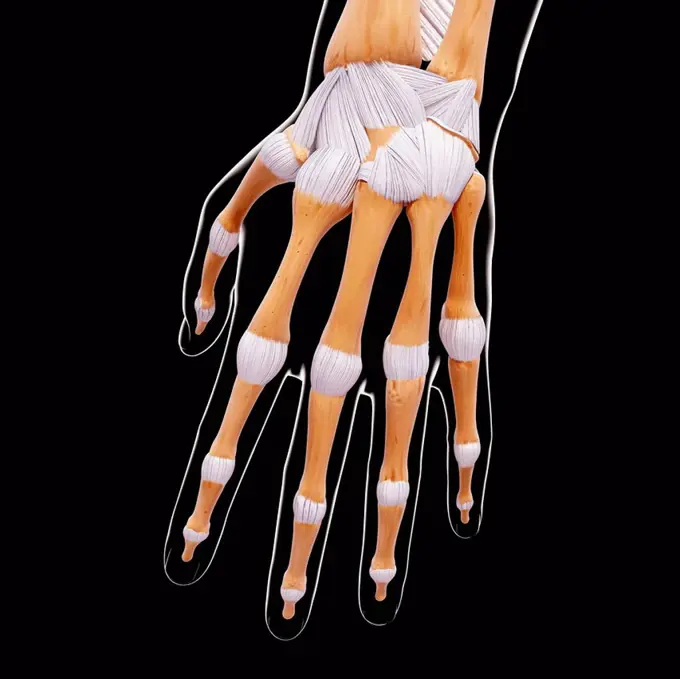
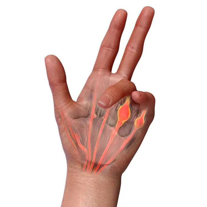
My Boards
View Pictures
  • My Boards
← Science

Human Hand and Foot Anatomy

Illustrations and renderings of human hand and foot bones and muscles, highlighting anatomical details in various artistic styles.

Foot Bones
Foot Bones
141 assets in this story
4128-19248776
Spine, illustration.
4128R-11477834
Human hand bones, computer artwork.
4128-19248778
Spine, illustration.
4128-V58567665
4128-19056468
Human ankle joint, illustration
4128-V58567234
4128-19056292
Knee joints, illustration
1428R-312
Skeleton of the human hand using a computer mouse showing carpal tunnel syndrome
4378-2794
Wire frame anatomical model showing the vertebral column.
4128R-12572086
Human hand bones, computer illustration.
1899-66340
X-ray image of female hand
4128R-11322821
Human scapula and humerus, computer artwork.
1899-53509063
Anatomy of the palm of the hand, bones, muscle, tendons, nerves.
4128-15825260
Robotic arm, illustration.
4128-V58567253
1428-67034183
Human Vertebral Column, Side View on Black Background, 3D rendering
1849-866
Man's hand with muscle and tendons showing
4128-16223980
Strapless fashion dress, X-ray
4128-30422439
Dupuytren's contracture, illustration
4128-30421509
Anatomy of the hand, illustration
4128-30422440
Dupuytren's contracture, illustration
1848-50239985
One of the first X-ray images of a human hand with a ring, ca 1900, digitally restored reproduction of an original from the 19th century
4128R-13022515
Human tendons of the elbow (bicipital aponeurosis), illustration.
4128R-32088
Human hand bones, computer artwork.
5507-31546475
Man skeletal system for anatomy learning. Human body biology
1525-28067639
Skeleton of the anterior and posterior members of the salamander, vintage engraved illustration. From the Universe and Humanity, 1910.
4128R-11938
Molar tooth
4128-30422443
Dupuytren's contracture, illustration
4409-23181
INVENTION OF X-RAYS IN 1895 - RADIOGRAPHY OF ONE HAND. NOBEL PRIZE FOR PHYSICS IN 1901. Author: ROENTGEN WILHEM.
4128R-13572599
Illustration of the human skeleton.
4378-3330
Front and rear view of the skeletal structures of the thoracic cage or rib cage.
4378-2768
Multiple view of the skeletal structure of the rib cage.
4128R-11297226
Coloured light interference pattern of knives and forks. When photographed through cross-polarised white light some transparent plastics display colou...
4128R-13572744
Illustration of the extensor carpi radialis longus muscle.
4128R-11312480
Human spine, computer artwork.
4239-69154121
Skeletal system of Tyrannosaurus rex, front view.
1788-39595
Medicine - Anatomy - Musculoskeletal (locomotor) system - Skeleton - Upper limb - Forearm. Drawing
4128R-26075
Calf muscle. Computer artwork showing the gastrocnemius muscle.
4128-16224080
Caduceus, X-ray
4128-30420347
Healthy joints, illustration
1525-75859970
Illustration of a human femur versus hip intra-articular comparison of normal bone symptoms with osteoporosis. For medical, educational and commercial use.
PREVIOUS
of 2
NEXT
2401 S. Ervay, Suite 206
Dallas, Texas 75215
United States
Get Started
Free ResearchMy BoardsMy Cart
For Creators
How To License Your ContentContributor PortalFrame of Mind
Resources
API accessPricing
Contact
+1 866 236 0087help@viewpictures.co.uk Contact form
©2026 View Pictures. All Rights Reserved. -A
This site is protected by reCAPTCHA and Google Privacy Policy and Terms of Service apply.