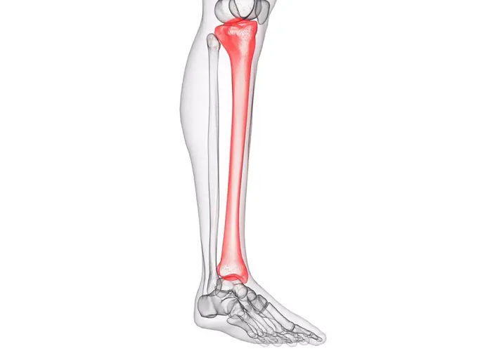
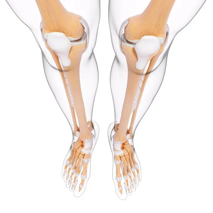
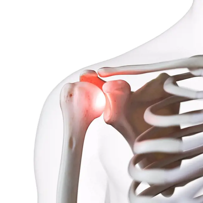
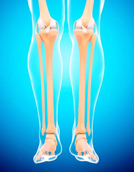
My Boards
View Pictures
  • My Boards
← Science

Human Muscle and Bone Anatomy

Illustrations focusing on human leg muscles and bones, highlighting anatomical structures in detail with vibrant color distinctions.

Tibia bone, illustration
282 assets in this story
4128R-11311138
Human knee joint, computer artwork.
4128R-11316399
Human leg bones, computer artwork.
1525-27775468
science anatomy scan of human blood vessels. Blood Vessels of Human Body
4128R-12580772
Human leg anatomy, computer illustration.
4128-15665769
Ulna bone, illustration
4128R-15536688
Illustration of a painful shoulder joint
4128R-30129
Ankle pain, conceptual computer artwork.
4128R-32262
Human foot anatomy, computer artwork.
4128-15981110
Knee joint, computer illustration.
4128R-36038
Human leg bones, computer artwork.
4128R-16628
Vascular system, computer artwork.
4128R-30134
Elbow pain, conceptual computer artwork.
1899-53509259
The muscles of the upper right limb in posterior view.
4128R-12577008
Human back anatomy, computer illustration.
6188-58135812
human bones radiography scan image human bones radiography scan image Copyright: xZoonar.com/OxanaxRishnyakx 8008165
4252-4193
Achille's tendon
6188-58107661
Transparent Human Body with Visible Bones medical science image of human skeleton bones Copyright: xZoonar.com/StanislavxRishnyakx 17141718
4128R-11324348
Human elbow joint, computer artwork.
4128R-32198
Human leg musculature, computer artwork.
1525-19745585
3D render of female medical skeleton legs running
4128R-36047
Human foot musculature, computer artwork.
4128R-26563
Thigh muscle. Computer artwork showing the vastus lateralis muscle.
4128R-28110
Knee joint. Coloured X-ray of a healthy knee joint, seen from the front. The knee is the largest joint in the body. It is formed by the articulation o...
4128-16223853
Skeleton feet with one foot in boot, X-ray
4128R-32314
Human leg musculature, computer artwork.
4128R-31663
Human arm bones, computer artwork.
4378-1431
Anatomical model showing part of the human muscular system.
4128R-13022517
Human muscle (coccygeus), illustration.
4128R-16819
Vascular system, computer artwork.
4128R-13388801
Human foot anatomy, illustration.
4128R-29004
Lower body anatomy, computer artwork.
4239R-8076
Illustration of a flexed arm showing bicep muscle.
4128R-32794
Human foot anatomy, computer artwork.
6188-55565478
ELLENBOGEN ELLENBOGEN Copyright: xZoonar.com/curryx 2482881
4128R-13572761
Illustration of the triceps muscle.
4128R-13572747
Illustration of the extensor digiti minimi muscle.
1525-56854534
RA is caused by immune cells attacking the body’s own synovial membranes, leading to joint swelling and the formation of auto-antibodies. 3D rendering. RA is caused by immune cells attacking the body’s own synovial membranes, leading to joint swelling and the formation of auto-antibodies.
4269-27204
Knee. Sagittal (side view) Magnetic Resonance Image (MRI) of the knee showing in orange the patellar tendon (between the patella and the tibia above), and the flexing quadriceps tendon (between the patella and the femur below). The distal femur (above), the proximal tibia (below), and the patella or knee cap (left) are in red.
824-72488771
Knee osteoarthritis (gonarthrosis). Osteoarthritis can affect one or more of the knee joints. This is global osteoarthritis with involvement of the internal tibiofemoral, external tibiofemoral and patellofemoral joints. In addition, the menisci are also affected by osteoarthritis, meniscosis is added to the gonarthrosis, forming meniscarthrosis. The cartilage of the joints is worn, causing the bones to rub against each other. Osteophytes (bony growths) have formed. Bone tissue is also remodeled: in the friction regions, bone condensation (osteocondensation) occurs while geodes (cavities) form in the adjacent areas.
4128R-13572546
Illustration of the medial gastrocnemius bursa.
6188-58105960
Blood Vessels of Human Body science anatomy scan of human blood vessels Copyright: xZoonar.com/StanislavxRishnyakx 14993748
4378-2291
Model showing the human knee joint and its connecting bones.
1525-26275123
The thigh bone is the large upper leg bone that connects the lower leg bones to the pelvic bone 3D illustration. Leg skeletal anatomy.Leg joint
4128R-13572748
Illustration of the extensor digitorum muscle.
4128R-24319
Knee anatomy, computer artwork.
4128-19052721
Muscles of the upper leg, illustration
4128R-13376383
Vascular system of knee
4128-30416844
Seminal vesicles, illustration
1525-56854441
RA is caused by immune cells attacking the body’s own synovial membranes, leading to joint swelling and the formation of auto-antibodies. 3D rendering. RA is caused by immune cells attacking the body’s own synovial membranes, leading to joint swelling and the formation of auto-antibodies.
4128R-13372249
Human foot anatomy, illustration.
4128-20042515
Sciatic nerve, illustration
4269-27169
Osteoclastoma. Colored X-ray of the knee front view (left) and side view (right) showing a giant cell bone tumor (osteoclastoma) on the apophysis of the femur (orange and pink round shape). The lysis of the bone is evidenced here by the grey aspect of the bone and the expansion of the soft tissues (swelling at right on the left X-ray).
6188-58105571
Transparent Human Body with Visible Bones medical science image of human skeleton bones Copyright: xZoonar.com/StanislavxRishnyakx 14703259
4128R-26074
Calf muscle. Computer artwork showing the gastrocnemius muscle.
4378-3709
Close up of hip joint, a synovial joint formed by the articulation of the rounded head of the femur and the cup-like acetabulum of the pelvis.
1439-57940631
An Xray image showcasing a human knee joint in detail.
4128R-15515313
Illustration of the lower leg muscles
1428R-472
X-ray view of a human's leg in running position
4128-30422356
Knee bones, illustration
4128R-23870
Knee anatomy, computer artwork.
4128R-11310329
Human knee joint, computer artwork.
4128-30422361
Lateral meniscus, illustration
1848-53366192
X-ray film of a childes femur and knee showing the formation of the joint, the cartilage, the femur and articulation with the tibia, taken by a radiologist for an orthopaedic diagnosis
4128-20042439
Quadriceps muscle anatomy, illustration
6188-55565477
Human elbows of humans
4128R-21991
Lower leg muscle. Computer artwork of the gastrocnemius muscle of the lower leg.
1439-57940366
Xray image of a human knee joint
4128R-12572084
Human elbow joint, computer illustration.
4128R-33136
Human leg bones, computer artwork.
824-63194998
Illustration of the spinal cord from the 8th dorsal vertebra to the 1st lumbar vertebra.
4128R-12964540
Female muscles, Illustration
1525-56190008
Knee joint anatomy, different structures in and around the knee 3D illustration. Knee joint anatomy, different structures in and around the knee.
4128R-15515044
Illustration of the lateral meniscus
4128R-11472757
Human calf muscles, computer artwork.
4128R-33018
Human leg musculature, computer artwork.
1525-27418251
Human Anatomy - Male Muscles made in 3d software
4128R-12576959
Human foot anatomy, computer illustration.
1439-57940632
Xray image showing a human knee joint
4128R-16647
Trapezius muscle, computer artwork.
4128-30420435
Knee bones, illustration
4128R-19176
Knee anatomy, computer artwork.
4128-20042459
Leg anatomy, illustration
PREVIOUS
of 3
NEXT
2401 S. Ervay, Suite 206
Dallas, Texas 75215
United States
Get Started
Free ResearchMy BoardsMy Cart
For Creators
How To License Your ContentContributor PortalFrame of Mind
Resources
API accessPricing
Contact
+1 866 236 0087help@viewpictures.co.uk Contact form
©2026 View Pictures. All Rights Reserved. -A
This site is protected by reCAPTCHA and Google Privacy Policy and Terms of Service apply.