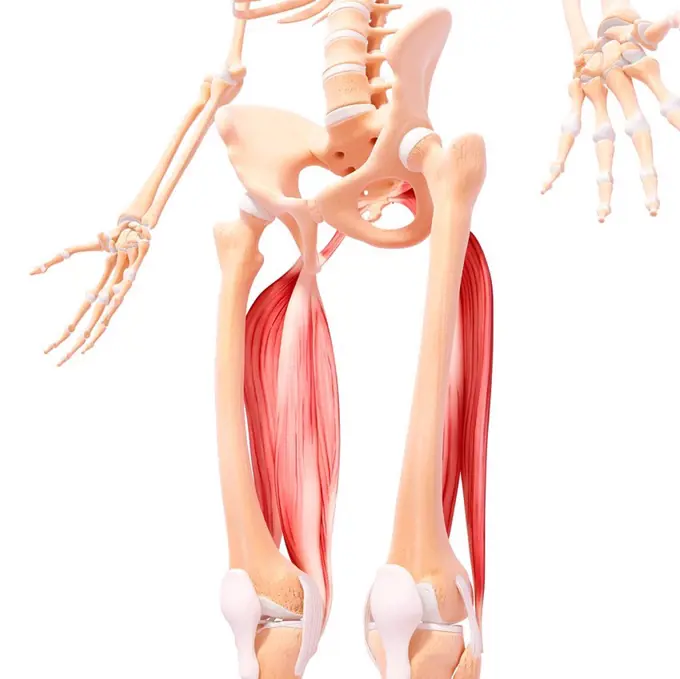
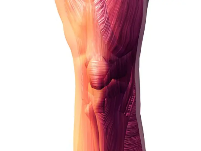
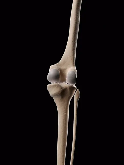
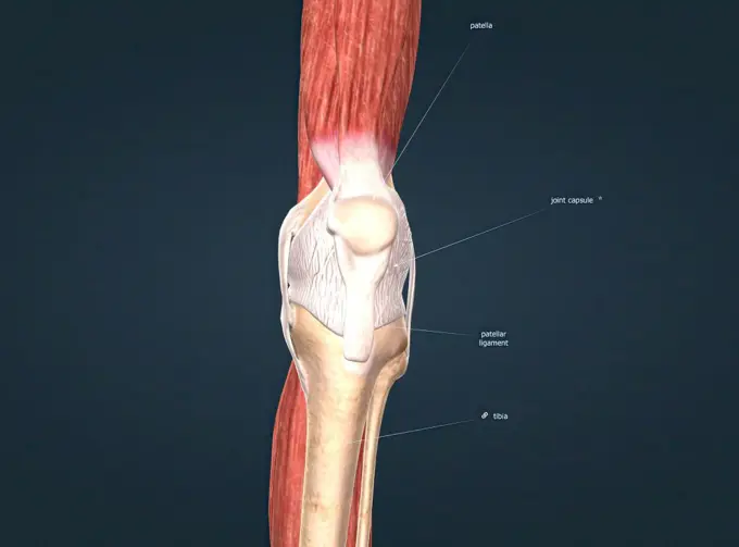
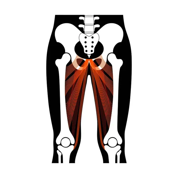
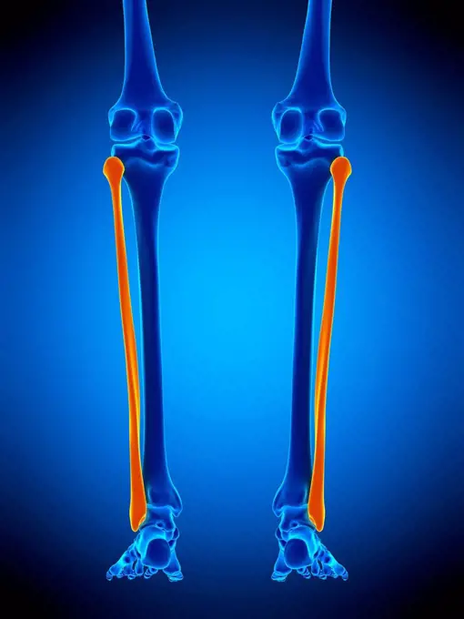
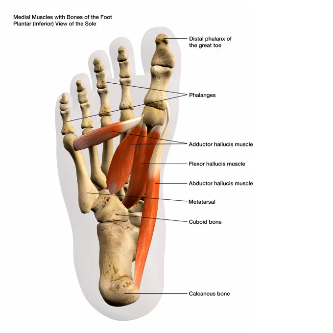
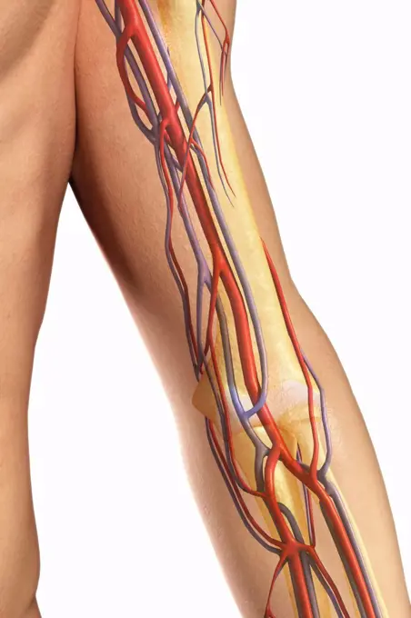
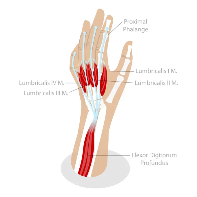
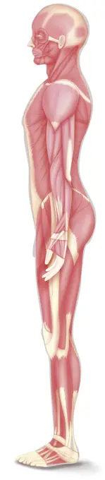
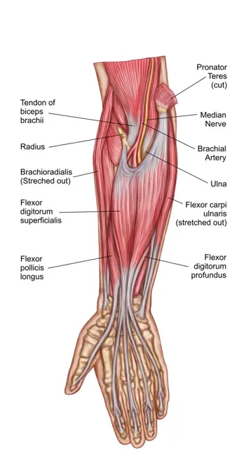
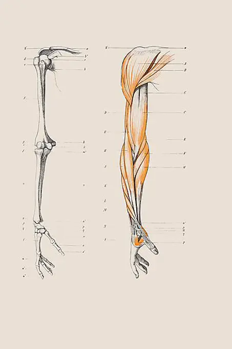
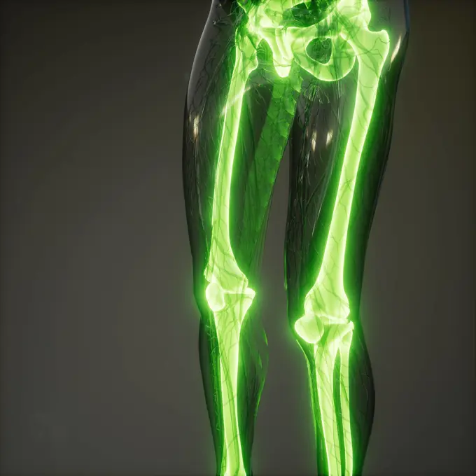
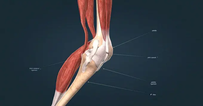
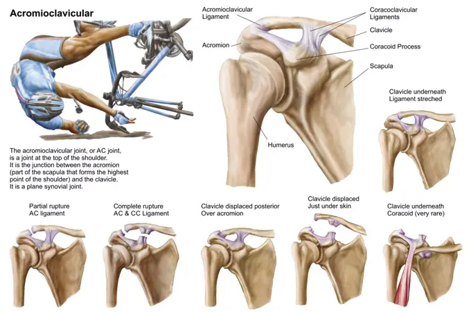
My Boards
View Pictures
  • My Boards

Human Muscle Structures

Illustrative representations of various human muscles, emphasizing the arm and thigh muscles with a focus on anatomical details.

Human leg musculature, computer artwork.
247 assets in this story
4128R-13574598
Illustration of the gastrocnemius muscle.
1574R-018831
Human Knee Muscles
1899-61459178
Medicine, human anatomy, the circulatory system, blood circulation: section of the front of the lower leg and ankle, leg, foot, with arteries shown in red and blue. Colored lithograph by J. Maclise, 1841.
4128R-11545072
Human knee bones, computer illustration.
1525-56183859
Knee joint anatomy, different structures in and around the knee 3D illustration. Knee joint anatomy, different structures in and around the knee.
4128-20042468
Leg anatomy, illustration
4128R-13572626
Illustration of the fibular.
4128R-15515316
Illustration of the leg muscles
4378-3013
Model showing the human knee joint and its connecting bones.
4128R-21948
Lower leg muscle. Computer artwork of the flexor hallucis muscle of the lower leg.
1788-21803424
Leg muscles, human body, drawing.
4378-2883
Model showing the human knee joint and its connecting bones.
1428-67304565
Medial Muscles and Bones of the Foot Labeled, 3D Rendering on white background
4239-69152008
Human foot depicting the plantar intermediate and deep muscles with bone structures.
4378-20014047
Thoracic Spinal Bones
4239R-8267
Medical illustration of human arm muscles, four side views.
4378-465
Front view of the upper arm showing the major blood vessels relative to the skeleton.
6188-62377689
3D Isometric Flat Conceptual Illustration of Lumbricals Muscles, Anatomy of Muscular Systembr3D Isometric Flat Conceptual Illustration of Lumbricals Muscles, Anatomy of Muscular System 3D Isometric Flat Conceptual Illustration of Lumbricals Muscles, Anatomy of Muscular System 3D Isometric Flat Conceptual Illustration of Lumbricals Muscles, Anatomy of Muscular System Copyright: xZoonar.com/AnastasiiaxTorianykx 22124275
4128R-11298294
Foot anatomy, computer artwork.
4128R-11291342
Human femur, computer artwork.
824-63191355
Illustration of the various types of fracture in the proximal part of the femur. From top to bottom : -subcapital fracture -transcervical fracture -petrochanteric fracture -subtrochanteric fracture
1525-28043457
Stem with many beech canker, vintage engraved illustration.
4128-19490400
Intervertebral hernia, illustration
1746-21105756
anatomical illustrations by Sigismond Balicki, 1858-1916 from 'Anatomie normale du corps humain' by Sigismond Laskowski, 1900
4128-30416920
Brain and nerves, illustration
6145-29267640
Femur bone, posterior view. Colour wood engraving with letterpress, 1860/1900
4128-20042487
Pelvis anatomy, illustration
1899-54027769
Left side illustration of a skinned anatomical figure. The full musculature of the various limbs, bust, abdomen and back is visible.
4128-20042291
Arm anatomy, illustration
4239R-8513
Anatomy of forearm muscles, anterior view, middle.
4239-69152043
Anterior and posterior view of the tibia and fibula with labeling.
1899-53513446
Structure of bone in children, bone development, cartilage structure, pediatrics. This drawing represents a child's humerus. the child the epiphyses (bone ends) are already made up of spongy bones, They are covered with articular cartilage as in adults. But they are still separated from the diaphysis (central part of the bone) by an area of epiphyseal cartilage. This area is shown in pink. This allows the child's bone to continue to grow in order to reach maturity in adulthood. A cut section is indicated at the level of the shaft of the bone. It is shown top view at the bottom of the illustration. This makes it possible to see the structure of the bone in horizontal section. The bone is covered with a membrane, the periosteum. Inside the periosteum is new bone tissue, and an area of resorbed bone tissue. Here the arrows represent the direction of bone resorption. Finally in the center is the medullary cavity. (Photo by: JACOPIN/BSIP/UIG)
1525-28067995
Skeleton of the lower thigh and foot of a Japanese seen externally, vintage engraved illustration. From the Universe and Humanity, 1910.
1525-26034199
Hand drawn illustration of the leg muscles isolated on white, artistic anatomy graphic study
4378-2342
Bones and joint that form the skeletal structure of the human leg
4128R-11472688
Human knee joint, computer artwork.
4128-38524451
Osteoporosis, conceptual illustration
1848-54506285
Skeleton of a human arm, anatomical illustration
1525-26034198
Hand drawn illustration of the foot bones isolated on white, artistic anatomy graphic study
6188-58103847
Transparent Human Body with Visible Bones medical science image of human skeleton bones Copyright: xZoonar.com/StanislavxRishnyakx 13688129
6188-58112288
science anatomy of human body in x-ray with glow back bones science anatomy of human body in x-ray with glow back bones Copyright: xZoonar.com/StanislavxRishnyakx 9690260
1525-56190204
Knee joint anatomy, different structures in and around the knee 3D illustration. Knee joint anatomy, different structures in and around the knee.
4128-20042335
Arm anatomy, illustration
4128-20042388
Arm anatomy, illustration
4239-18641413
Anatomy of acromioclavicular joint rupture and displacement.
4239-69151971
Gross anatomy of a long bone, using a femur with annotations.
4128-20042518
Sciatica, illustration
PREVIOUS
of 3
NEXT
2401 S. Ervay, Suite 206
Dallas, Texas 75215
United States
Get Started
Free ResearchMy BoardsMy Cart
For Creators
How To License Your ContentContributor PortalFrame of Mind
Resources
API accessPricing
Contact
+1 866 236 0087help@viewpictures.co.uk Contact form
©2026 View Pictures. All Rights Reserved. -A
This site is protected by reCAPTCHA and Google Privacy Policy and Terms of Service apply.