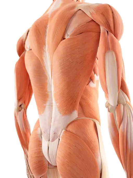
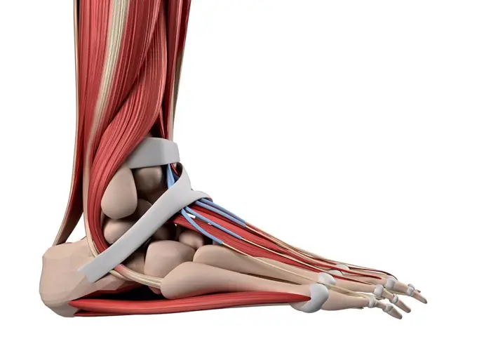
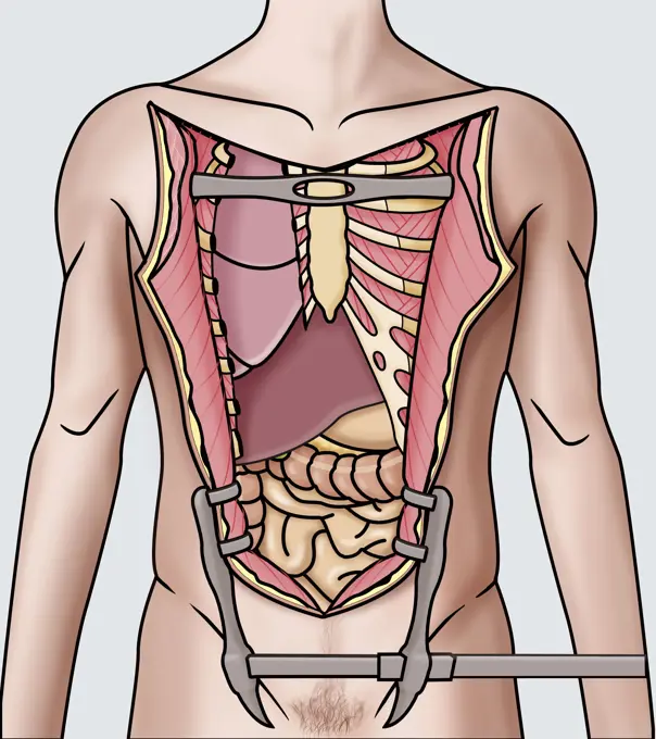
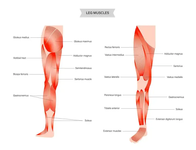
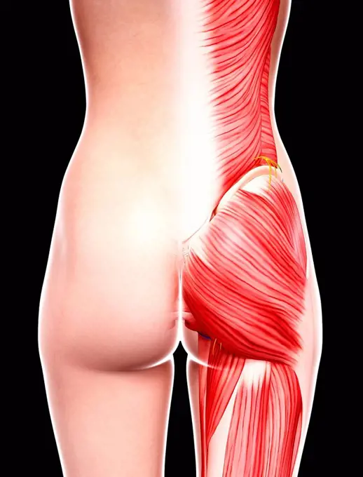
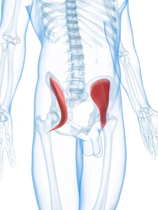
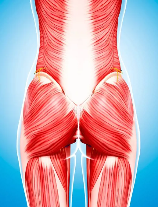
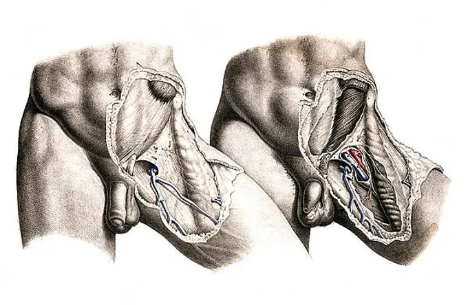
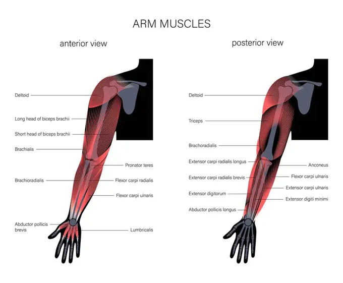
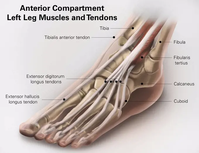
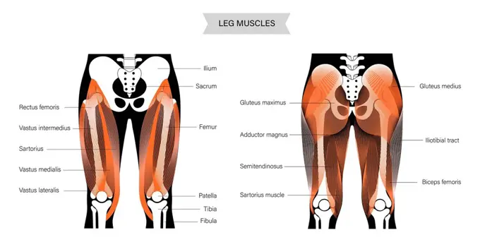
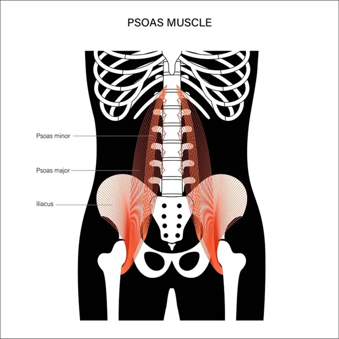
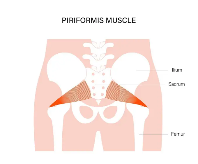
My Boards
View Pictures
  • My Boards
← Science

Human Musculature Anatomy

Detailed computer illustrations of human leg and body musculature, showcasing muscles and structures in vibrant colors.

Anatomical model showing the lower abdominal muscles.
160 assets in this story
4128R-13385262
Human back muscles, illustration.
4128R-11478306
Human hand, computer artwork.
4128R-11311068
Human foot anatomy, computer artwork.
1899-54028667
Illustration showing the opening of the thorax and abdomen during an autopsy in order to reach the various organs to be studied. (Photo by: BSIP/UIG via Getty Images)
4128-20042297
Biceps muscle anatomy, illustration
824-63195460
Illustration of the knee joint, showing a healthy knee (left) and a knee suffering from rheumatoid arthritis (right), with a deterioration of the bone surface and cartilage, thickening of the synovial membrane, effusion of the synovial fluid and inflammation.
4128-30422746
Muscles of the abdomen and pelvis, illustration
4239R-8511
Anatomy of human forearm muscles, deep anterior view.
4239-69152011
3D model of the foot depicting the plantar deep muscles and bone structures.
4128-20042465
Leg anatomy, illustration
4128R-36642
Human arm bones, computer artwork.
1899-13235
Dissection of the carpal tunnel III
4128R-15515034
Illustration of the human knee
824-63163986
Superior view of the foot and the different bones: calcaneus, talus.
4128R-32676
Female musculature, computer artwork.
1525-56190218
The larynx is an organ in the upper part of the neck that protects the trachea against breathing, sound production, and food aspiration. 3D illustration. The larynx is an organ located in the anterior neck.
4128R-11473040
Human upper leg muscles, computer artwork.
4128-20042339
Arm anatomy, illustration
4128R-22000
Hip muscles. Computer artwork of the iliacus muscles.
1525-56722735
Several different viruses cause gastroenteritis, including norovirus, rotavirus, and adenovirus. 3D rendering. Several different viruses cause gastroenteritis, including norovirus, rotavirus, and adenovirus.
4128-20042341
Arm anatomy, illustration
1525-23843165
Ngf - Nerve Growth Factor 3d medical 3d illustration. Ngf - Nerve Growth Factor 3d medical
4128-20042395
Muscular dystrophy, illustration
824-63188526
Illustration of Charcots disease. This disease is the result of motor neuron degeneration. Motor neurons are nerve cells that work as control units and communication links between the nervous system and voluntary muscles. Motor neurons located in the brain (superior motor neurons) transmit nerve impulses to motor neurons in the spinal cord (inferior motor neurons) , as well as to particular muscles. With Charcots disease, the superior motor neurons (bulbar) and inferior motor neurons (spinal) degenerate or die, and stop sending impulses to the muscles. The muscles are incapable of functioning and gradually weaken and atrophy.
4128-20042343
Arm anatomy, illustration
824-63205856
Illustration of a hand suffering from rheumatoid arthritis, with a deterioration of the surface of the bone and cartilage, thickening of the synovial membrane, effusion of synovial fluid and swelling.
4128R-35903
Female musculature, computer artwork.
4239R-20483514
Human anatomy showing deep muscles in the neck and upper back.
4409-17349023
Thyroid gland.
4128R-11323810
Human shoulder anatomy, computer artwork.
4128-20042345
Arm anatomy, illustration
1525-56190191
Knee joint anatomy, different structures in and around the knee 3D illustration. Knee joint anatomy, different structures in and around the knee.
4239-69152000
3D model of the foot depicting the dorsal deep muscles and bone structures.
4239-18641166
Diigital illustration of muscles of the forearm, anterior view.
4378-1440
Anatomical model showing the deltoid muscles.
824-63190466
Illustration of the muscular, arterial and nervous anatomy of the inferior part of the elbow (crook of the arm).
4128-20042466
Leg anatomy, illustration
6188-62376983
3D Isometric Flat Conceptual Illustration of Supinator Muscle, Labeled Anatomical Schemebr3D Isometric Flat Conceptual Illustration of Supinator Muscle, Labeled Anatomical Scheme 3D Isometric Flat Conceptual Illustration of Supinator Muscle, Labeled Anatomical Scheme 3D Isometric Flat Conceptual Illustration of Supinator Muscle, Labeled Anatomical Scheme Copyright: xZoonar.com/AnastasiiaxTorianykx 22123909
1525-25129431
Dissection of muscles on the front of the upper arm,from an early 20th century anatomy textbook, out of copyright
6188-64789755
3D Isometric Flat Conceptual Illustration of Valgus Deformity , Cosmetic Pathologybr3D Isometric Flat Conceptual Illustration of Valgus Deformity , Cosmetic Pathology 3D Isometric Flat Conceptual Illustration of Valgus Deformity , Cosmetic Pathology 3D Isometric Flat Conceptual Illustration of Valgus Deformity , Cosmetic Pathology Copyright: xZoonar.com/AnastasiiaxTorianykx 22296003
6188-62377696
3D Isometric Flat Conceptual Illustration of Longus Colli Muscle, Neck Anatomybr3D Isometric Flat Conceptual Illustration of Longus Colli Muscle, Neck Anatomy 3D Isometric Flat Conceptual Illustration of Longus Colli Muscle, Neck Anatomy 3D Isometric Flat Conceptual Illustration of Longus Colli Muscle, Neck Anatomy Copyright: xZoonar.com/AnastasiiaxTorianykx 22124293
4239-18642012
Lower leg showing normal and compression of lateral and anterior compartments.
1848-49200632
Medicine, human anatomy, dissection of a man's left groin, vein, vein, penis. Coloured lithograph by J. Maclise, 1841, 1844, Historic, digitally restored reproduction from a 19th century original
4239-18641174
Digital illustration of muscles of the human torso, anterior view.
4128-20042348
Arm anatomy, illustration
4239R-20482709
Anterior compartment anatomy of left leg muscles and tendons.
4128-20042433
Leg anatomy, illustration
1525-26274972
Cross section through a typical synovial joint showing the bone synovial membrane synovial fluid cartilage and ligament 3d illustration. Cross section through a typical synovial joint showing the bone synovial membran
1525-27535825
Human Anatomy Leg Bones Vector Illustration
4409-17349038
Ossification. Bone formation.
4443-21168011
excision of the shoulder, medical equipment, surgical instrument, history of medicine
4128-20042443
Quadriceps muscle anatomy, illustration
824-57656268
Hand anatomy. Representation of the anatomy of the palmar face of the hand highlighting the carpal tunnel closed in front by the anterior annular ligament and its contents: the median nerve, the tendons of the flexor muscle of the hand, the radial artery.
4128-30420346
Healthy joints, illustration
4128-20042483
Psoas muscles, illustration
4128-20042413
Tennis elbow, illustration
4128-20042479
Piriformis muscle, illustration
824-63189402
Illustration of lymph node involvement in prostate cancer. On the left there is no lymph node involvement, on the right there is regional lymph node involvement.
4128-19358620
Elbow bursitis, illustration.
4409-28580084
Infography on the human larynx. Adobe InDesign (.indd); 4795x3543.
PREVIOUS
of 2
NEXT
2401 S. Ervay, Suite 206
Dallas, Texas 75215
United States
Get Started
Free ResearchMy BoardsMy Cart
For Creators
How To License Your ContentContributor PortalFrame of Mind
Resources
API accessPricing
Contact
+1 866 236 0087help@viewpictures.co.uk Contact form
©2026 View Pictures. All Rights Reserved. -B
This site is protected by reCAPTCHA and Google Privacy Policy and Terms of Service apply.