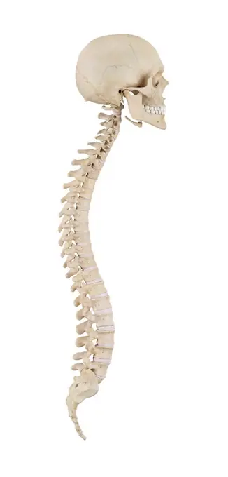
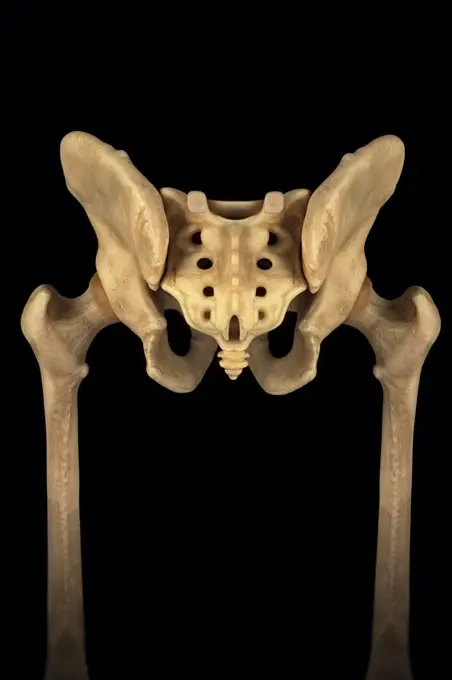
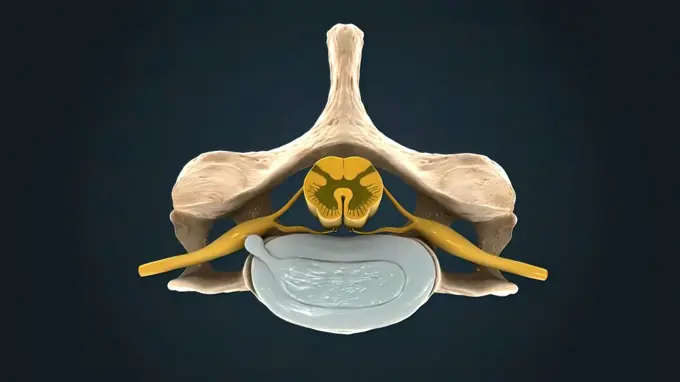
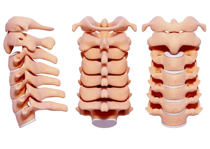
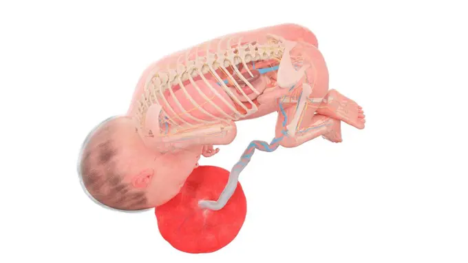
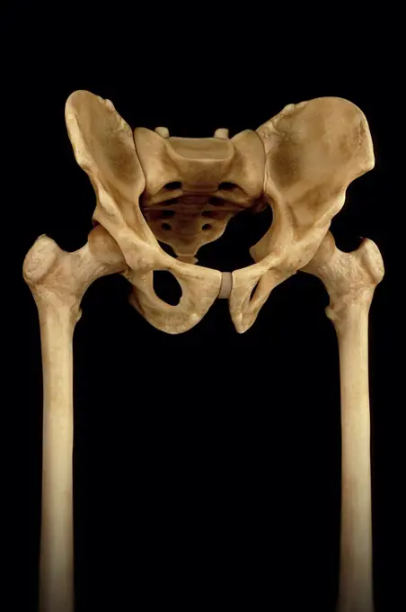
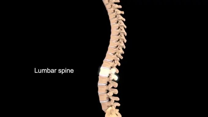
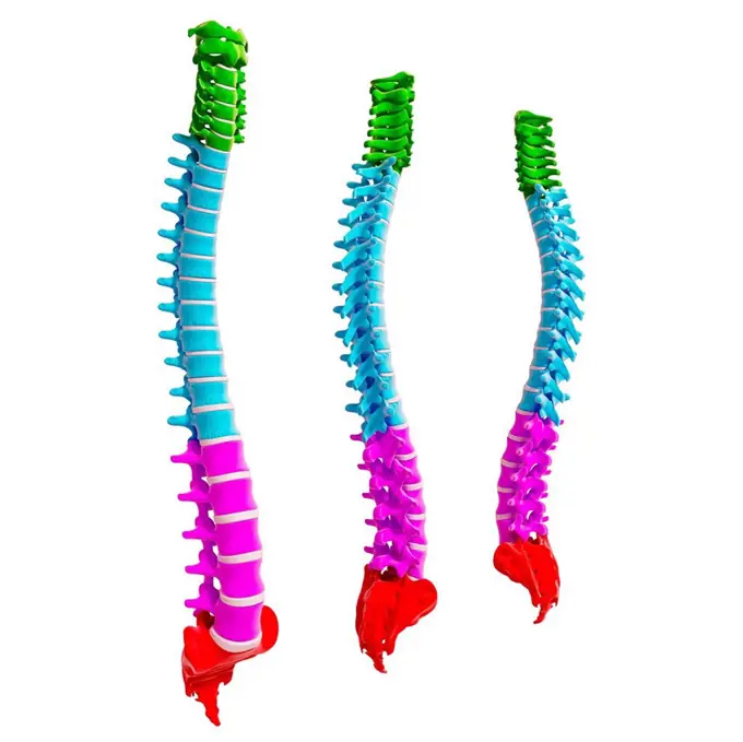
My Boards
View Pictures
  • My Boards
← Science

Human Pelvic Anatomy Illustrations

Detailed computer artwork showcasing human pelvic bones, including views of the spine, abdominal muscles, and associated structures in a scientific style.

Human muscles (psoas major), illustration.
242 assets in this story
824-63195642
Frontal and rear view illustration of the spinal column.
4128R-12580826
Human pelvis bones, computer illustration.
4128R-12577458
Human spine, computer illustration.
4239R-8217
Three dimensional view of female urinary system, close-up.
1525-24324630
Human spine
4128R-11472495
Human hip bone, computer artwork.
4128R-14637864
Human spine, illustration
4378-3093
Rear view of the male pelvis, sacrum and hip joints.
1525-23843042
Human Spine In Details: Vertebra, Bone Marrow, Disc And Nerves 3d illustration. Human spine in details: Vertebra, bone marrow, disc and nerves
1899-54027776
Illustration of spinal stenosis in L4 and L5. Spinal stenosis is a pathology located in the lumbar spinal column where the cauda equina nerves pass. It is caused by an intrinsically narrow vertebral canal or a degenerative narrow canal related to a herniated disc, an increase in the volume of rear joints and ligaments that join the vertebrae together, or an instability in the vertebrae with vertebral displacement (or degenerative spondylolisthesis), which decreases the diameter of the canal, compressing the nerves, causing lumbar pain in the lower limbs.
4128-19250082
Pig anatomy, illustration.
1899-54027777
Illustration of spinal stenosis in L4 and L5. Spinal stenosis is a pathology located in the lumbar spinal column where the cauda equina nerves pass. It is caused by an intrinsically narrow vertebral canal or a degenerative narrow canal related to a herniated disc, an increase in the volume of rear joints and ligaments that join the vertebrae together, or an instability in the vertebrae with vertebral displacement (or degenerative spondylolisthesis), which decreases the diameter of the canal, compressing the nerves, causing lumbar pain in the lower limbs.
4128R-33641
Human spine, computer artwork.
4378-5682
Thoracic Spinal Bones
4128-19247403
Human foetus anatomy at week 27, illustration.
4378-3898
Three-quarter view of the male pelvis, sacrum and hip joints.
824-63195644
Frontal and rear view illustration of the spinal column.
5507-30999224
Closeup of human pelvis bone of an anatomical human body skeleton
1525-23135370
science anatomy of human body in x-ray with glow back bones
1525-26275217
Your lumbar spine is the lower back region of your spinal column or backbone. It consists of five bones 3D illustration. Lumbar Spine Anatomy .Pain in the Lumbar Spine
4128R-11544298
Chest muscles of a weightlifter, computer illustration.
4128-28968806
Anatomy of the pelvis bones, illustration
6188-58112258
science anatomy of human body in x-ray with glow back bones science anatomy of human body in x-ray with glow back bones Copyright: xZoonar.com/StanislavxRishnyakx 9690217
4128R-31867
Human spine, computer artwork.
4128-30415783
Sacrum, illustration
5507-51798002
Human spine with vertebrae, simple anatomy spinal silhouette
1899-54027775
Illustration of the spinal chord, in the vertebral canal, protected by the spinal column.
4128-19053236
Cervical vertebrae, illustration
4128R-11472488
Human hip bone, computer artwork.
1899-53513184
Spinal column, drawing.
4128-30419379
Prostate, illustration
4409-17348489
Pelvic bones.
4128-30417826
Fish anatomy, illustration
4128-28682480
Spinal cord anatomy, illustration
1848-77418079
3D Illustration Concept of Human Skeleton System Rib Cage Described with Labels Anatomy
4409-28658642
Anatomy of the human body, written by Henry Gray, illustrated by Henry Vandyke Carter. Fig. 111 lateral view of the spine.
6188-55611179
The location of the thyroid gland The location of the Copyright thyroid: xzoonar.com/curryx 2447884
1848-77405021
3D Illustration Concept of Human Skeleton System Upper Limbs Described with Labels Anatomy
1848-77392602
3D Illustration Concept of Human Skeleton System Upper Limbs Described with Labels Anatomy
1525-24402732
Human bones sketch. Hand drawn anatomy joints or body skeleton parts. Spine with vertebrae and femur. Isolated engraving skull. Scapula and thorax with ribs. Vector orthopedic medical skeletal set. Human bones sketch. Hand drawn anatomy joints or skeleton parts. Spine with vertebrae and femur. Isolated engraving skull. Scapula and thorax. Vector orthopedic medical skeletal set
4128-20042441
Quadriceps muscle anatomy, illustration
1848-77391545
3D Illustration Concept of Vertebral Column of Human Skeleton System Described with Labels Anatomy
PREVIOUS
of 3
NEXT
2401 S. Ervay, Suite 206
Dallas, Texas 75215
United States
Get Started
Free ResearchMy BoardsMy Cart
For Creators
How To License Your ContentContributor PortalFrame of Mind
Resources
API accessPricing
Contact
+1 866 236 0087help@viewpictures.co.uk Contact form
©2026 View Pictures. All Rights Reserved. -B
This site is protected by reCAPTCHA and Google Privacy Policy and Terms of Service apply.