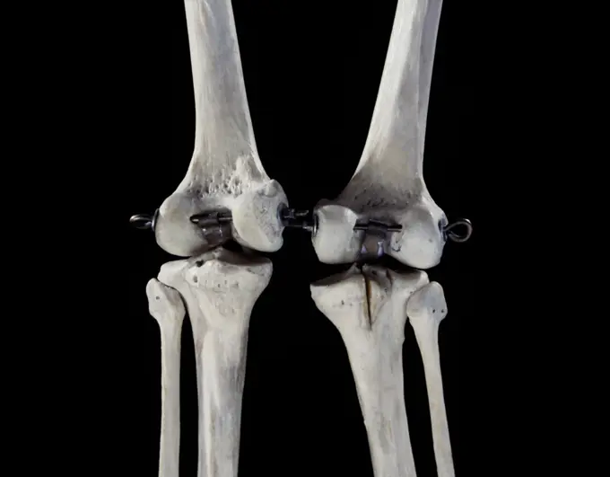
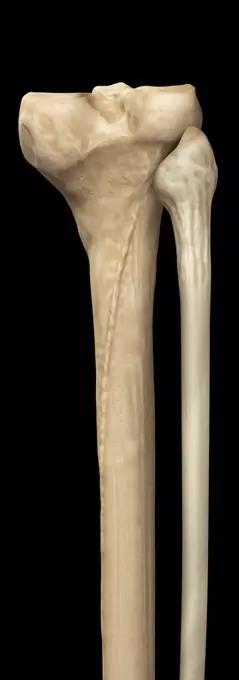
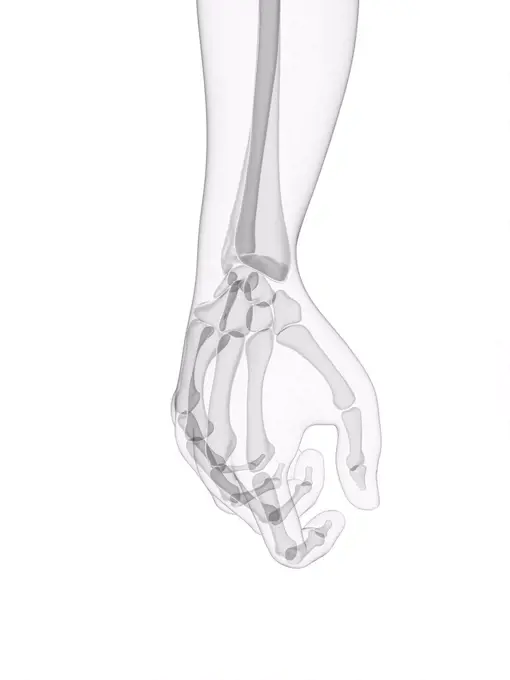
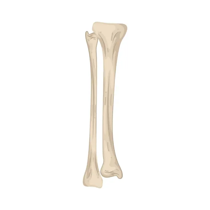
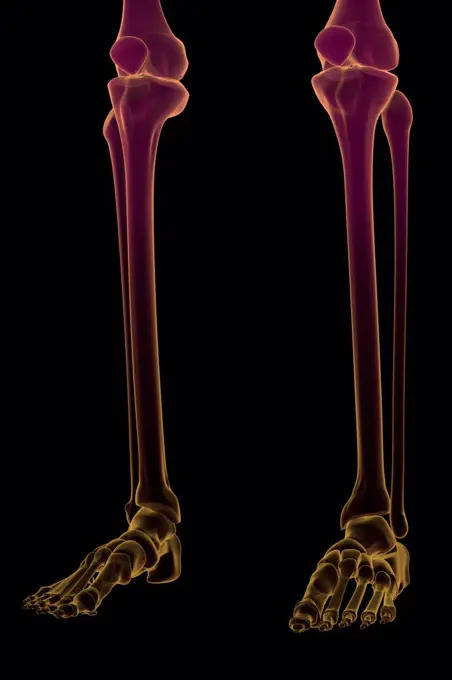
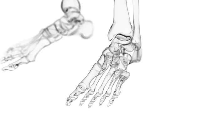
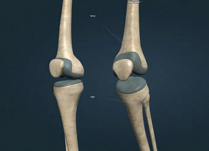
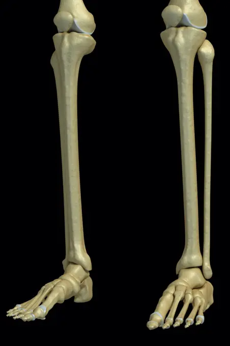
My Boards
View Pictures
  • My Boards

Human Skeletal and Muscle Illustrations

Detailed computer illustrations of human bones and muscles in various body parts, highlighting anatomical structures.

Human knee ligaments, computer artwork.
142 assets in this story
4128R-11544880
Human hand anatomy, computer illustration.
4220-21334621
Human Skeleton.     Date:
4128R-11542558
Human foot bones, computer illustration
4128R-11477828
Human hand bones, computer artwork.
4378-2891
Model of the tibia and fibula bones that form part of the human leg.
4128-19247937
Skeletal ankle, illustration.
4128R-30122
Hand bones, computer artwork.
4128R-12964190
Human foot bones, Illustration
4128R-36554
Human leg bones, computer artwork.
4128R-11298415
Leg anatomy, computer artwork.
4128-V58575026
4239-18642011
Rheumatoid arthritis in hip joint. Detail showing inflammation of the capsule and tissues.
1525-71581536
human bone
4378-275
Three-quarter view of stylized bones of the lower legs, ankle joints and feet.
4128-19056462
Human ankle joint, illustration
4128R-15515346
Illustration of the skeletal knee
4128R-12576963
Human foot anatomy, computer illustration.
4128R-33131
Human skeleton, computer artwork.
1525-56190041
It is the weight-bearing component of the knee joint. 3d illustration. A hinge joint is a type of synovial joint that exists in the body and serves to allow motion primarily in one plane.
4128R-11323751
Human tensor fascia lata muscle, computer artwork.
4128R-36050
Human leg bones, computer artwork.
4239-69152005
3D model of the foot depicting the plantar superficial muscles and bone structures.
4128-V58567612
4128R-12665
Normal lower leg. X_ray of the lower leg of a 24 year old woman.
4378-445
Three-quarter view of the bones of the lower legs, ankle joints and feet.
4409-17349059
Lower extremities.
1525-75962416
Human Skeleton Hand (Bones). Vector Illustration.
1525-56184105
The foot is the terminal portion of the lower limb, whose primary function is to bear weight and facilitate locomotion 3D illustration. The foot is conventionally divided into three zones. hindfoot, midfoot, and forefoot
1899-66328
X-ray image of female scapula and upper humerous
1848-54506286
Skeleton of a human arm, anatomical illustration
4128R-15196
Normal wrist, X_ray.
4128R-11323197
Human ilium and sacrum bones, computer artwork.
4128R-11311142
Human arm bone (radius), computer artwork.
4128-48285504
Healthy hand, X-ray
4378-2850
Model showing the human knee joint and its connecting bones.
4128R-12576953
Human foot anatomy, computer illustration.
4128R-11310874
Human knee joint, computer artwork.
4128-V58567336
1788-39679
Medicine - Anatomy - Musculoskeletal (locomotor) system - Skeleton - Diaphysis of long bones. Drawing.
6145-29270362
The bones of a hand, with a ring on one finger, viewed through x-ray. Photoprint from radiograph after Sir Arthur Schuster, 1896.
4048-6000
Konrad Roentgen's X-ray of the hand of Professor Albert von Kolliker, showing bones and the ring he was wearing. The x-ray was made at a public lecture to demonstrate the new discovery.  Published in PHYSIKALISCH-MEDICINISCHE GESELLSCHAFT, Wurzburg, 1895.
1525-27726550
Arm parts poster with explanation in beautiful font, human body piece made of bones, humerus and ulna radius vector illustration isolated on blue. Arm Parts Poster Explanation Vector Illustration
PREVIOUS
of 2
NEXT
2401 S. Ervay, Suite 206
Dallas, Texas 75215
United States
Get Started
Free ResearchMy BoardsMy Cart
For Creators
How To License Your ContentContributor PortalFrame of Mind
Resources
API accessPricing
Contact
+1 866 236 0087help@viewpictures.co.uk Contact form
©2026 View Pictures. All Rights Reserved. -B
This site is protected by reCAPTCHA and Google Privacy Policy and Terms of Service apply.