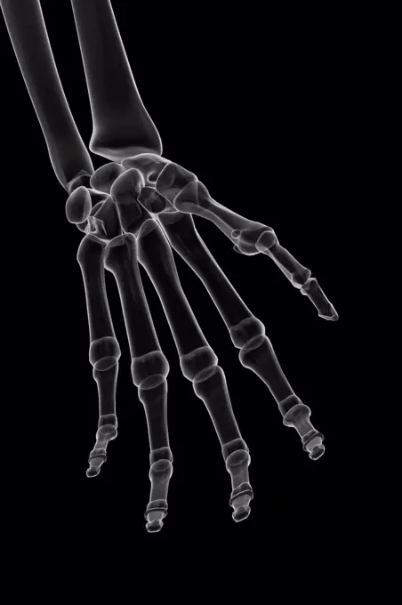
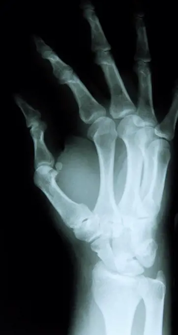
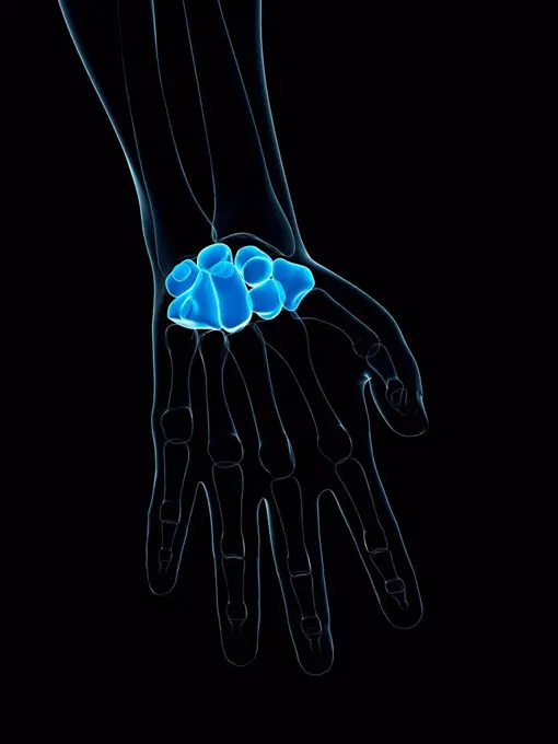
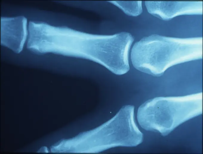
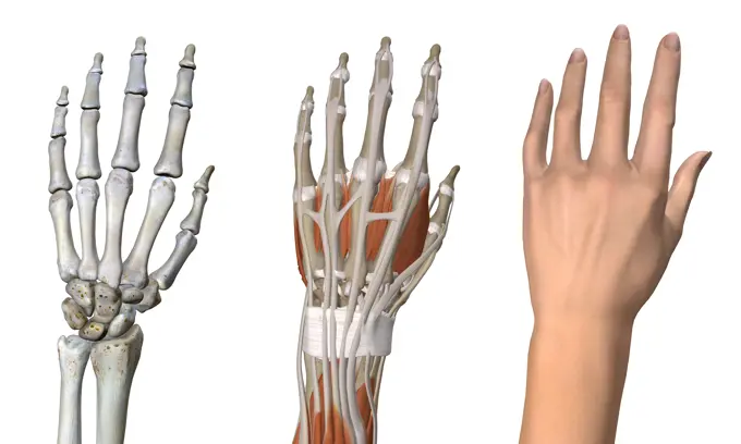
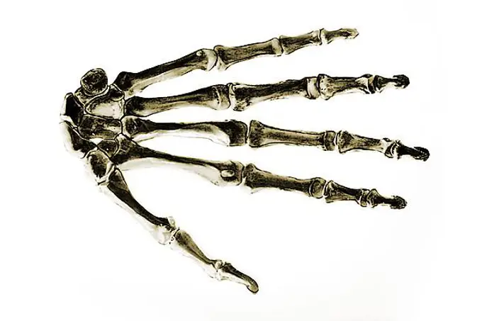
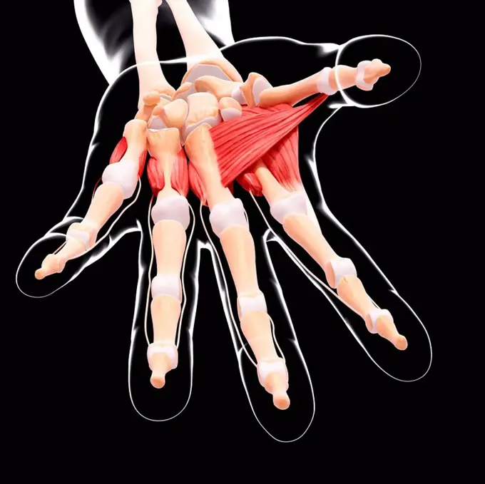
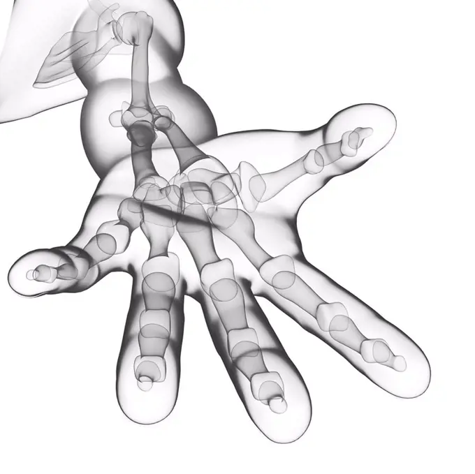
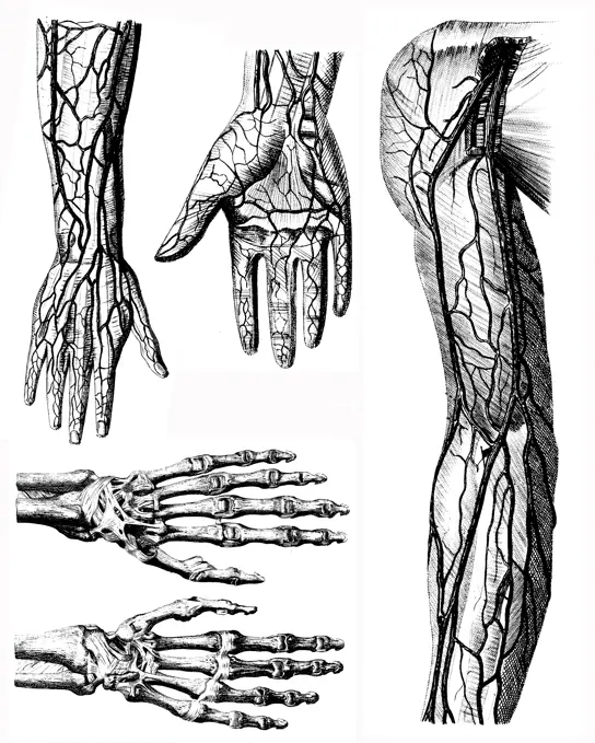
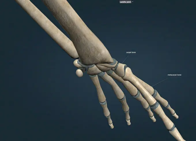
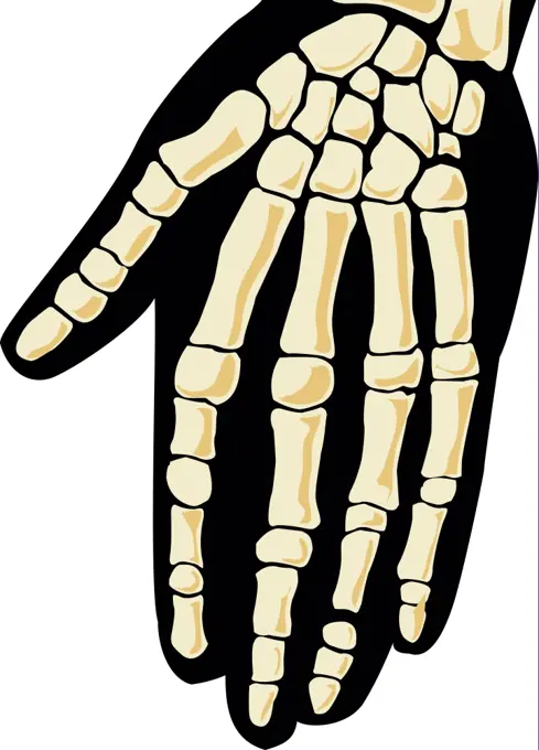
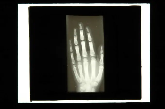
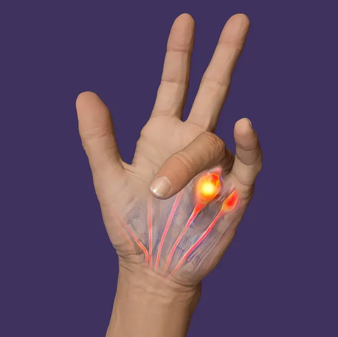
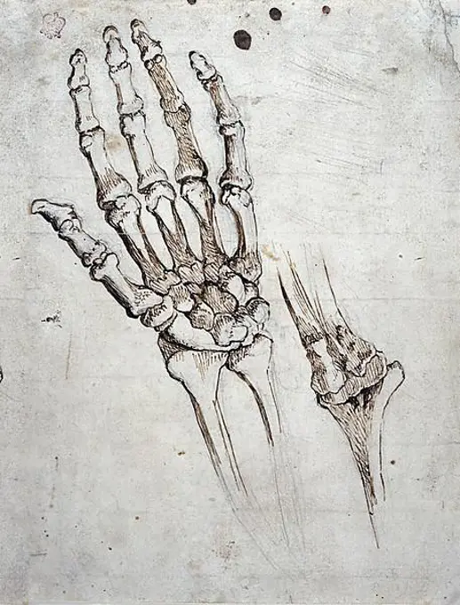
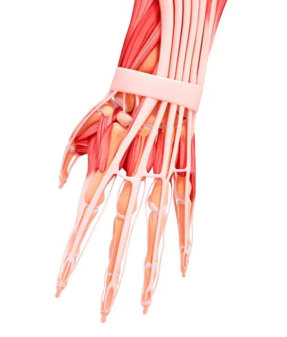
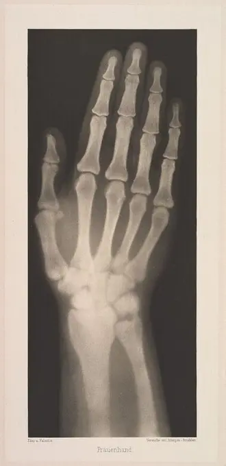
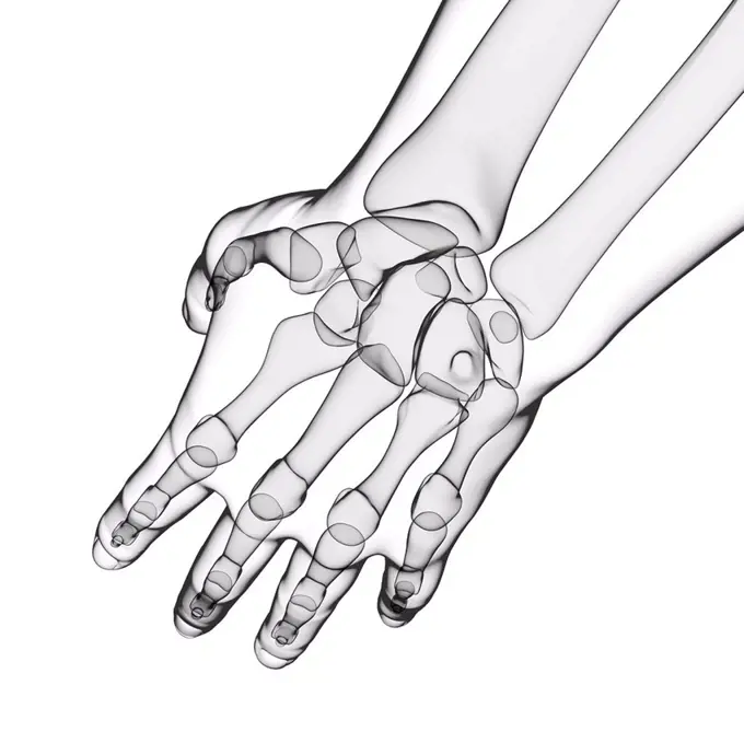
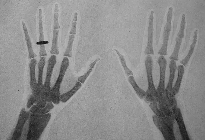
My Boards
View Pictures
  • My Boards

Human Skeletal System Art

Stylized images of human hand and foot bones, presented in contrasting colors against black, demonstrating skeletal anatomy and structure.

X-ray stylized bones of the left hand and wrist.
158 assets in this story
4128-30418395
Nerves of the hand, illustration
4128R-11541441
Human skeleton, computer illustration.
1899-66331
X-ray image of female hand
4128R-26230
Hand bones. Computer artwork showing the carpal bones.
4128R-13572706
Illustration of the talus bone.
4128-V58567350
6188-55645553
X -ray image of fingerbones with joints X -ray image of finger bones with joints
1428-67034146
Three Anatomical Dorsal Views of Female Hand:  1) skeletal, 2) tendons and muscles, 3) skin
6145-52738489
The X-ray shows the left hand of a man with a ball in the wrist. The image is printed on glossy bromide paper in a box format from 1896.
1848-54506305
Bones of the human hand, anatomical illustration
5507-32175081
Front view of female body sketch with minimalistic lines. Vector illustration
4128R-31876
Human hand musculature, computer artwork.
4128R-31656
Human hand bones, computer artwork.
4128R-11324338
Human foot bones, computer artwork.
6243-72452453
Medical - Victorian Anatomical Illustrations - isolated on a white background for cut out.
4269-24797
Rheumatoid polyarthritis. X-ray of hand with Rheumatoid polyarthritis. Rheumatoid arthritis is a chronic joint disease that can cause cartilage destruction, bone erosions, and tendon inflammation and rupture. It can lead to polyarthritis which happens when inflammation spread to multiple joints by.
1899-54027173
Rheumatoid arthritis seen on an x-ray of the hand.
4128R-11542216
Human foot anatomy, computer illustration.
4239-18641140
Posterior view of left hand, with labels.
4128R-15460
Normal foot. Coloured 3D computed tomography CT scan of the healthy foot of a 23 year old patient.
4128-30420418
Foot bones, illustration
1525-56211365
A saddle joint is a type of synovial joint in which the opposing surfaces are reciprocally concave and convex. 3d illustration. A saddle joint is a type of synovial joint in which the opposing surfaces are reciprocally concave and convex.
4128R-31684
Human hand musculature, computer artwork.
1525-25137390
Human anatomy. Hand. Vector illustration. Human skeleton. Hand
6145-52890213
X-ray glass diapositive by Josef Maria Eder from 25 January 1896 showing the hand of an 8-year-old girl with traces of rachitis for medical research.
4128R-11477630
Human hand passing a relay baton, computer artwork.
4128-30422438
Dupuytren's contracture, illustration
1848-49464542
Medicine, Anatomy, Bones of the human hand and wrist and elbow joint, Michelangelo Buonarroti, Historical, digitally restored reproduction from a 19th century original
4128R-11545004
Human hand muscles, computer illustration.
4141-8021
fossil turtle (toxochelys sp.) flipper late cretaceous 83 million years old kansas
4128R-11476924
Human leg and foot bones, computer artwork.
1788-39718
Medicine: Human anatomy, leg and hand muscles, synovial membrane, or synovium. Drawing
4128R-34640
Human hand musculature, computer artwork.
4409-17390225
Frauenhand. Artist: Josef Maria Eder (Austrian, Krems an der Donau, 1855-1944 Kitzbühel); Eduard Valenta (Austrian, 1857-1937). Dimensions: Image: 10 3/16 × 4 1/4 in. (25.9 × 10.8 cm)Plate: 11 7/16 × 5 5/16 in. (29 × 13.5 cm)Sheet: 19 5/16 in. × 14 in. (49 × 35.5 cm). Date: 1896.Eder was the director of an institute for graphic processes and the author of an early history of photography. With the photochemist Valenta, he produced a portfolio in January 1896, less than a month after Wilhelm Conrad Röntgen published his discovery of X-rays. Eder and Valenta's volume, from which this plate derives, demonstrated the X-ray's magical ability to reveal the hidden structure of living things. Human hands and feet, fish, frogs, a snake, a chameleon, a lizard, a rat, and a newborn rabbit are all presented in exquisitely printed photo-gravures, as are carved cameos and an assortment of natural materials. In an era when photography's ability to accurately depict the visible world had become comm
824-63184400
Three-dimensional reconstruction of the right foot, from a series of cross-section scans.
4128R-35513
Human hand bones, computer artwork.
4128-30421510
Human hand, illustration
4128-16224441
Nutcrackers, X-ray
1899-53513989
one of the first X-ray images of a human hand with ring, ca 1900 / eines der ersten Röntgenbilder einer menschlichen Hand mit Ring, ca 1900, Historisch, historical, digital improved reproduction of an original from the 19th century / digitale Reproduktion einer Originalvorlage aus dem 19. Jahrhundert.
4128-20045426
Calcaneus bone, illustration
4128-30422441
Dupuytren's contracture, illustration
4128R-14323953
Skin bacteria, illustration
4128R-12580454
Human hand bones, computer illustration.
6145-29247229
The bones of a hand, with the tip of the index finger missing, viewed through x-ray. Photoprint from radiograph after Sir Arthur Schuster, 1896.
4128-30415790
Skeletal foot, illustration
6188-66486438
3D-Illustration von gebrochenen oder defekten weiß gefärbten X- und Y-Chromosomen 3D Illustration of broken or defected white colored x and y chromosomes Copyright: xZoonar.com/ChristophxBurgstedtx 22603187
1746-21106156
The hand of Mrs. Wilhelm Roentgen: the first X-ray image, 1895
5514-58777417
X-ray of a human hand.
1570R-21137239
X-ray image of human hand
5507-31058092
Science and educational process concept.
1746-57661735
X-ray of human hand
4409-17432466
Fuss eines 17 jährigen Jünglings. Artist: Josef Maria Eder (Austrian, Krems an der Donau, 1855-1944 Kitzbühel); Eduard Valenta (Austrian, 1857-1937). Dimensions: Image: 8 1/4 in. × 7 in. (21 × 17.8 cm)Plate: 11 1/4 × 8 1/16 in. (28.5 × 20.4 cm)Sheet: 19 5/8 × 13 3/4 in. (49.9 × 35 cm). Date: 1896.Eder was the director of an institute for graphic processes and the author of an early history of photography. With the photochemist Valenta, he produced a portfolio in January 1896, less than a month after Wilhelm Conrad Röntgen published his discovery of X-rays. Eder and Valenta's volume, from which this plate derives, demonstrated the X-ray's magical ability to reveal the hidden structure of living things. Human hands and feet, fish, frogs, a snake, a chameleon, a lizard, a rat, and a newborn rabbit are all presented in exquisitely printed photo-gravures, as are carved cameos and an assortment of natural materials. In an era when photography's ability to accurately depict the visible world
1525-22348620
Close-up of a human hand
4128-20045500
Bunion, illustration
1746-21105800
anatomical illustration from 'Antropologium de hominis dignitate, natura et proprietatibus, de elementis, partibus et membris humani corporis' (Leipzig: 1501) by Magnus Hundt
6176-67069455
X-ray photograph of a hand, 1926. Artist: Unknown
1848-54762595
Close-up of a human foot from above, focussing on the toes and skin texture
1746-57661734
X-ray of human foot
PREVIOUS
of 2
NEXT
2401 S. Ervay, Suite 206
Dallas, Texas 75215
United States
Get Started
Free ResearchMy BoardsMy Cart
For Creators
How To License Your ContentContributor PortalFrame of Mind
Resources
API accessPricing
Contact
+1 866 236 0087help@viewpictures.co.uk Contact form
©2026 View Pictures. All Rights Reserved. -B
This site is protected by reCAPTCHA and Google Privacy Policy and Terms of Service apply.