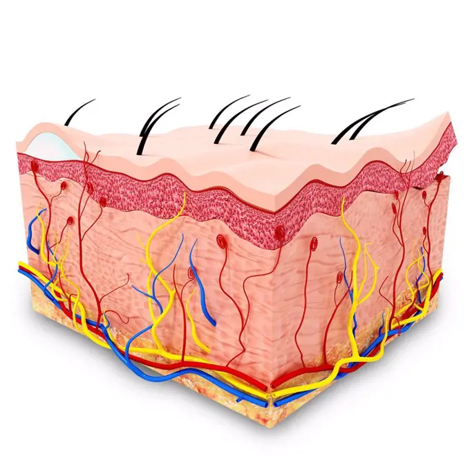
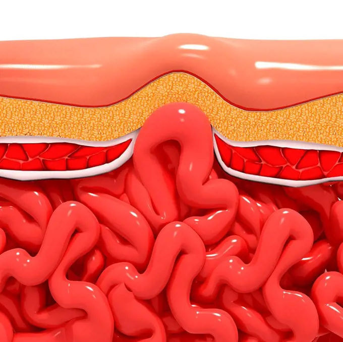
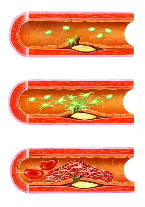
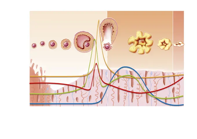
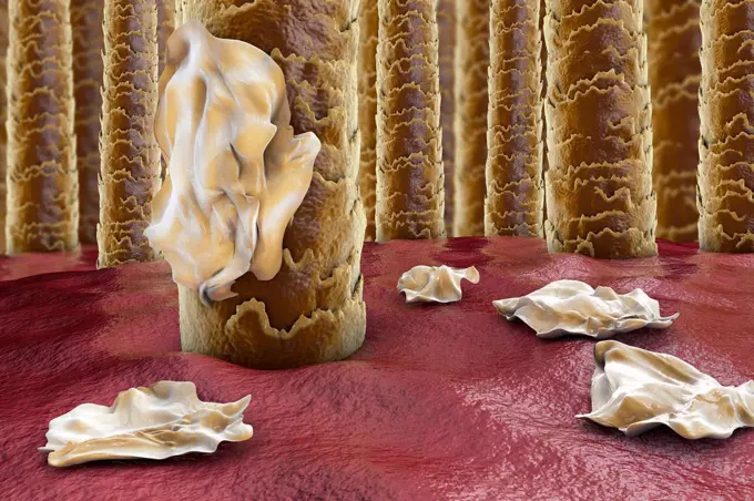
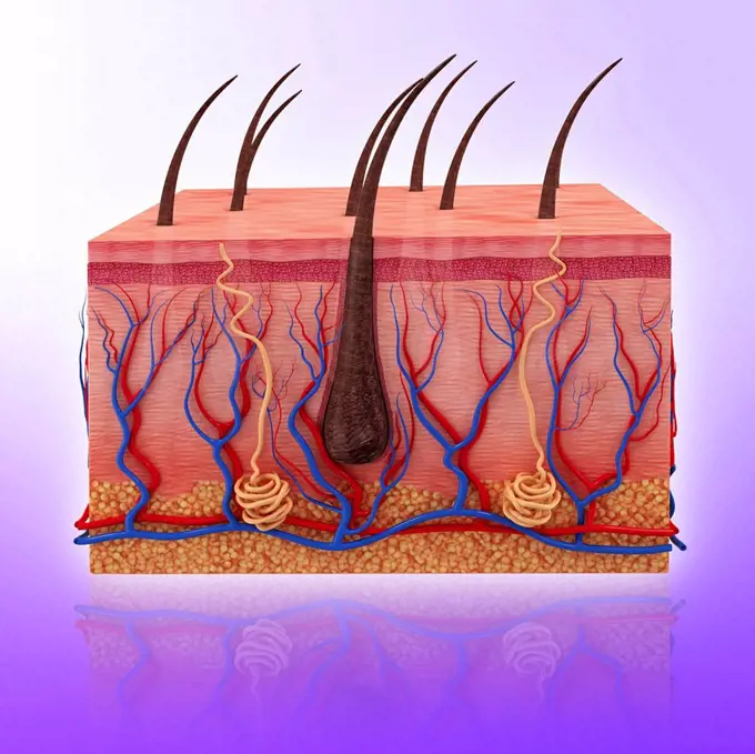
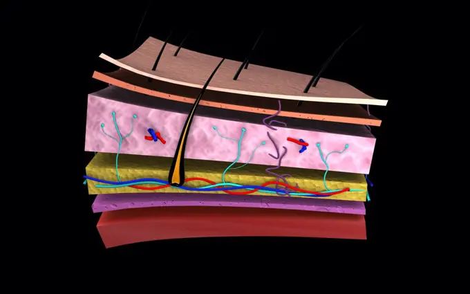
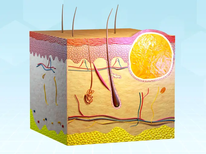
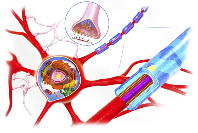
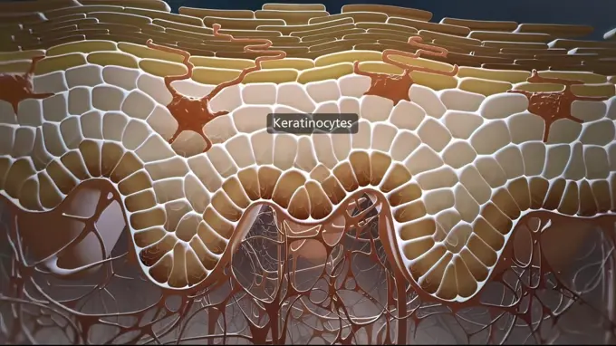
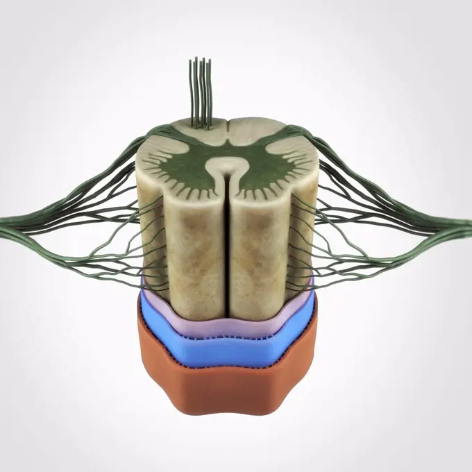
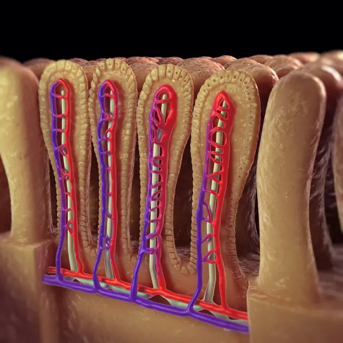
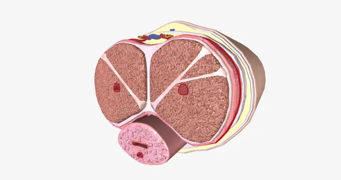
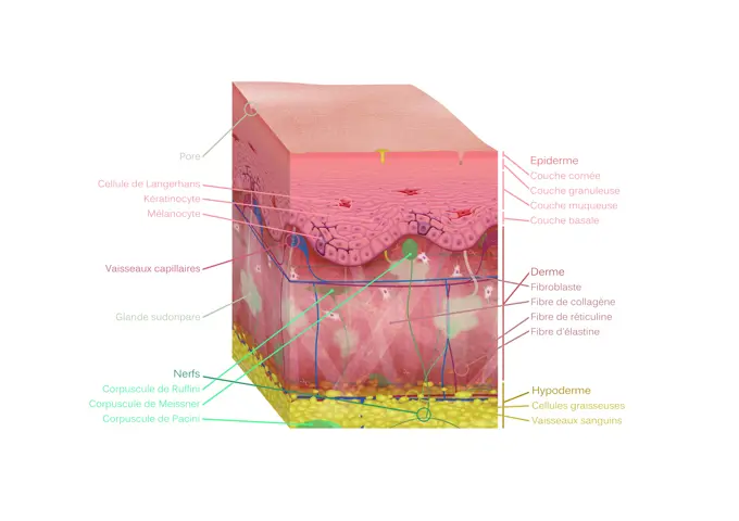
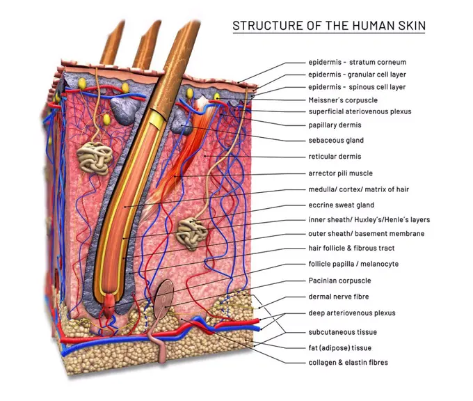
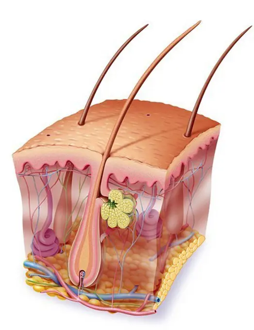
My Boards
View Pictures
  • My Boards

Human Skin Anatomy

Illustrations depicting the complex layers of human skin, including structures like hair follicles, blood vessels, and glands.

Human skin anatomy, computer artwork.
190 assets in this story
824-63183447
Illustration of the ciliated cells of the sinus. These cells drain mucus that traps external particles.
4128R-11291005
Ventral hernia, computer artwork.
1899-53513246
Atheroma, drawing.
4128R-11315745
Human skin, computer artwork.
824-63214089
Illustration of the menstrual cycle over 28 days without fertilization after ovulation. From top to bottom: -Illustration of the ovarian cycle. -Graph of pituitary and ovarian hormone dosages: LH (yellow line), FSH (red line), estrogen (green line), progesterone (blue line). -Illustration of the endometrium.
824-63167669
Atheroma, drawing.
5507-43617617
Fatigue factors accumulate . Flat illustration of muscle and vessel.
1525-56722646
A first degree burn is the most common, and one of the most common household injuries. 3D rendering. A first degree burn is the most common, and one of the most common household injuries.
4128R-15366479
Human scalp with dandruff, illustration
4128R-11312421
Human skin, computer artwork.
1525-56149169
intestinal villi (singular: villus) are small, finger-like projections that extend into the lumen of the small intestine 3d illustration. intestinal villi (singular: villus) are small, finger-like projections that extend into the lumen of the small intestine
1525-26305308
Nicotine raises blood pressure 3d illustration. Nicotine raises blood pressure
824-63208855
Illustration of hair cells, hearing cells. With the movement of liquid in which the hair cells (pink) are immersed in the cochlea channel, the hairs (white) move and send an electric signal to the auditory nerve (yellow), which carries the message to the brain.
4128R-11291712
Human skin anatomy, computer artwork.
4128-19490388
Celiac disease inflammation, illustration
6188-65537708
3d-Illustration einer Stent-Implantation zur Unterstützung der Blutzirkulation in den Blutgefäßen 3d illustration of stent implantation for supporting blood circulation into blood vessels Copyright: xZoonar.com/ChristophxBurgstedtx 22561029
4128R-11312416
Human skin, computer artwork.
4239R-20483408
Conceptual image of the layers of human skin.
4128R-11314493
Human artery, cut-away computer artwork.
5507-37480020
Transdermal drugs delivery system
6188-67152171
Talgdrüsenzyste in der menschlichen Haut Cross section of human skin showing a sebaceous cyst. Digital illustration, 3D render. Copyright: xZoonar.com/AndreaxDantix 22828897
6188-65540333
Querschnitt eines Neurons, Funktion und Zellaufbau - 3d Illustration Cross section of a neuron, function and cell-building - 3d illustration Copyright: xZoonar.com/ChristophxBurgstedtx 22579330
824-63164296
Comparison between a healthy ankle and hemophilic arthropathy (hemarthrosis).
1899-53509319
Section of skin with hair and all types of sensory receptors.
1525-26182248
In the layer adjacent to the dermis, the keratinocytes are divided and thrown into the upper layers. 3D illustration. In the layer adjacent to the dermis, the keratinocytes are divided and thrown into the upper layers.
824-72488738
Section of the skin, showing the epidermis, the dermis (and the basal layer of melanocytes) and the subcutaneous tissue (hypodermis).
1525-28295596
Human skin. Layered epidermis with hair follicle, sweat and sebaceous glands. Healthy skin anatomy medical vector illustration. Dermis and epidermis skin, hypodermis. Human skin. Layered epidermis with hair follicle, sweat and sebaceous glands. Healthy skin anatomy medical vector illustration
4128R-12923140
Small intestine, illustration
1525-56854756
The Penis Transverse Cross Section 3D rendering. The Penis Transverse Cross Section
824-63164020
From muscle to muscle fiber: tendon, muscle, muscle fiber.
1525-26182250
In the layer adjacent to the dermis, the keratinocytes are divided and thrown into the upper layers. 3D illustration. In the layer adjacent to the dermis, the keratinocytes are divided and thrown into the upper layers.
824-63180621
Illustration of the nasal mucous membrane showing the epithelial cells (dark pink), the chorion (lighter pink), glands (purple) that secrete the mucus (yellow) and numerous blood vessels.
1899-61461900
The menstrual cycle and the periods of fertility. Top: genitalia during fertilization. Bottom: ovarian cycle, hormonal cycle, uterine cycle, temperature curve
1899-53513168
Cross-section of a blood vessel, with platelets in purple.
5507-43876962
Absorption of nutrients in the small intestine
1525-56184323
Anatomy of the skin, showing the epidermis, dermis, and subcutaneous tissue. 3D illustration. Anatomy of the skin, showing the epidermis, dermis, and subcutaneous tissue.
1899-54027780
Illustration of the provenance of cholesterol, where it is stored and its elimination. One third of cholesterol is provided by food and the remaining two thirds by the liver. It is transported through the bloodstream by two types of protein: -LDL which carries it to the tissue but which, when too great in quantity for the blood vessels, blocks them up forming an atheromatous plaque which can burst and form a thrombus (clot). This is known as bad cholesterol. -HDH, which carries the cholesterol to the liver where it will be recycled and eliminated. This is known as good cholesterol.
4409-17348785
Digestive system of a ruminant.
1899-61461915
Anatomy of the tooth: from top to bottom - enamel in white - dentin in orange - pulp in dark orange containing the vessels and nerves - two roots surrounded by the cementum in gray, - then surrounded by the alveolo-dental ligament in pink clear - which joins the gum between each tooth at the top - the roots are enclosed tooth the alveolar bone in yellow
4128-V58614916
824-63220052
Infectious conjunctivitis and its treatments. View of an eye with inflammation of the conjunctiva. Zoom on the conjunctiva with from top to bottom 1 bacterial infection (green) and 1 viral infection (red-orange). In yellow, purulent discharge due to the presence of bacteria (green), the cells of the conjunctiva are destroyed. Goblet cells secreting mucus and edema of the conjunctiva in the area infected with the virus. Right from top to bottom, a bacteria seen in section, a window with an infected connective cell (Herpes Simplex virus) detailing the process of viral replication transcription of viral DNA, DNA polymerase, viral replication
4128-28575239
Lymphatic vessel, conceptual illustration
4378-3565
A section of the spinal cord isolated on the a white background. Note the connecting dorsal and ventral root filaments.
4128-19249267
Atherosclerosis, illustration.
4378-3145
Sectional view of intestinal villi revealing the network of blood vessels involved in the transport of absorbed nutrients to the rest of the body.
1525-28493354
Shows the moisture absorbed into the skin, cosmetic type, lotion, cream, serum. Vector Realistic file.
824-63164516
Dark phototype skin in which black melanin is important.
1525-56854774
The Penis Transverse Cross Section 3D rendering. The Penis Transverse Cross Section
824-63207112
3D cross-section illustration of skin : epidermis, dermis and hypodermis. With Langheran cells, keratinocytes and melanocytes, capillaries, pores, sweat glands, Meissner, Ruffinian and Pacinian corpuscles, fibroblasts, reticulin, collagen and elastin.
1525-28093687
cholesterol plaque in artery vessels vector illustration
4239-18641683
Cross-section of artery showing angioplasty and stent deployment.
1525-56854714
The Penis Transverse Cross Section 3D rendering. The Penis Transverse Cross Section
4128-19249219
Mesenteric ischaemic, illustration.
824-63164268
Tooth: formation and development of dental caries.
824-63167594
Epidermis, dermis, hypodermis, nerve endings, sebaceous glands and hair bulbs.
824-63164390
Section of skin with the layers of the epidermis.
824-63165117
Different walls constituting an artery: collagen and elastic tissues, smooth muscle.
1899-53509380
Structure of an eye with zoom on the retina and fovea.
824-63164843
Structure of an eye with zoom on the retina and fovea.
1899-53509447
Different walls constituting an artery, collagen and elastic tissues, smooth muscle.
4128-19490869
Nasal epithelium, illustration
824-63220053
Infectious conjunctivitis and its treatments. View of an eye with inflammation of the conjunctiva. Zoom on the conjunctiva with from top to bottom 1 bacterial infection (green) and 1 viral infection (red-orange). In yellow, purulent discharge due to the presence of bacteria (green), the cells of the conjunctiva are destroyed. Goblet cells secreting mucus and edema of the conjunctiva in the area infected with the virus. Right from top to bottom, a bacteria seen in section, a window with an infected connective cell (Herpes Simplex virus) detailing the process of viral replication transcription of viral DNA, DNA polymerase, viral replication
1899-53513171
Epidermis, dermis, hypodermis, nerve endings, sebaceous glands and hair bulbs.
824-57656234
Path of sound from the ear to the brain. Anatomy of the ear, from left to right: - Outer ear and external auditory canal - Middle ear, eardrum (light purple), ossicles (beige), hammer, incus, stirrup, Eustachian tube (burgundy) - Inner ear, cochlea, vestibules and semicircular canals (purple) - Auditory nerve (yellow) With Zoom of the organ of hearing: - Cochlear canal (pink) containing the organ of Corti, including the hair cells (orange) - The cochlear nerve (yellow) transmits the electrical signal to the auditory area of the brain.
4239-18641679
Comparison of a healthy artery and an unhealthy artery with plaque build-up inside.
4128R-11291716
Human skin anatomy, computer artwork.
4239-18641769
Cutaway view of a tooth cavity.
4128-18573527
Hair loss, conceptual illustration
4128R-25487
Human skin. Computer artwork of a cross-section through human skin.
4128-19249287
Atherosclerosis, illustration.
4128-111494424
Cross-section of human skin with hair follicle, illustration.
4128R-11291383
Human skin anatomy, computer artwork.
5507-43624582
Type of obesity illustration (Japanese) . Abdominal sectional View (visceral fat )
5507-31758421
Causes of aging odors vector illustration
1525-56190177
Anatomy of the tooth and gums and the supporting structures surrounding the tooth 3D illustration. Anatomy of the tooth and gums and the supporting structures surrounding the tooth.
1899-61461937
Bone cut. Representation in 3/4 view of the structure of the bone with zoom showing the different cells that make up the bone tissue
1899-53509031
White skin where the melanocyte secretes little melanin.
6188-67191776
3D Isometrische flache konzeptionelle Illustration einer Talgzyste, medizinisches Diagrammbr3D-Isometrische flache konzeptionelle Illustration einer Talgzyste, medizinisches Diagramm 3D Isometric Flat Conceptual Illustration of Sebaceous Cyst, Medical Diagram 3D Isometric Flat Conceptual Illustration of Sebaceous Cyst, Medical Diagram Copyright: xZoonar.com/AnastasiiaxTorianykx 22904992
824-63164747
Skin section showing all the structures: the layers of the epidermis, the structure of the dermis.
5507-39240854
Acne Vulgaris
5507-37928270
Vaginal Epithelium
824-63164528
Leaf blade with stomata, mesophyll, a vein.
6188-67654323
Infektion mit Zehennagelpilz Diagram showing fungal pathogens in a toenail. Digital illustration. Copyright: xZoonar.com/AndreaxDantix 23003035
4128-20043806
Varicose veinss, illustration
1525-28134358
Human skin layers, healthcare and medical illustration about human skin
5507-46943457
Layer of human skin illustration
4128R-15366534
Simple squamous epithelium, illustration
824-66010
SKIN, ILLUSTRATION. SKIN, ILLUSTRATION Cutaway view of the skin. Representation in cutaway view of the skin anatomy with all its structures, and notably the three layers fropm the top to the bottom : _ epidermis, _ dermis : hair, sebaceous gland in green, next to the hair, sudoriferous gland in purple, erector pili muscle, nerve endings, blood vessels, _ hypodermis or cutaneous tissue.
4128-20043912
Skin pigmentation, illustration
1525-25871960
Nanotechnology based chemical and biological sensors isometric composition with carbon nano tubes detecting elements vector illustration . Nanotechnology isometric composition
PREVIOUS
of 2
NEXT
2401 S. Ervay, Suite 206
Dallas, Texas 75215
United States
Get Started
Free ResearchMy BoardsMy Cart
For Creators
How To License Your ContentContributor PortalFrame of Mind
Resources
API accessPricing
Contact
+1 866 236 0087help@viewpictures.co.uk Contact form
©2026 View Pictures. All Rights Reserved. -B
This site is protected by reCAPTCHA and Google Privacy Policy and Terms of Service apply.